


 8 / 21

Page 71
allied
academies
Journal of Neurology and Neurorehabilitation Research | Volume 3
November 26-27, 2018 | Dubai, UAE
Spine and Spine Disorders
Addiction Research and Therapy
3rd International Conference on
International Conference on
Joint Event
&
Mini Open Spinous Process splitting Laminectomy for Cervical Spondylotic Myelopathy
Hatem Hamdy
1
and
Ahmad Fouad Abdelbaki Allam
2
1
One Day Surgery Hospital, Egypt
2
Minia University, Egypt
Background Data:
Muscle dissection associated with posterior
approaches to the Cervical Spine usually results in local pain,
musclewasting and temporarily restricted neckmovement. Use
ofmusclesparingSpinousProcessSplittingApproachforCervical
Laminectomy allows decompression of the spinal cord and
neural foramen if needed, it does not require instrumentation
and fusion and it preserve Cervical Spine stability.
Purpose:
To assess the effectiveness of Spinous Process
Splitting Approach for Cervical Laminectomy in Cervical
Spondylotic Myelopathy.
Study Design:
Prospective study.
Patient Sample: Fifteen patients with Cervical Spondylotic
Myelopathy; the study included 11 males and 4 females; the
mean age at surgery was 66.4±6.6 (range 44-71) years.
Outcome Measures:
Operative time and blood loss were
recorded. Clinical outcome was assessed by the JOA score
and VAS. MRI was done 6 months postoperative to assess
decompression. Spinal stability and curvature index were
assessed on plain cervical radiographs.
Patients and Methods:
Fifteen patients underwent muscle
Sparing Spinous Process Splitting Cervical Laminectomy.
Results:
No case of wound dehiscence was recorded. There
was significant improvement of JOA scores and Brachialgia
VAS scores at 6 months, the mean JOA recovery rate was
56.2%. No patient had postoperative Kyphosis or instability
and 66.6% of patients had improved modified Ishihara Cervical
Curvature Index. No neurological deterioration was recorded
in the follow-up. No patient had newly developed axial pain.
MRI revealed adequate decompression of the spinal cord.
Conclusion:
The Spinous Process Splitting Laminectomy
allows good spinal cord decompression and preserves Cervical
Spine stability. The mini open approach and preservation of
interspinous ligaments could play a role in wound dehiscence
prevention.
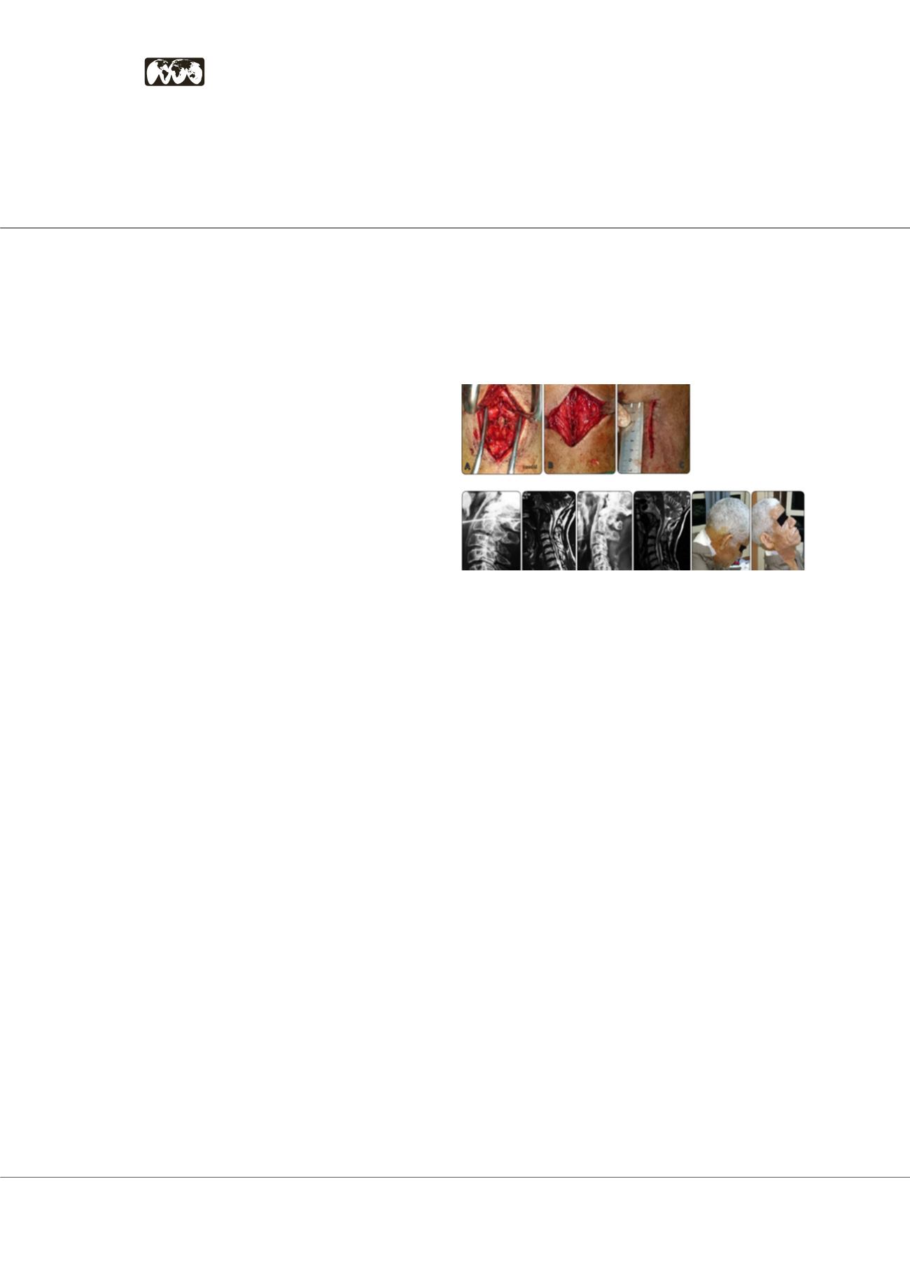
Figures1:Intraoperativephotograph:(A)thearrowshowsthespinousprocesswithattached
musclesbeforearrowshowsalreadysplitspinousprocess.(B)muscles lookscoaptedbefore
closureattheendofthesurgery.(C)thelengthofincisionwasabout6cmtodecompress3levels.
Figures 2: (A) Preoperative lateral radiograph of 68-year-old man shows lordotic cervical
curvature (CCI=34). (B) Preoperative T2-weighted sagittal MRI cervical spine with
multilevel canal stenosis from both posterior and anterior. (C) 6 months postoperative
lateral radiograph shows 4 levels laminectomy with preserved spinous processes (arrow)
and same preoperative CCI. (D) 6 months postoperative T2-weighted sagittal MRI
shows successful decompression of the spinal cord. (E) and (F) clinical photos for the
patient at the first postoperative day with good active flexion and extension movement.
Speaker Biography
Hatem Hamdy has completed his MBBCH from Kasr Elieny Medical School in 1995. He has
acquired hisMaster degree of Orthopedics in 2007. He completed European Spine Diploma
atFrancein2016.HehasdoneFellowshipatNanooriHospitalatKoreain2016.Atpresentheis
OrthopedicandSpineConsultantandHeadofSpineunitatOneDaySurgeryHospital,Egypt.
e:
dr.hatemhamdy00@yahoo.com