Case Report - Current Trends in Cardiology (2021) Volume 5, Issue 5
Unusual cause of chest pain: Congenital absence of pericardium.
Onkar B Auti1, Arul Narayanan2, Pandi Gurusamy Ashok kumar3, Vimal Raj4*
1Department of Radiology, Ruby Hall Clinic, Pune, Maharashtra, India
2Department of Cardiology, Narayana Health City, Bangalore, Karnataka, India
3Department of Pulmonology, St Martha’s Hospital, Bangalore, Karnataka, India
4Department of Radiology, Narayana Health City, Bangalore, Karnataka, India
- Corresponding Author:
- Vimal Raj
Department of Radiology
Narayana Hrudayalaya
Bangalore, India
Tel. +91 9538659316
E-mail: drvimalraj@gmail.com
Accepted date: 15 June, 2021
Citation: Auti OB, Narayanan A, Ashok kumar PG, et al. Unusual cause of chest pain: Congenital absence of pericardium. 2021;5(5):58-59.
Abstract
Congenital Absence of the Pericardium (CAP) is a rare anomaly which is either partial or complete. Complete CAP cases are usually asymptomatic and devoid of complications. We present a case with atypical chest pain evaluated with Cardiac Magnetic Resonance (CMR) imaging revealed features of complete CAP with adhesion along the lateral LV wall. Presence of atypical chest pain in complete CAP cases can be explained by adhesions of myocardium in absence of other complications.
Keywords
Congenital absence of pericardium, Chest pain, Absent pericardium
Introduction
Pericardial defects are a rare disorder which can be characterized as acquired or congenital. Acquired absence of the pericardium is related to pericardiectomy for the treatment of constrictive or recurrent pericarditis [1]. Congenital Absence of the Pericardium (CAP) is a rare anomaly. The prevalence of CAP is approximately 0.002%-0.004% including cases with other cardiopulmonary anomalies [2]. Congenital pericardial defects are classified into partial and complete defects. Complete left-sided defects are most common with a reported prevalence of 70% of all pericardial defects. Complete absence of the pericardium accounts for 9% and right-sided defects comprise 17% of all defects [3-5].
Cases with CAP are largely asymptomatic and mostly detected incidentally, however few cases may present with atypical chest pain, syncope and/or dizziness [6]. Complications of CAP include incarceration of cardiac tissue, myocardial ischemia, aortic dissection or valvular insufficiency [5,6].
We present a case presented with a typical chest pain and was detected to have a complete CAP on Cardiac Magnetic Resonance (CMR).
Case Report
A 46 year old man presented to the emergency department with non-specific chest pain. This was sudden in onset with no radiation or exertional exacerbation. ECG showed sinus rhythm with right bundle branch block. Chest radiograph showed malposition of heart in the left hemi thorax with absent normal right heart border. Blood markers were within normal limits. Echocardiogram could not assess the heart anatomy and morphology due to poor echo windows. Cardiac Magnetic Resonance (CMR) imaging was requested for further assessment of anatomy and morphology of the heart.
T1 and T2 weighted spin echo static images were performed to establish anatomy and assess the pericardium. Cine imaging was performed using Steady-state Free Precession (SSFP) sequences in standard cardiac imaging planes. Myocardial edema was assessed using T2 weighted triple inversion recovery sequence. MR angiography was also performed with contrast to exclude pulmonary emboli. Post contrast delayed enhancement imaging was also performed.
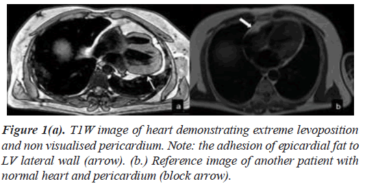
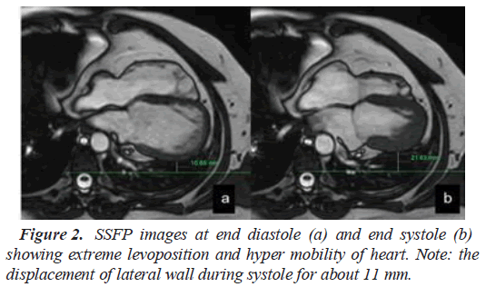
CMR demonstrated extreme levoposition of heart. Pericardium was not visualized in its entirety. The epicardia fat was adherent to the Left Ventricle (LV) lateral wall at mid cavity level (Figures 1a and 1b). No hernia or diverticulum was seen. There was hypermobility of heart (Figures 2a and 2b) with normal biventricular function. There was no myocardial edema or evidence of pulmonary emboli. There was no evidence of myocardial infarction or infiltration.
Discussion
Congenital Absence of the Pericardium (CAP) is a rare anomaly with prevalence of 0.002%-0.004% [2]. Congenital pericardial defects are either partial or complete. Partial CAP can present with herniation of heart and complications such as incarceration of cardiac tissue or myocardial ischemia. Complete CAP are mostly asymptomatic and incidental finding, however they can also result in complications. Therefore, it is important clinically to detect CAP [5,6].
Major function of pericardium is to stabilize and maintain the heart’s position within the thoracic cavity by virtue of its ligamentous attachments. Pericardial fluid acts as lubricant to reduce friction of cardiac surface throughout cardiac cycle. Complete absence of pericardium results in hypermobile heart which results in formation of pleuropericardial adhesions. This may lead to undue torsion or strain on the great vessels as they serve as the only anchor for the heart. This strain or tension results in atypical chest pain in these patients [7]. Some of the symptomatic patients who may be at risk of serious complications require surgical treatment. Surgical options vary depending on the degree of absence of pericardium and include pericardiotomy and pericardioplasty [6]. Surgical correction is aimed at reducing the mobility of the heart and correcting any focal adhesions/strangulation. Chest pain is often resolved in patient’s post-surgical correction [8].
In our case, the patient was treated symptomatically with analgesics and followed up as an outpatient. His symptoms had resolved in initial follow up and he had stopped taking analgesics. He is currently under annual review and has been asked to present to the hospital if the symptoms recur.
Conventional imaging modalities such as radiographs and echocardiogram are not often helpful in diagnosing this condition. As in our case, echocardiogram is often difficult to perform in CAP due to malposition of the heart and its inherent inability to visualize pericardium. CMR is considered the gold standard test [9] in such patients as it evaluates the anatomy of the pericardium and myocardium without the use of intravenous contrast or exposure to radiation. Addition of contrast examination will help in excluding other causes of chest pain, such as aortic dissection, pulmonary embolism, myocardial infarction and myocarditis. CMR has also been utilized to assess the functional impact of CAP and to ascertain a diagnostic criteria. It has been observed the difference between the total volume of the heart during systole and diastole is increased in patients with CAP compared to normal subjects [10]. The cardiac apex was also found to be hyper mobile compared to normal subjects.
Conclusion
Complete absence of pericardium is a rare anomaly and patients are often asymptomatic, especially those with CAP. Any patient suspected to have CAP should undergo CMR to establish the diagnosis and clearly delineate complications related to partial or complete absence of pericardium.
References
- Kronzon I, Srichai MB. Rare pericardial disorders. Dynam Echocardio. 2011;262–5.
- Yamano T, Sawada T, Sakamoto K, et al. Magnetic resonance imaging differentiated partial from complete absence of the left pericardium in a case of leftward displacement of the heart. Circ J. 2004;68(4):385-8.
- Centola M, Longo M, De Marco F, et al. Does echocardiography play a role in the clinical diagnosis of congenital absence of pericardium? A case presentation and a systematic review. J Cardiovasc Med. 2009;10(9):687-92.
- Vesely T, Julsrud PR. Congenital absence of the pericardium and its relationship to the ligamentum arteriosum. Surg Radiol Anat. 1989;11(2):171-5.
- Misthos P, Neofotistos K, Drosos P, et al. Paroxysmal atrial fibrillation due to left atrial appendage herniation and review of the literature. Int J Cardiol. 2009;133(3):122-4.
- Brulotte S, Roy L, Larose E. Congenital absence of the pericardium presenting as acute myocardial necrosis. Can J Cardiol. 2007;23(11):909-12.
- Wilson SR, Kronzon I, Machnicki SC, et al. A constrained heart: A case of sudden onset unrelenting chest pain. Circul. 2014;130(18):1625-31.
- Gatzoulis MA, Munk MD, Merchant N, et al. Isolated congenital absence of the pericardium: clinical presentation, diagnosis, and management. Ann Thorac Surg. 2000;69(4):1209-15.
- Cuccuini M, Lisi F, Consoli A, et al. Congenital defects of pericardium: Case reports and review of literature. Ital J Anat Embryol. 2013:136-50.
- Psychidis-Papakyritsis P, De Roos A, Kroft LJ. Functional MRI of congenital absence of the pericardium. Amer J Roentgeno. 2007;189(6):312-4

