Research Article - Biomedical Research (2017) Advances in Health Science and Biotechnology Application
Treatment of bone and joint injuries in athletes
Sun Peng and Liu Xiaohai*
Department of Physical Education, Dalian Medical University, Dalian 116044, Liaoning, PR China
- *Corresponding Author:
- Liu Xiaohai
Department of Physical Education
Dalian Medical University, PR China
Accepted date: October 23, 2017
Abstract
In recent years, the development of competitive sports in China has entered a new stage of historical development. In order to achieve better competitive results, athletes have to increase the intensity of training and technical difficulties, which inevitably causes all kinds of sports injuries. In these injuries, the greatest impact on athletic athletes is bone and joint injury. In the existing clinical studies, there are few studies on cartilage injuries of bone and joint. The patients with joint pain were treated by micro fracture in a hospital of Beijing during January 2016-December 2016, in which a total of 76 patients were screened. The effect of micro fracture in the treatment of cartilage injuries was judged through Tegner exercise rating and the preoperative and postoperative comparative analysis of clinical symptoms. The results show that the treatment effect of micro fracture surgery on athletes’ bone and joint cartilage injuries is obvious, which can be applied in specific clinical practice.
Keywords
Cartilage injuries, Athletes, Micro fracture surgery, Bone and joint
Introduction
The success of the 2008 Olympic Games in Beijing has greatly raised the average sports level of our country and improved the consciousness and quality of the national sports. With the continuous improvement of China's economic level, people pay more and more attention to physical exercise, so as to continuously improve the quality of life. At the same time, people pay more attention to competitive sports [1]. Competitive sports not only can promote the quality of our people, but also can enhance national pride and self-esteem. In pursuit of higher athletic performance, athletes have to do a lot of intense training to pursue greater strength, faster speed, and more difficult skills. This increases the risk of athletic trauma in the course of training and competition [2]. Common injuries in sports injuries include sprains, fractures, muscle strain, etc. Although the improvement of modern medical technology provides better and better treatment for athletes, it still difficult to change the status quo of modern athletes' injuries [3]. However, the injuries suffered by athletes during the training process will not only affect the athletes' own competitive performance, but also may even end the athletic career of athletes. Even effective protective measures can reduce athletes' injuries, athletes' injuries are unavoidable. Therefore, we must pay more attention to the injury treatment of athletic sports.
However, more attention has been paid to the study of injury therapy for athletes with chronic injuries caused by acute sports injuries or long-term sports [4]. Because the main strength support of athletes depends on the lower limbs, the joint injuries of the lower limbs are very common. The current research focuses on the study of acute injury of bone and joint fracture. Insufficient attention has been paid to cartilage injuries in bone and joint injuries. In fact, according to the existing clinical diagnosis experience, the athlete's cartilage damage is a common disease. Because the daily attention is small, it has not been able to carry on the cartilage injury promptly the treatment. The investigation was carried out on the articular cartilage injuries of athletes in a hospital in Beijing during January 2016-December 2016. The patients were treated with micro fracture. Through the preoperative and postoperative comparative study, the effect of micro fracture treatment was discussed. Through this study, we can further improve the treatment methods of bone and joint injuries in athletic athletes, and reduce the injuries caused by bone and joint cartilage to athletes. This is of great significance for promoting the competitive level of our country's competitive sports and improving the physical quality of athletes. It is also valuable for the clinical treatment of cartilage trauma.
Study on the Injury and Treatment of Bone and Joint in Athletes
Bone and joint injuries in athletes
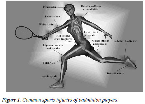
Sports can not only improve the physical quality of athletes, but also have high appreciation value. Since the founding of new China, the level of competitive sports in China has been continuously improved. In the international sports events, we have refreshed our achievements. With the continuous improvement of China's competitive sports and the development of modern science and technology, more and more athletic athletes have come into being, and gradually form a powerful sports power in our country [5]. In the constant pursuit of higher, faster and stronger at the same time, many athletes constantly improve the training intensity of their own with more difficult movements to constantly improve and break their own achievements, which makes them more vulnerable to injury. Today, a large proportion of professional athletes are often accompanied by injuries. Many good athletes even retire early because of their injuries, and they still suffer from injuries after they retire [6]. Competitive sports have very strong antagonism, and have high requirements for strength, speed, flexibility and so on. In the specific sports process, athletes’ injury may be caused by too high speed, strong impact, heavy load and so on. Take tennis players during training or competition may be injured as an example, as shown in Figure 1. As can be seen from the diagram, tennis players may suffer injuries throughout all parts of the body. The head may have a severe concussion due to an impact. Due to long time exercise and training, it may form a tennis elbow, or tendinitis of rotator cuff tear. And because of excessive force or impact and other external reasons, hip joint stress fracture, ankle sprain, and muscle pull superior condition may be caused [7]. As competitive sports are often related to the mobilization of the whole body function, when affected by external forces, all parts of the body may be hurt.
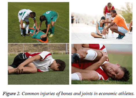
Athletes suffer from sports injuries, which has a serious impact on their sports status, even their sports career. Although the athletes' protection measures and training methods are more and more scientific and advanced, many athletes will suffer more injuries during training and competition due to the increasing intensity of training [8]. Among these injuries, one of the most common injuries is bone and joint injury. This kind of injury is more likely to happen, and it is very harmful to athletes. There are many kinds and causes of bone and joint injuries. Once there is damage, the treatment or rehabilitation process will be relatively long. If the serious damage to body tissues, it may ruin the player’s career directly [9]. In most competitive sports, as the lower limb takes the most important part of the body, it plays an important supporting role, so the injury of bone and joint is more concentrated in the knee joint and ankle joint of the lower limb, as shown in Figure 2. Lower extremity injuries are more common in athletic sports. Of course, there are some competitive sports caused due to the concentration of power in the hands, such as gymnastics and other projects, which also cause fractures of the upper limb. Weightlifting events are prone to spinal damage. The possible injuries caused by different sports have their own characteristics. In specific sports, it is necessary to take targeted protective measures and corresponding training techniques to avoid possible joint injuries [10].
As athletes have certain competitive skills and protective awareness, there are relatively few cases of fractures, and more of them are injuries of bone, joint and cartilage. In the bone and joint, articular cartilage is an important component to effectively reduce the friction between the joints, and it can absorb and disperse the weight-bearing, and can avoid joint damage to a certain extent [11]. However, because of the high load training of the athletes, it makes the articular cartilage vulnerable to injury. Especially for older athletes, it is easier to develop cumulative knee joint cartilage damage. After the joint cartilage is damaged, cartilage tissue is extremely difficult to recover from self-repair because there are no nerves, blood vessels and lymph, and the cells are restricted to the extracellular matrix and unable to move [12]. After injuries to the articular cartilage, athletes continue training before they have fully recovered. This leads to increased damage to the articular cartilage and eventually results in cumulative cartilage damage. If this kind of injury fails to take effective measures for treatment, it will have a long-term impact on athletes, and make the quality of life of athletes decrease significantly [13].
Treatment of joint injuries in athletes
The joint damage caused by training affects athletes participating in the event. Even in the event of participation, many elite athletes are also difficult to play their true level. According to the relevant statistics, we can see that most of the athletes have more or less all kinds of physical damage. The injury of bone and joint has the greatest impact on athletes in all kinds of injuries, so attention should be paid to the treatment of athletes with bone and joint injuries [14]. According to the characteristics of different sports injuries, targeted therapies should be adopted so as to ensure that athletes can continue systematic training. On the one hand, it can reduce the possible joint injuries. And on the other hand, it can also prolong the athletic life of athletes. In the case of severe bone and joint injury, especially in the case of limb fracture, it must be divided into two stages to achieve rehabilitation treatment [15]. The first stage is the fixation of the fracture. This stage is mainly to remove the swelling caused by injury, relieve the patient's pain and avoid possible complications [16]. At this stage, it is necessary to lift the affected limb and maintain normal activities of the trunk and limbs as much as possible. The second stage is the bone fracture healing period. The main objective is to completely eliminate the swelling of the injured part, enhance the range of motion, increase muscle strength and muscle coordination. Although there are relatively mature techniques for fracture damage, the treatment of cartilage injuries is still at an exploratory stage [17].
The prevention and treatment of joint injuries in Chinese medicine is also a very important way of treatment, including acupuncture, Chinese medicine and massage and other traditional Chinese medicine treatment methods. Chinese medicine not only has more applications in the treatment of joint injuries of athletes in China, but also has been recognized and loved by foreign athletes. This shows that Chinese medicine treatment of athletes’ joint damage has played a positive role. Traditional Chinese medicine treatment has been widely recognized because of its advantages of safety and low cost [18]. The application of acupuncture in sports medicine has been gradually deepened. After scientific research, more breakthroughs and developments have been made on the basis of traditional medicine. Through the research, the preventive and health care function of acupuncture has been scientifically confirmed. Acupuncture can fully stimulate the body through the system, adjust the physiological functions of the human body, help to enhance the body's ability to lower the disease and self-repair. In the above joint injury, using acupuncture can stimulate the meridians, improve blood flow, and stimulate tissue to repair itself. Therefore, in actual clinical applications, acupuncture is often combined with other therapies to treat joint injuries [19].
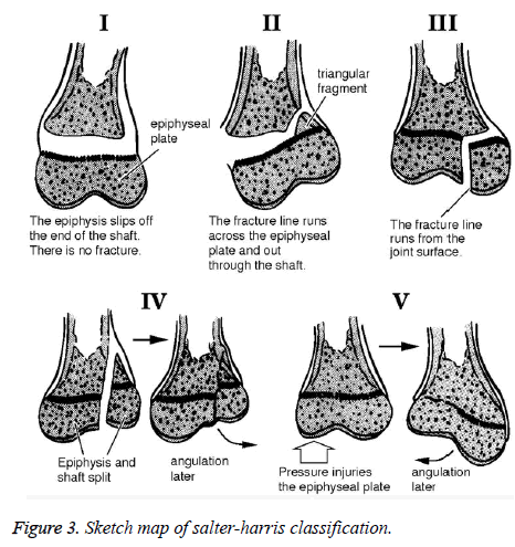
In the treatment of bone and joint, the repair of cartilage injury is a difficult problem to be solved. Although some of the existing treatments for cartilage injuries can restore part of the joint function and relieve pain to some extent, it is difficult to fundamentally solve the problem. The resulting fibrocartilage is not suitable for athletes with high strength and high pressure. Taking the epiphyseal Salter-Harris type for example, it can be divided into V types, which are epiphyseal separation, epiphyseal separation with metaphyseal fracture, epiphyseal fracture, epiphyseal and metaphyseal fracture, epiphyseal extrusion injury, and the lack of epiphyseal plate cartilage ring caused by cutting edge, as shown in Figure 3. Among them, there are relatively few treatments for cartilage damage and loss. On the other hand, people are not paying enough attention to the treatment of cartilage injuries [20]. In the traditional treatment, cartilage, bone graft and micro fracture are mostly used to repair the cartilage surface defect. In fact, the problem of articular cartilage regeneration has not been solved in essence.
Study on the Treatment of Athletes' Articular Cartilage Injuries Based on Micro Fracture
General information
Patients with joint pain symptoms were selected from a hospital in Beijing in January 2016-December 2016. A total of 76 cases belonged to the competition athletes were selected out. There were 48 males and 28 females, with a male to female ratio of 1.71:1. Through statistical analysis, among these patients, there were16 gymnasts, accounting for 21.05% of the total number; there were 5 fencing athletes, accounting for 6.58%; there were 12 gymnasts, accounting for 15.79%; there were 7 wrestlers, accounting for 9.21%; there were 9 football players, accounting for11.84%; there were 27 other athletes, accounting for 35.53% of the total number. These athletes belonged to the provincial or National Team. They were professional athletes who participated in various sports competitions and had great intensity of daily training. The average age was 21.3 years with the oldest of 35 years old and the youngest of 17 years old. The inclusion criteria and exclusion criteria refer to this literature [21]. This research was approved by the Ethical Committee of Dalian Medical University according to the declaration of Helsinki promulgated in 1964 as amended in 1996, the approval number is 2016002.
MRI examinations (MAGNETOM Aera, SIEMENS, Germany) were performed on all patients before surgery, so as to diagnose the cartilage damage in the patients. The cartilage damage in all patients was consistent with the diagnostic criteria for full-thickness cartilage injuries. In clinical manifestations, joint swelling and pain were observed, and the activity was limited. Articular cartilage injuries were concentrated in the femoral, patellar and tibial joints and platform positions of the lower extremities. The criteria for cartilage damage were divided according to the Outerbridge classification criteria (Table 1). The rating of cartilage damage was evaluated by Outerbridge classification criteria and divided into 0, I, II, III and IV, as shown in Table 2. According to the degree of damage of cartilage, the corresponding evaluation was made. A total of 124 cartilage lesions were identified in 76 patients, with an average of 1.63 cartilage lesions in each patient. Among them, there were 14 in class I, 21 in class II, 31 in class III, and 58 in class IV. The average area of cartilage injury was 3.1 cm2, and the damage area was 0.9 cm2-4.4 cm2.
| Classification | Injury of articular cartilage |
|---|---|
| 0 | Normal articular cartilage |
| I | Cartilage softened or localized swelling |
| II | The cartilage surface showed mild fibrosis, and the thickness of cartilage defect was <50% |
| III | Severe fibrosis on the cartilage surface, cracking, cartilage defect thickness of >50% |
| IV | Complete cartilage defect, and subchondral bone exposure |
Table 1. Outerbridge grading standard.
| Fraction | Able to work and sport to the maximum extent | |
|---|---|---|
| Work | Sport | |
| 1 | Sit down and work | Walk on the smooth road |
| 2 | A light manual labor | You can't walk in the forest |
| 3 | A light manual labor | You can swim and walk in the forest |
| 4 | Do manual labour such as heavy domestic work | Cycling, skiing, jogging, etc |
| 5 | Heavy physical labor can be carried out | Cycling, cross country skiing |
| 6 | Heavy physical labor can be carried out | Badminton, tennis, basketball, jogging, etc |
| 7 | Heavy physical labor can be carried out | Tennis, football, handball, basketball, high jump, cross-country and so on |
| 8 | Heavy physical labor can be carried out | Softball, badminton, alpine skiing, etc. |
| 9 | Heavy physical labor can be carried out | Ice hockey, wrestling, gymnastics |
| 10 | Heavy physical labor can be carried out | Football |
Table 2. Tegner athletic ability scoring criteria.
Operation method
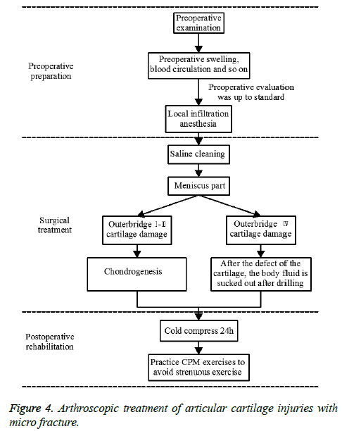
Different surgical treatments were adopted according to different Outerbridge grades, as shown in Figure 4. The whole operation process consisted of three stages, preoperative preparation stage, operation treatment and postoperative rehabilitation. After the completion of the operation, local infiltration and anesthesia were performed on the inlet and joint cavity of the operation. The anesthetic drugs were epinephrine (Kaifeng Pharmaceutical, China) (0.1%, 0.1 ml), lidocaine (Bosen Bio Pharm, China) (2%, 10 ml) and procaine (Sunflower Pharmaceutical, China) (40 ml, 0.1%). In order to facilitate the operation, a mixture of normal saline and Adrealine injection was used to clean. After successful anesthesia, routine examinations were performed with arthroscopy. The free body of the joint cavity was removed by the blue forceps and partial meniscus plasty was performed. Further surgery was needed to determine the Outerbridge classification. If Outerbridge was classified as grade I-III cartilage damage, the non-smooth surface of the cartilage was trimmed with a plasma knife, and cartilage formation was performed. When Outerbridge was classified as a IV class of cartilage damage, it was necessary to first use the curette to correct and separate the unstable cartilage fragments until the normal cartilage margin. The edges of the cartilage were trimmed with a plasma knife and trimmed into a slope. Drilling was performed on the cartilage defect area with the micro fracture tip vertebra, for which the hole diameter, hole spacing and depth was 3 mm. When the pressure in the joint cavity was adjusted to bleed blood or visible fat droplets in the hole, all the liquid within the joint was sucked out.
After the bleeding of the wound eventually forms clots, it was observed whether there was an untreated missing zone. If the omission was found and must be dealt with, then additional drilling could be made. After the operation was completed, the postoperative rehabilitation was performed. Ice compress was carried out for the affected area for 24 h. For much endosmosis, puncture was needed. In order to speed up the recovery of the affected area, the CPM exercises of the joints were necessary. But in the absence of full recovery, strenuous exercise should be avoided as much as possible.
Evaluation criteria and data statistics
The study of the effect of micro fracture on articular cartilage injuries required a comparative analysis of the preoperative and postoperative outcomes. The indicators of contrast were mainly from two aspects, namely, Tegner exercise rating and clinical symptoms. Tegner sport rating is a kind of objective judgment of the human body that can engage in the greatest degree of work and movement. According to the degree of possible labor and movement, it was valued from low to high by 1 point-10 points, as detailed in Table 2. Among them, if athletes could only sit to do simple work, or could only walk on the flat road, then it was rated for 1 point. If athletes could do heavy physical labor related work, all sports events, represented by football, sports events could be carried out, then it was rated by 10 points. Tegner sports rating could be used to evaluate the patient's physical condition. Through quantitative analysis, it could be more intuitive for analysis and research.
The standard of clinical symptom is shown in Table 3. According to the movement of joints, it could be divided into four grades: difference, average, good and excellent. Among them, the worst clinical symptoms were poor, manifested as nocturnal pain, or long range of joints, and limited range of activities.
| Standard | Symptom manifestation |
|---|---|
| Poor | Nocturnal pain or long range of joints; restricted motion |
| Average | During the day, it is difficult to control the use of non-steroidal antibiotics |
| Good | During the movement, joint pain will occur |
| Excellent | No further symptoms occurred |
Table 3. Clinical symptom standard.
All the data was analyzed by using SPSS software, and ͞x ± s was used to represent the measurement data. All data statistics must ensure accuracy and check over and over again.
Treatment outcome and discussion
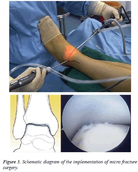
Continuous follow-up was performed on all patients after the operation. Through the preoperative and postoperative grading of Tegner and clinical symptoms, the effect of micro fracture on the cartilage injury of athletes was understood. During the operation, the articular cartilage injuries were observed by arthroscopy. One case was taken as the example, as shown in Figure 5. Arthroscopic debridement and drilling of cartilage injuries of the ankle joint were performed using arthroscopy.
Preoperative and postoperative Tegner exercise ability score contrast is shown in Table 4. As can be seen from Table 4, the average preoperative Tegner exercise ability was 2.7, and the postoperative average score was 6.4. Compared with the preoperative, it was improved by 137.04%, and the exercise ability was improved significantly. According to the postoperative average score, it can be seen that most of the athletes can do heavy physical work, and the most possible sports include badminton, tennis, basketball, jogging and so on. Thus, most of the patients recover successfully after exercise. With further postoperative rehabilitation training, athletes' athletic ability is gradually improved and systematic training can be carried out.
| n | Preoperative | After the operation | t | P |
|---|---|---|---|---|
| 76 | 2.7 ± 0.7 | 6.4 ± 1.4 | 6.88 | <0.01 |
Table 4. Preoperative and postoperative Tegner exercise ability score contrast table.
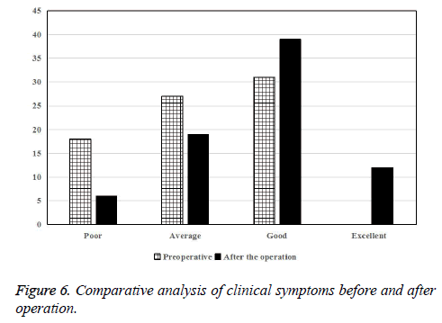
The clinical symptoms before and after operation are shown in Table 5. It can be seen from the table that the clinical symptoms of the surgery have obvious improvement, and the operation effect is obvious. Preoperative patients feel joint swelling, pain accounts for 59.2% of the total number.
| Standard | Preoperative | After the operation | The postoperative ratio was increased |
|---|---|---|---|
| Poor | 18 | 6 | -66.67% |
| Average | 27 | 19 | -29.63% |
| Good | 31 | 39 | 25.81% |
| Excellent | 0 | 12 | - |
Table 5. The level of clinical symptoms before and after operation.
The clinical symptoms of the patients before and after the operation were compared and analyzed (Figure 6). There were no further symptoms in the 12 patients after the operation, and the patients recovered well after the operation.
Arthroscopic micro fracture surgery has a good clinical effect on bone and joint cartilage injuries in athletic sports. Postoperative clinical observation also shows that there are fewer complications in the treatment of articular cartilage injuries. As a new type of minimally invasive surgery for articular cartilage, micro fracture can effectively relieve the pain and swelling of the joint, and effectively repair the cartilage damage. The principle is to remove the necrotic tissue and form a cavity. At the site of the injury, the liver cells in the bone marrow exude blood clots and repair themselves to produce new cartilage replacement tissue. In the specific application process, the most critical thing is to diagnose the patient's cartilage damage before surgery and to master the operation of arthroscopic technique, so that the established surgical target can be achieved.
Conclusions
Athletes need to do a lot of systematic and high-intensity training, which may cause athletic injuries. This not only seriously affects the health of athletes, but also may end the athletic career ahead of schedule. Although the treatment of modern athletes has gradually improved, insufficient attention has been paid to the injury of athletic athletes. Neither the athletes nor the treatments have been able to treat the bone and cartilage injuries effectively. Although there are many treatments for cartilage injuries, the relief and treatment of pain patients are limited. A total of 76 athletes with joint pain symptoms were selected from a hospital in Beijing during January 2016-December 2016. The patients were treated with arthroscopic micro fracture surgery. Tegner exercise ratings and clinical symptoms were used to evaluate preoperatively and postoperatively. After the operation, the average exercise phase of Tegner was increased by 137.04% compared with that before operation, and the clinical symptoms were relatively good after operation. Through clinical observation, the micro fracture complication of articular cartilage was relatively few. This study is a comparative analysis of preoperative and postoperative, and it is not compared with other similar operations.
References
- Tang G, Li J. Regression analysis-based chinese olympic games competitive sports strength evaluation model research. Open Cybernet Syst J 2015; 9: 2729-2735.
- Denker J, Iii MC. Acromioclavicular joint injuries overhead athletes. Operat Tech in Sports Med 2016; 24: 213-222.
- Mitchell J, Graham W, Best TM. Epidemiology of meniscal injuries in US high school athletes between 2007 and 2013. Knee Surg Sports Traumatol Arthroscopy 2015; 2015: 1-8.
- Lam GW, Park EJ, Lee KK. Shoe collar height effect on athletic performance, ankle joint kinematics and kinetics during unanticipated maximum-effort side-cutting performance. J Sports Sci 2015; 33: 1738-1749.
- Wang D, De VG, Ditroilo M. A comparison of muscle stiffness and musculoarticular stiffness of the knee joint in young athletic males and females. J Electromyography Kinesiol 2015; 25: 495-500.
- Lynch AD, Logerstedt DS, Grindem H. Consensus criteria for defining ‘successful outcome’ after ACL injury and reconstruction: a Delaware-Oslo ACL cohort investigation. Br J Sports Med 2015; 49: 335-342.
- Bates N A, Nesbitt RJ, Shearn JT. A Novel methodology for the simulation of athletic tasks on cadaveric knee joints with respect to in vivo, kinematics. Annal Biomed Eng 2015; 43: 2456.
- Beitzel PMK, Jung C, Imhoff AB. Return to sports “nach schulterstabilisation”. Arthroskopie 2016; 29: 1-6.
- Seybold JD, Coetzee JC. Lisfranc Injuries: When to Observe, Fix, or Fuse. Clin Sports Med 2015; 34: 705-723.
- Cronström A, Creaby MW, Nae J. Modifiable factors associated with knee abduction during weight-bearing activities: A systematic review and meta-analysis. Sports Med 2016; 46: 1-16.
- Bere T, Kruczynski J, Veintimilla N. Injury risk is low among world-class volleyball players: 4-year data from the FIVB injury surveillance system. Br J Sport Med 2015; 49: 1132-1137.
- Prucz RB, Friedrich JB. Finger joint injuries. Clin Sports Med 2015; 34: 99-116.
- Chachula LA, Cameron KL, Svoboda SJ. Association of prior injury with the report of new injuries sustained during crossfit training. Athletic Train Sports Health Care 2016; 8: 28-34.
- Şahin N, Bianco A, Patti A. Evaluation of knee joint proprioception and balance of young female volleyball players: a pilot study. J Physical Ther Sci 2015; 27: 437.
- Myer GD, Ford KR, Stasi SLD. High knee abduction moments are common risk factors for patellofemoral pain (PFP) and anterior cruciate ligament (ACL) injury in girls: Is PFP itself a predictor for subsequent ACL injury? Br J Sports Med 2015; 49: 118-22.
- El-Liethy N, Kamal H, Elsayed RF. Role of conventional MRI and MR arthrography in evaluating shoulder joint capsulolabral-ligamentous injuries in athletic versus non-athletic population. Egyptian J Radiol Nuclear Med 2016; 47: 969-984.
- Carruthers K H, Skie M, Jain M. Jam Injuries of the Finger: Diagnosis and management of injuries to the interphalangeal joints across multiple sports and levels of experience. Sports Health 2016; 8: 469.
- Hurtubise JM, Beech C, Macpherson A. Comparing severe injuries by sex and sport in collegiate-level athletes: A descriptive epidemiologic study. Int J Athletic Ther Train 2015; 20: 44-50.
- Rex C, Vignesh R, Javed M. Safe corridors for K-wiring in phalangeal fractures. Indian J Orthopaedics 2015; 49: 388-392.
- Kai M, Peterson L, Zenobiwong M. Cartilage issues in football-today's problems and tomorrow's solutions. Br J Sports Med 2015; 49: 590-596.
- Rangger C, Rogmans S. Bone marrow edema and joint injuries. Der Unfallchirurg 2015; 118: 206-212.
