Image Article - Ophthalmology Case Reports (2020) Volume 4, Issue 2
The ring that resounds on the ultrasound.
Ankita Shrivastav, Vikram Vinayak Koundanya*, Shalini Singh, Chanda Gupta, Sumit Kumar
Vitreoretina Services, Dr. Shroff?s Charity Eye Hospital, New Delhi, India
- *Corresponding Author:
- Vikram Vinayak Koundanya
Vitreoretina Services
Dr. Shroff’s Charity Eye Hospital
Kedarnath Road, New Delhi, India
Tel: +91-9500135421
E-mail: vikramkoundanya@gmail.com
Abstract
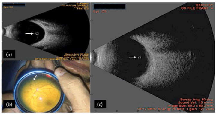
Figure 1 shows B-scan of a 75 year old lady prior to penetrating keratoplasty. A cyst like structure which we presumed to be weiss ring (white arrow) (Figure 1a) was noted. Differentials include pigmented vitreous cyst and parasites like toxoplasma, echinococcus and cysticercosis. To test our hypothesis we performed B-scan on another gentleman in whom weiss ring was seen clinically (Figure 1b). A similar picture appeared on ultrasound (Figure 1c). Fundus examination post operatively confirmed our hypothesis that it was indeed the weiss ring. We hereby propose that Weiss ring should be included in differentials for cystic lesion on ultrasound.
