Case Report - Biomedical Research (2018) Volume 29, Issue 13
The radiology and cerebral vascular insufficiency subject-A case report and literature review
Kuan-Yung Chen1, Tsung-Fu Yu2 and Hua-Tzu Huang3*
1Department of Radiology, Chang-Bing Show-Chwan Memorial Hospital, Taiwan
2Institute of Biomedical Sciences, Academia Sinica, Nankang, Taipei, Taiwan
3Department of Medical Imaging, Show Chwan Memorial Hospital, Changhua, Taiwan
- *Corresponding Author:
- Hua-Tzu Huang
Department of Medical Imaging
Show Chwan Memorial Hospital
Changhua Taiwan
Accepted date: June 21, 2018
DOI: 10.4066/biomedicalresearch.29-18-775
Visit for more related articles at Biomedical ResearchAbstract
Magnetic Resonance (MR) uses many exam of medical image field. In this case report, we found an interesting case. We did not call it as patient because she did not present any symptoms, thus we call it as subject. Natural cerebral vascular deficiency was seldom to be reported due to subject does not present any obvious symptoms when subject is health or young. Hope that this case report would increase we know more about the congenital part cerebral vascular deficiency. A 36 y old female, who come to our hospital for advanced medical health examination. On the regular radiology exam, the MRI showed she has a congenital part cerebral vascular deficiency on December in 2013. In general, main cerebral artery deficiency would cause some impact on physiological, however, its seemly does not affect her in daily or intelligent working, or we still not know would it affect to human. This case report showed a young women persistent hypoglossal artery was missing. We should continuous follow the subject by regular medical health examination. For her, we can prevent further congenital caused injuries. It also added a rare case of Taiwanese in medical imaging academic field.
Keywords
Medical imaging, Congenital deficiency, Cerebral vascular, Natural born deficiency, MRI
Introduction
Magnetic resonance (MR) uses many exam of medical image field. MRI is a non-invasive and versatile imaging modality that allows longitudinal and three-dimensional assessment of tissue morphology, metabolism, physiology, and function.
The combination of different MRI methods (e.g., diffusion-weighted MRI, perfusion MRI, functional MRI, cell-specific MRI, and molecular MRI) allows for the in vivo multi-parameter assessment of the pathophysiology, recovery mechanisms, and treatment strategies in stroke experimental models.
Neoplasms, multiple sclerosis, neurodegenerative diseases, traumatic brain injury, epilepsy and other brain diseases [1]. In this case report, we found an interesting case. We did not call it as patient because she did not present any symptoms, thus we call it as subject.
It is clearly critical to elucidate the mechanisms that mechanisms of VD from the human brain, as well as understand how to the heterogeneity of cerebrovascular disease, yet there causing it challenging to elucidate in a human context [2]. As they participate in cerebral ischemia, various cell signaling and regulation mechanisms (including apoptosis, autophagy, oxidative stress, and inflammation) are associated with VD [3].
Belkehelfa showed that cognitive impairment is caused by vascular disease and neuro-inflammation [4]. Natural cerebral vascular deficiency was seldom to be reported due to subject does not present any obvious symptoms when subject is health or young. Hope that this case report would increase we know more about the congenital part cerebral vascular deficiency.
Case Presentation
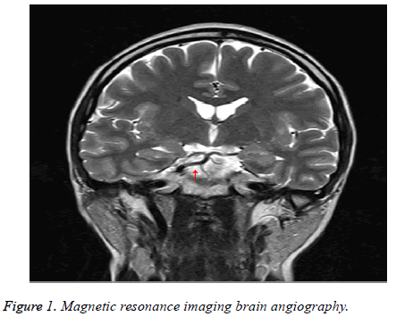
A 36 y old female, who come to our hospital for advanced medical health examination. On the regular radiology exam, the MRI showed she has a congenital part cerebral vascular deficiency in December 2013. In Figure 1, it was MRI brain angiography.
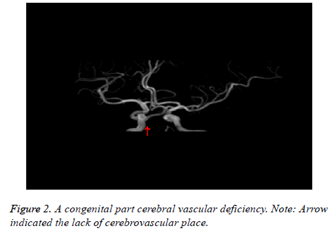
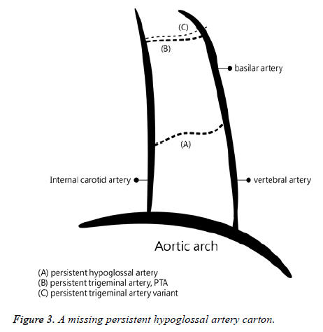
We enlarged part of the brain stem. Figure 2 showed a congenital part cerebral vascular deficiency, a main persistent hypoglossal artery was missing. This study plotted the cartoon in Figure 3.
In general, main cerebral artery deficiency would cause some impact on physiological, however, its seemly does not affect her in daily or intelligent working, or we still not know would it affect to human.
Literature Review
MRI to know blood flow started since very early age in the twenty century. Saltzman reported a case, a woman who 41 y old had suffered from migraine since childhood [5]. Through radiology exam, Saltzman found some vascular deficiency would now produce symptoms.
Steiner et al. ever reported a 23 y old female, who had congenital part cerebral vascular deficiency [6]. The female death was due to weakness of brain vessel walls. Weissman et al. used MR scan two patients’ brain, who presented sudden hearing loss and vertigo [7]. One of them was a 57 y old man presented with right-sided hearing loss and vertigo of several days duration. The other one was a 34 y old man experienced 4 to 5 d of left-sided hearing loss. Vestibular testing was not performed. It also showed high signal from otic labyrinth. They supposed the possibility that the high signal was caused by hemorrhage but that clinical significance need further study. The cerebral vascular deficiency would also be possibly related to hemorrhage symptom.
Athale et al. reported two women (46 and 19 y old), finding the classic appearance of persistent trigeminal artery on MR imaging [8]. This finding also was used as an ancillary sign of this anomaly in subtle cases of persistent fetal carotid-basilar artery anastomoses. Two cases showed a hypoplastic basilar trunk flow void in the caudal posterior fossa that enlarged to a more normal caliber rostrally above the persistent trigeminal artery anastomosis. The actual anastomosis was also demonstrated in both cases. It showed that the cerebral vascular deficiency would be checked by MRI.
Hirai et al. evaluated the MR angiographic findings of the persistent trigeminal artery variant (PTAV) and discussed its clinical implications [9]. They reviewed time-of-flight MR angiography, which were including individual source imaging in three patients with PTAV, which had been recognized as an incidental finding on conventional arteriography. They stated imaging of MR angiography helped demonstrate PTAV and its adjacent structure. Knowledge of the presence and course of this anomalous vessel was useful for various treatment strategies, and do the further study.
Hagino et al. stated that vascular deficiency was related to aging [10]. This would cause deficiency of recognition and reference memory during the processes of aging. In man in Alzheimer's disease, one might speculate that formation and accumulation produce cerebral amyloid angiopathy and it bring about hypoglycemia and hypoxia in the hippocampal pyramidal neurons. They said vascular deficiency related to the vascular change of the retinal capillaries, it became a prominent feature during the processes of aging, and it had a positive correlation with the vascular change of hippocampal capillary. Maybe our subject is healthy in young, but get worse when she was old.
Uchino et al. stated that anterior cerebral artery (ACA) study from anatomical and angiographic was well known [11]. However, ACA variations had rarely been studied by magnetic resonance (MR) angiography. Their study purpose was to investigate not only the type, location, configuration, and incidence of ACA variations, but also coexisting arterial pathology, such as aneurysms detected by cranial MR angiography. Our case report was also similar to their study. They retrospectively reviewed 891 cranial MR angiography images, all images were obtained with one of two 1.5-T scanners using the three-dimensional time-of-flight technique. They found although the clinical significance of ACA variations was usually minor, an associated aneurysm was found relatively frequently, recognizing ACA variations during the interpretation of cranial MR angiograms was important.
Munteanu et al. proposed biomechanical model of cerebral aneurysm generation [12]. The model was the conditions and steps in this vascular deficiency generation. The mathematical model showed the internal carotid artery and blood vessel compliance. In their model, we would know some characteristics due to hydraulic vascular parietal resistance if vascular deficiency. Shankar et al. studied time-resolved CT angiography compared to DSA in cerebral vascular malformations [13]. They prospectively tested the hypothesis that time-resolved CT angiography (TRCTA) on a Toshiba 320-slice CT scanner enables the same characterization of cerebral vascular malformation (CVM) including arteriovenous malformation (AVM), dural arteriovenous fistula (DAVF), pial arteriovenous fistula (PAVF) and developmental venous anomaly (DVA) compared to digital subtraction angiography (DSA). They found image techniques would be noninvasive substituted for conventional DSA in certain clinical situations. Li et al. stated that chronic hypertension altered cerebral vascular morphology [14]. Cerebral blood flow, cerebrovascular reactivity and neurological disorders; they used spontaneously hypertensive rats to do experiments. Using magnetic resonance angiography would be exam to cerebral and downstream arteries.
Murai et al. stated that the intraoperative confirmation of blood flow direction is necessary in cerebral vascular surgery [15]. Using indocyanine green video angiography (ICG-VAG) with the FLOW 800 system, they examined the transit time of the blood vessel. Video angiography is still important to nowadays for patients’ cure and operation. Gao et al. studied the feasibility of low injection rate and low contrast agent dose in three-dimensional rotational digital subtraction angiography (3D DSA) of the intracranial aneurysm [16].
Although there were numerous MR papers related to exam cerebral vascular exam; however there was only one paper, which was Steiner et al. [6], we found in this case report study about cerebral vascular deficiency.
Discussion
MRI uses many exam of medical image field. This case report showed a young women persistent hypoglossal artery was missing. We should continuous follow the subject by regular medical health examination. For her, we can prevent further congenital caused injuries. The related case report in PubMed Literature Database was seldom to be reported. It also added a rare case of Taiwanese in medical imaging academic field.
Acknowledgment
We would like to acknowledge the medical doctor Chun-Yi Lin, Dr. PH. and Rong-Fu Chen, PhD of Directors of Research Assistant Center, Show Chwan Health Care System. Thanks to their administrative support.
Competing Interests
The authors declare that there is no conflict of interests regarding the publication of this article.
References
- Dijkhuizen RM, Nicolay K. Magnetic resonance imaging in experimental models of brain disorders. J Cereb Blood Flow Metab 2003; 23: 1383-1402.
- Kalaria RN. Neuropathological diagnosis of vascular cognitive impairment and vascular dementia with implications for Alzheimer’s disease. Acta Neuropathol 2016; 131: 659-685.
- Mulugeta E, Molina-Holgado F, Elliott MS. Inflammatory mediators in the frontal lobe of patients with mixed and vascular dementia. Dement Geriatr Cogn Disord 2008; 25: 278-286.
- Belkhelfa M, Beder N, Mouhoub D. The involvement of neuroinflammation and necroptosis in the hippocampus during vascular dementia. J Neuroimmunol 2018; 320: 48-57.
- Saltzman GF. Patent primitive trigeminal artery studied by cerebral angiography. Acta Radiologica 1959; 51: 329-336.
- Steiner H, Lammer J, Kleinert R, Schreyer H. Dissecting aneurysm of cerebral arteries in congenital vascular deficiency. Neuroradiology 1986; 28: 331-334.
- Weissman JL, Curtin HD, Hirsch BE, Hirsch Jr WL. High signal from the otic labyrinth on unenhanced magnetic resonance imaging. Am J Neuroradiol 1992; 13: 1183-1187.
- Athale SD, Jinkins JR. MRI of persistent trigeminal artery. J Computer Assisted Tomography 1993; 17: 551-554.
- Hirai T, Korogi Y, Sakamoto Y. MR angiography of the persistent trigeminal artery variant. J Computer Assisted Tomography 1995; 19: 495-497.
- Hagino N, Kobayashi S, Tsutsumi T. Vascular change of hippocampal capillary is associated with vascular change of retinal capillary in aging. Brain Res Bulletin 2004; 62: 537-547.
- Uchino A, Nomiyama K, Takase Y, Kudo S. Anterior cerebral artery variations detected by MR angiography. Neuroradiology 2006; 48: 647-652.
- Munteanu F, Poeată I. Biomechanical modelling of cerebral aneurysm generation. Revista medico-chirurgicală̆ a Societă̆ţ̜ii de Medici ş̧i Naturaliş̧ti din Iaş̧i 2009; 113: 293-298.
- Shankar JJ, Lum C, Chakraborty S, Dos Santos M. Cerebral vascular malformations: Time-resolved CT angiography compared to DSA. Neuroradiol J 2015; 28: 310-315.
- Li Y, Shen Q, Huang S. Cerebral angiography, blood flow and vascular reactivity in progressive hypertension. Neuroimage 2015; 111: 329-337.
- Murai Y, Nakagawa S, Matano F, Shirokane K, Teramoto A, Morita A. The feasibility of detecting cerebral blood flow direction using the indocyanine green video angiography. Neurosurg Rev 2016; 39: 685-690.
- Gao Z, Zeng Y, Sun J. Application of low injection rate and low contrast agent dose in three-dimensional rotational digital subtraction angiography of the intracranial aneurysm. Interventional Neuroradiol 2016; 22: 287-292.


