Research Paper - Archives of General Internal Medicine (2018) Volume 2, Issue 1
The Hemodynamics of Inferior Vena Cava Caliber Changes from Rest to Exercise.
Vincent JB Robinson1, Siva M Krothapalli1, Umer Saleem1, Mahendra Mandawat1,2, Jim H Corley1, James D Halbert1, Harry Davis1 and Gaston K Kapuku1*
1Medical College of Georgia, Augusta University, Augusta, USA
2Veteran Affairs Medical Center, Augusta, USA
- *Corresponding Author:
- Vincent Robinson, MD
Augusta University
Medical College of Georgia
Division of Cardiology
1120 15th Street, Augusta, GA 30912, USA
E-mail: vrobinso@augusta.edu
Accepted on January 19, 2018
Citation: Robinson VJB, Krothapalli SM, Saleem U, et al. The hemodynamics of inferior vena cava caliber changes from rest to exercise. Arch Gen Intern Med. 2018;2(1):20-25.
Abstract
Objective: Venous Return is a major determinant of Cardiac Output and is modulated in a large part by conduit vein hemodynamics. We aimed to study changes in Inferior Vena Cava (IVC) cross-sectional area morphology, which is a major conduit vein, from rest to exercise with the hypothesis that sympathetic reflex mechanisms cause IVC dilation during exercise, thereby preventing an increase in venous resistance.
Design: Uncontrolled longitudinal study. Setting: Single urban academic medical center.
Subjects: Adult subjects free of any known cardiovascular disease, without a history of abdominal surgery that might interfere with CT scan measurements of IVC size and no contraindications to performing near-maximal levels of exercise.
Interventions: We studied 14 healthy subjects (age 31 ± 10 y., BMI: 26 ± 4 kg/m2) at supine rest, passive leg-raising, and supine exercise (50% and 90% maximal effort) and measured blood pressure, heart rate (HR), peripheral and central venous pressures (PVP and CVP), and cardiac output (CO). Venous resistance (VR) calculated as VR=(PVP-CVP)/CO and cross-sectional areas (CSA) of the aorta and IVC were measured at each stage.
Measurements and Main Results: Measurement instruments consisted of exercise equipment, hemodynamics, a thoracic bioimpedance monitor, Computed Tomography, Body Mass Index, and blood pressure. CVP increased with leg-raising from baseline 3.2 ± 2.3 to 5.2 ± 2.4 cm H2O (p<0.001). IVC CSA increased from 386 ± 216 mm2 at baseline to 449± 231 mm2 with passive legraising (p<0.001), and to 459 ± 171 mm2 at 50% exercise (p<0.05). VR did not change with leg raising or exercise. BMI showed a negative correlation with IVC CSA at baseline that tended to persist with leg raising and exercise (r=-0.35 to -0.52, p=0.028 to 0.109).
Conclusions: The unchanged VR with exercise suggests that IVC dilation stabilizes VR despite increased CO. IVC size negatively correlates with BMI inferring that the IVC caliber is modulated by obesity and/or cardiovascular fitness.
Keywords
Venous return, Venous resistance, Exercise, Vena cava, Aorta size.
Introduction
Modulation of venous return by the low-pressure compartment and its impact on cardiac output has been a concept of critical importance to cardiovascular physiologists since Guyton’s pioneering original experiments [1-3]. However, most clinicians focus on the left ventricular function when analyzing cardiovascular function. This is in large part due to the burden of ischemic heart disease, which predominantly affects left ventricular function. Because of this, the importance of the right heart function and venous return in modulating cardiac output is underestimated. While such an approach might be appropriate for cardiologists given their focus on myocardial infarction and congestive heart failure, intensivists deal with a broader array of cardiovascular perturbations including shock states in which vascular dysfunction and other extra-cardiac disturbances may dominate the clinical picture. An approach to understanding cardiovascular physiology that incorporates cardiac and vascular elements may be more useful to intensivists than one that focuses exclusively on left ventricular physiology. Most of our present understanding in this topic has been from Guyton’s instrumented animal model [4]. However, Guyton used an anesthetized canine model, which cannot be used to predict the venous physiology in a conscious human body [5-10]. In our present study, we describe an experiment designed to assess venous return in healthy young adults both at rest and with exercise. We attempt to understand the mechanism behind the ability of the body to handle a 2-3 fold increase in cardiac output during exercise by studying the conduit veins at rest and during supine exercise. This initial investigation may lead us to a better understanding of the acute hemodynamic responses to exercise in the low-pressure compartment.
Materials and Methods
After approval by the IRB, informed consent was obtained from healthy volunteers (n=14, age range 21-41). There were 9 males and 5 females healthy normotensive individuals, either workers in our hospital or medical students. Subjects were free of any known cardiovascular disease, had no history of abdominal surgery that might interfere with CT scan measurements of IVC size and no contraindications to performing near-maximal levels of exercise.
Exercise equipment
Exercise was performed by all subjects using an exercise cycle ergometer specifically designed for supine exercise (Biodex Medical Systems, New York, NY), which was mounted over the CT imaging bed. Shoulder pads were employed to prevent subjects from sliding upwards on the bed while pedaling. The bicycle ergometer had adjustable resistance to increase workload. The exercise was performed in 3-minute stages to achieve a steady state during exercise, as workload is linearly correlated to cardiac output [11]. Subjects were rapidly titrated to maximum workload while maintaining a constant pedaling rate of 60 revolutions per minute. The resistance was increased to a point where the subject could no longer pedal; this was defined as 100% or maximal exercise [12].
Hemodynamics
A plastic, single-lumen, central line with a drum cartridge catheter (Venisystems, Abbott, Sligo, Ireland) was placed in each subject via the basilic vein by a trained cardiac anesthetist. Central venous pressure was recorded using the central line and peripheral venous pressure was recorded by a side hole in the sheath of the same intravenous line. Cardiac output was measured by thoracic bioimpedance monitor (IQ system, Renaissance Technologies Inc., Newtown, Pennsylvania). The thoracic bioimpedance monitor utilizes four pairs of mylar electrode strips, which were applied on opposite sides of the neck and chest. The monitor displayed impedance and electrocardiographic signal and the beat-to-beat stroke volume was computed using the Nyboer equation, as modified by Kubicek [13]. Thoracic bioimpedance has been shown to correlate well both with Fick and thermodilution cardiac output measurements [14]. An index of venous resistance (VR=PVP-CVP/ CO), was calculated at baseline, with passive leg elevation in the stirrups, after returning legs to the supine position, and for both levels of exercise (at 50% and 90%).
Computed tomography
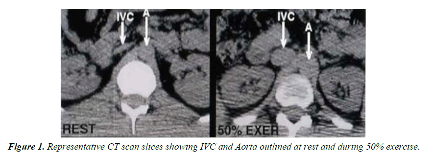
Non-contrast CT image slices were obtained at the predefined sub-hepatic level at baseline, with passive leg elevation, after returning legs to the supine position, and at 50% and 90% exercise. CT scanning (High Speed Advantage, General Electric, Schenectady, NY) was performed utilizing a slice to measure IVC and aortic CSA at the sub-hepatic level. Here, the entire circumference of the IVC could be identified clearly separated from the surrounding soft tissue. CT images were recorded during quiet held expiration to prevent a false increase in IVC CSA with the Valsalva maneuver. Three separate measurements of the IVC and aortic CSA were made for each image and their average was calculated (Figure 1).
Exercise protocol
After obtaining informed consent, the subject was brought into the imaging room, and asked to lie on the CT imaging table. The subject was then left in the quiet room with lights dimmed for 10-15 minutes to achieve baseline-resting state, minimizing any sympathetic stimulation. Following this, baseline hemodynamic readings were recorded. Computerized tomographic images were then obtained. After a rest period of three minutes, the subject’s legs were then elevated passively utilizing stirrups and computerized tomographic images and echocardiographic readings were recorded. The subject’s legs were then brought down to the supine position and following a rest period of three minutes, identical measurements were again recorded. The measurement protocol was similarly repeated immediately after the cessation of both 50% and 90% maximal exercise using the supine bicycle ergometer. After the study, the subjects were taken from the radiology suite, lines were removed, and adequate hemostasis was ensured.
Statistics
All data are displayed as means and standard deviations. Systolic blood pressure, diastolic blood pressure, mean arterial pressure (MAP), CO, BMI, and VR were analyzed against IVC CSA. Comparisons with the adjacent abdominal aorta served as control within each subject. A mixed- model, repeated-measures analysis of variance was used to evaluate differences in the hemodynamic variables at various exercise levels. A priori contrasts were made of baseline vs. passive leg raising, supine, and each level of incremental exercise for each variable.
Results
Fourteen subjects (9 males, 5 females) were enrolled in the study. The mean age of the subjects was 31 ± 10 years. The mean BMI was 26 ± 3.6 kg/m2.
Hemodynamic changes
The hemodynamic data at various interventions of the study protocol are shown in Table 1. Heart rate, MAP, CO, and PVP increased significantly from baseline through incremental exercise (p<0.01 for all variables). HR was significantly higher at 50% exercise (p<0.01) and at 90% exercise (p<0.001) relative to baseline but was unaffected by passive leg raising. MAP increased significantly with passive leg raising (p<0.05) and with 90% exercise (p<0.001) relative to baseline but did not show a significant change with 50% exercise. CO increased significantly from baseline to 50% and 90% exercise (p<0.001 for both conditions).
| Measures | Baseline | Passive Leg Raising | Supine | 50% Maximal Exercise | 90% Maximal Exercise | p– value |
|---|---|---|---|---|---|---|
| HR bpm | 61 ± 10 | 63 ± 11 | 63 ± 10 | 90 ± 28** | 110 ± 29*** | 0.001 |
| MAP mmHg | 75.9 + 8.8 | 78.8 + 9.6* | 78.4 + 11.7 | 79.3 + 24.6 | 90.4 + 14.0*** | 0.009 |
| PVP cm H20 | 9.3 + 6.1 | 8.7 + 3.4 | 6.6 + 3.2 | 10.3 + 4.8 | 13.7 + 7.7* | 0.001 |
| CVP cm H20 | 3.2 + 2.3 | 5.2 + 2.4*** | 2.8 + 2.5 | 3.8 + 2.6 | 4.6 + 5.4 | 0.134 |
| CO L/Min | 5.16 + 1.07 | 5.36 + 1.14 | 4.97 + 0.97 | 7.55 + 1.46*** | 9.35 + 0.95*** | 0.001 |
| AO mm2 | 259 + 81 | 263 + 73 | 262 + 80 | 262 +75 | 273 + 88* | 0.043 |
| IVC mm2 | 386 + 216 | 449 + 231*** | 405 + 202 | 459 + 171* | 405 + 137 | 0.027 |
| VR cm H2O. min/L | 1.29 + 1.07 | 0.80 + 0.90 | 0.84 + 0.54 | 0.96 + 0.84 | 0.91 + 0.57 | 0.233 |
*p<0.05, **p<0.01, ***p<0.00.
Table 1. Baseline hemodynamic variables at each intervention. The last column gives the p value for the overall F-test of homogenous means for the various interventions for each of the hemodynamic variables. (HR heart rate, MAP mean arterial pressure, PVP peripheral venous pressure, CVP central venous pressure, CO cardiac output, AO aorta, IVC inferior vena cava, VR index of venous resistance).
Effects of passive leg raising and exercise
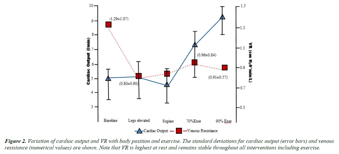
PVP increased with passive leg raising and with exercise, but this increase was statistically significant only at 90% exercise (p<0.05). CVP rose significantly with passive leg elevation from baseline (p<0.001). However, no significant change in CVP was noted with exercise. All parameters returned nearly to baseline levels upon resumption of the supine position following leg raising. VR showed non-significant decreases with all conditions relative to baseline. Figure 2 shows the changes in CO and VR with each test condition.
Effects of interventions on IVC and aorta
IVC CSA increased significantly with passive leg elevation (p<0.001) and with 50% exercise (p<0.05) relative to baseline. There was; however, a decrease in IVC CSA with 90% exercise compared to 50% exercise, but this change was not significant.
Aortic CSA increased significantly at 90% workload relative to baseline (p<0.05).
IVC and Aorta changes as function of BMI
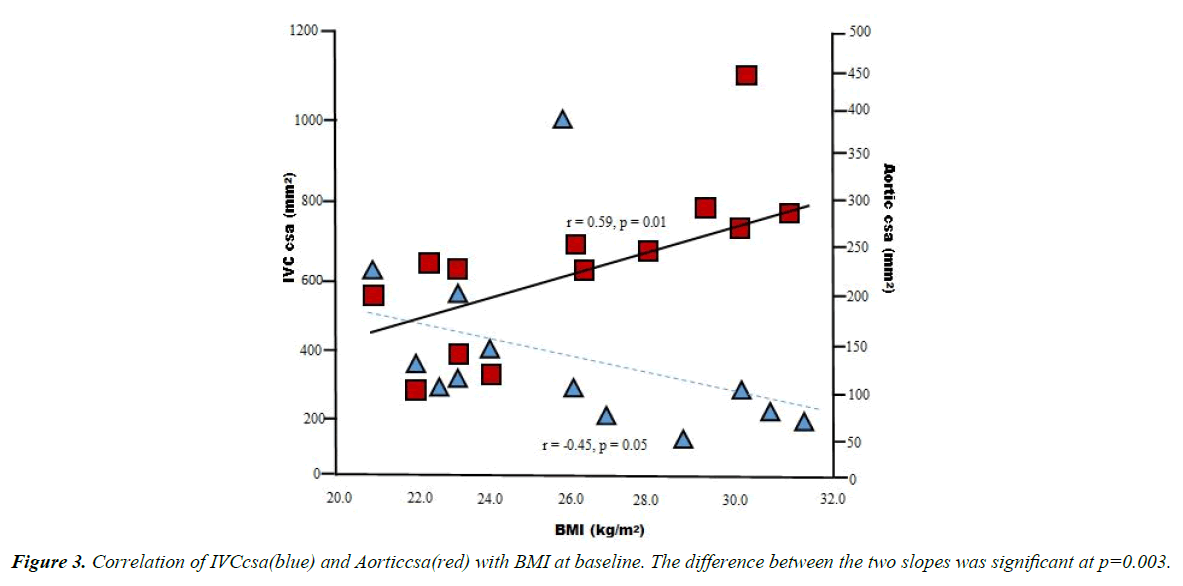
Table 2 shows the correlation between BMI and IVC CSA at various interventions. BMI showed a negative correlation with IVC CSA at baseline (r=-0.46, p=0.05) and at supine position (r=-0.52, p=0.02). Aortic CSA showed a positive correlation with BMI at all experimental interventions (r=0.53-0.59, p=0.01- 0.02). Figure 3 is a linear regression analysis plot of IVC and aortic CSA vs. BMI that shows opposing trends of the IVC and aortic CSA relative to BMI. The difference between the slopes of these two relationships was highly significant (p=0.003).
| Analysis | Baseline | Passive Leg Raising | Supine | 50% Exercise | 90% Exercise |
|---|---|---|---|---|---|
| Pearson Correlation | -0.459 | -0.389 | -0.522 | -0.415 | -0.351 |
| Significance 1-tailed | 0.049 | 0.085 | 0.028 | 0.07 | 0.109 |
| Sample (N) | 14 | 14 | 14 | 14 | 14 |
Table 2. Correlation between IVC diameter and BMI at various interventions.
Discussion
Venous return and CO increased 2-3 fold from resting level during vigorous exercise states. The IVC, by virtue of its anatomic position as a major conduit into the right atrium, needs to adapt to this increase in blood flow. This adaptation occurs by one of two methods implicit in the Poiseulle equation which describes the relation between pressure and flow in cylindrical tubes. According to this equation, higher flows could be achieved either by increasing the pressure gradient, or by decreasing the resistance to flow. A decrease in resistance would help in accommodating large increases in flow with minimal increase in the pressure gradient. Such a decrease in resistance could be achieved by dilation of the conduit veins, such as the IVC. Our investigation looked at the acute morphological changes occurring in the IVC in healthy volunteers at baseline, with passive leg raising, and two levels of supine exercise to determine the active versus passive nature of these IVC changes. We also correlated these changes to simultaneous hemodynamic measures, including an index of venous resistance.
Hemodynamic changes
Passive leg rising was associated with a statistically significant increase (~3 mm Hg) in MAP. This was thought to be due to either auto-transfusion of blood into the central compartment or sympathetic stimulation. The absence of changes in HR with this maneuver favors auto- transfusion as the underlying mechanism. PVP, which was recorded in the left basilic vein in a non- exercising limb, showed no significant change with passive leg rising. However, CVP increased with this maneuver by almost 2 cm H2O, due to the translocation of blood from the periphery to the central compartment. The observed increases in HR, MAP, and CO with exercise are as expected and in concert with those found by other investigators [15-18]. An increase in PVP was also seen with exercise.
This increase in PVP and MAP would lead us to assume a similar increase in CVP with exercise. However, CVP did not change with supine exercise. Although an auto-transfusion of blood into the central compartment occurs with both passive leg raising and supine exercise, the absence of a rise in CVP with exercise is interesting. This may be related to a corresponding increase in MAP, HR, and CO, resulting in translocation of some blood from the low-pressure compartment into the arterial system. In addition, exercise in humans appears to be associated with a 50% increase in pulmonary blood volume, which may not have occurred with passive leg raising alone [19]. Unfortunately, we have no measurements of pulmonary blood volume in the present study.
Changes in venous resistance
Guyton et al. have suggested the importance of resistances in the venous system in determining overall impedance to venous return [20]. Using an analogy of a bathtub, Magder described the role of drainage characteristics of the tube draining the tub in determining the resistance to flow and downstream pressure [21,22]. Conduit veins, by virtue of their position in the mainstream of blood flow to the heart in the two-compartment model play an important role in determining venous resistance [15].
We calculated an index of venous resistance, VR=(PVP-CVP)/ CO as a modification of the formula (MCFP-Pra)/ Rv, where MCFP is the mean circulatory filling pressure, Pra is the right atrial pressure, and Rv is the venous return [1,15,23]. A significant change in VR was not observed with our interventions despite changes in the pressure gradient and CO at 50% exercise (PVP-CVP=+7.6%, CO=+46.3%) and 90% exercise (PVP-CVP=+50.3%, CO=+ 81.2%) relative to baseline. These findings are consistent with the circulatory system changes needed to maintain cardiovascular homeostasis during supine exercise. If the VR were to increase during exercise the pressure gradient, i.e. (PVP-CVP), needed to accommodate the increased venous return and maintain CO would need to increase substantially.
In a classic experiment designed by Guyton to study the relative impact of venous resistance compared to arterial resistance on cardiac output, he showed that by increasing peripheral resistance through injection of glass beads into the arterial system, peripheral vascular resistance increased by 70% whereas cardiac output only decreased by 12%. On the other hand, when peripheral vascular resistance was increased by the same amount using compression of the large veins returning blood to the heart, CO was decreased by an average of 75%. This experiment helped Guyton explain the importance of venous resistance in modulating venous return and thereby CO [20]. Venous resistance was further found to have a 19-fold greater impact on cardiac output than arterial resistance [3]. Hence, the stabilization of VR through IVC dilation (noted specifically at 50% exercise in our study) helps in maintaining cardiovascular homeostasis during increased CO states.
IVC morphologic changes
The similar extent of IVC dilation observed with 50% exercise (18.8%) and with passive leg raising (16.2%) confirms the passive nature of IVC dilation during exercise. When exercise workload was increased to 90%, there was a non-significant decrease in IVC CSA from that at 50% exercise. This could be secondary to increase sympathetic stimulation causing some degree of conduit vein vasoconstriction [24].
Relationship of IVC and aorta with BMI
Goldhammer et al. [25] noted that IVC sizes were increased in athletes at rest who performed chronic higher levels of aerobic exercise. Abergel et al. similarly noted that athletes had abnormally dilated IVC. As a result, we looked at the correlation of IVC CSA with BMI as a surrogate of cardiovascular fitness in our young healthy volunteers. We documented an inverse and moderate correlation between BMI, IVC CSA at baseline, in supine position, and post leg raising. This relationship persisted with other maneuvers but did not attain statistical significance. On the other hand, aortic CSA increase was directly proportional to BMI at all experimental interventions. The difference between the slopes of these two relationships (BMI vs. IVC and aortic CSA) was highly significant. Taken together, the large IVCs noted in athletes and our finding of a negative correlation of IVC CSA with BMI suggests a possible plastic remodeling of the IVC occurring in individuals based on either cardiovascular fitness or BMI. This, of course, will require further study. The equal dilation of IVC with leg raising and with supine exercise suggests that the same passive dilation of a remodeled IVC is the mechanism by which the body reduces venous resistance in active exercising states. The decreased venous resistance would thus require decreased pressure gradients to move blood across the venous portion of the cardiovascular circuit.
Limitations
This study should be interpreted with its limitations in mind. First, This study has a small sample size. Second, the training status could influence our findings. Unfortunately, physical activity or cardiovascular fitness measures were not systematically recorded. This precluded to do the needed propensity score to contract findings from "normal patient" to those of "highly trained patients". Third, we measured CT scan measurements immediately after the cessation of exercise rather than during exercise. This may hypothetically result in changes in the IVC dimensions, which may have affected our results. Finally, supine exercise also relates to a relatively small segment of the human exercise spectrum. The problems associated with reproducing hemodynamic and morphologic measurements in the erect position is the interference and compensation for the effect of gravitational forces on cardiac output, central conduit vein morphology, and the circulatory system in general. Such an interrogation would require different techniques, but is certainly of paramount significance in elucidating functional changes in conduit veins during upright human exercise.
Conclusion
In summary, the change in IVC CSA during supine exercise appears to occur passively, as indicated by a similar extent of IVC dilation with passive leg raising and 50% exercise. IVC dilates with supine exercise at a low workload but dilates to a lesser extent with a high workload likely due to increased sympathetic tone. There is a strong negative correlation between BMI and IVC size, which tends to persist during supine exercise. This paradoxical increase in IVC size with smaller BMI may be related to remodeling based on body habitus and/or cardiovascular fitness. More detailed investigations into the genesis of this relationship between body size and IVC size is required.
References
- Beard DA, Feigl EO. Understanding guyton's venous return curves. American Journal of Physiology- Heart and Circulatory Physiology. 2011;301:H629-33.
- Furst B. The heart: Pressure-propulsion pump or organ of impedance? Journal of Cardiothoracic and Vascular Anesthesia. 2015;29:1688-701.
- Guyton AC, CE Coleman T. Circulatory physiology: Cardiac output and its regulation. 1973.
- Guyton A, Taylor A, Drake R, et al. Regulation of cardiac output by stroke volume and heart rate in conscious dogs. Circulation Research. 1978;42.
- Brengelmann GL. A critical analysis of the view that right atrial pressure determines venous return. Journal of Applied Physiology. 2003;94:849.
- Brengelmann GL. Steady-state venous return: Residue in a recent model analysis of the notion that it is driven by elastic recoil of the venous system. J Applied Physiology 2009;107:369.
- Levy MN. The cardiac and vascular factors that determine systemic blood flow. Circulation Research. 1979;44:739-47.
- Tyberg JV. How changes in venous capacitance modulate cardiac output. Pflugers Archiv: Eur J Physiology. 2002;445:10-17.
- Sunagawa K. Guyton's venous return curves should be taught at medical schools (complete english translation of japanese version). The J Physiological Sci 2017.
- Magder S. Volume and its relationship to cardiac output and venous return. Crit Care. 2016;20:271.
- Karpman VL. Cardiovascular system and physical exercise. CRC Press; 1987.
- Borg GA. Psychophysical bases of perceived exertion. Med Sci Sports Exerc 1982;14:377-81.
- Ovsyshcher I, Zimlichman R, Katz A, et al. Measurements of cardiac output by impedance cardiography in pacemaker patients at rest: Effects of various atrioventricular delays. J American College of Cardio. 1993;21:761-7.
- Moore FA, Haenel JB, Moore EE. Alternatives to swan-ganz cardiac output monitoring. Surgical Clinics of North America. 1991;71:699-721.
- Gelman S. Venous function and central venous pressurea physiologic story. The Journal of the American Society of Anesthesiologists. 2008;108:735-48.
- Wigertz O. Dynamics of respiratory and circulatory adaptation to muscular exercise in man. A systems analysis approach. Acta Physiol Scand Suppl. 1971;363:1-32.
- Astrand PO, Cuddy TE, Saltin B, et al. Cardiac output during submaximal and maximal work. J Appl Physiol. 1964;19:268-274.
- Stohr EJ, Gonzalez-Alonso J, Shave R. Left ventricular mechanical limitations to stroke volume in healthy humans during incremental exercise. Am J Physiol Heart Circ Physiol. 2011;301:H478-87.
- Flamm SD, Taki J, Moore R, et al. Redistribution of regional and organ blood volume and effect on cardiac function in relation to upright exercise intensity in healthy human subjects. Circulation. 1990;81:1550-9.
- Magder S, De Varennes B. Clinical death and the measurement of stressed vascular volume. Crit Care Med. 1998;26:1061-4.
- Scharf SM, Pinsky MR, Magder S. Respiratory-circulatory interactions in health and disease. CRC Press; 2001.
- Rose W. The classical guyton view that mean systemic pressure, right atrial pressure, and venous resistance govern venous return is/is not correct. J Applied Physiology (Bethesda, Md.: 1985). 2007;102:1243.
- Folkow B. Regulation of the peripheral circulation. British Heart J. 1971;33:27-31.
- Goldhammer E, Mesnick N, Abinader EG, et al. Dilated inferior vena cava: A common echocardiographic finding in highly trained elite athletes. J Am Soc Echocardiogr. 1999;12:988-93.
- Abergel E, Oblak A. Arch Mal Coeur Vaiss. 2006;99:969-74.


