Research Article - Biomedical Research (2017) Volume 28, Issue 7
The expression level of gene MAP4K4 and its clinical effect in cancerous tissue of gastric carcinoma
Jian Huo1, Jing-gui Qiao2, Kun Han3*
1Xi’an Medical University, Xi’an, China
2Department of Gastroenterology, Xi’an Gaoxin Hospital, Xi’an, China
3Department of Gastroenterology, the Central Hospital of Xian City, Xi’an, China
- *Corresponding Author:
- Kun Han
Department of Gastroenterology
The Central Hospital of Xian City
China
Accepted date: November 26, 2016
Abstract
Objective: To clarify the expression of MAP4K4 in gastric carcinoma and its relationship with clinical pathological features of gastric carcinoma and further reveal the role of MAP4K4 in the development of gastric cancer.
Methods: We randomly selected 20 patients diagnosed with GC by clinical signs and symptoms, imaging diagnosis and laboratory diagnosis in department of general surgery in University Medical department Affiliated Hospital from January 2014 to December 2015. Gastric cancer tissues and adjacent tissues were studied with immuno histochemistry staining and the expression level of MAP4K4 level was detected postoperatively. The relationship between the MAP4K4 expression level and the clinical stage, pathological grade relationship was analysed. The Kaplan-Meier method was used to analyse the effect of MAP4K4 expression level on clinical outcome.
Result: The expression level of MAP4K4 gene was related to lymph node metastasis and pathological staging of gastric cancer (P<0.05), but it was not related to the tumor size and recurrence and metastasis of gastric cancer (P>0.05). In the -~+ group, the median progression free survival was (4.98 ± 14.42) months; the median progression of free survival was (34.00 ± 27.56) months. In ++~+++ group, the median progression free survival was (19.57 ± 4.76) months, and the median progression free survival was (13.00 ± 1.25) months. The difference was statistically significant (2=13.167; P=0.005). In-~+ group, the average survival time was (65 ± 116.70) months, the median overall survival was (50 ± 39.90) months. The difference was statistically significant (2=10.084; P=0.003). Conclusion: The expression level of MAP4K4 in gastric cancer patients was significantly increased, and it was negatively correlated with the pathological grade of gastric cancer.
Keywords
Gastric cancer, MAP4K4, PFS, OS
Introduction
Gastric Cancer (GC) is one of the most common types of cancer and the second reason for cancer-related high mortality worldwide [1], as well as the fifth most common cancer [1]. The radical surgery usually has a poor prognosis coupled with unsatisfying treatment effect of molecular target [2], which also make the gastric cancer become one of the three major reasons for cancer related deaths in global [1-3].
MAP4K4 is another risk factor associated with gastric cancer discovered in recent years [4-6]. As a MAPK kinase isoform 4, it belongs to the Mitogen Activated Protein Kinase (MAPK) family. MAPK family is a conserved serine/threonine protein kinase system and plays a very important role in the process of extracellular signal delivering to cytoplasm [7]. MAPKs also play significant role in regulating and controlling the whole process of cell proliferation and differentiation, the main function of which includes the regulation of cell proliferation and apoptosis [8-10]. In addition, MAPKs can also take part in cell motility regulation, cytoskeleton, proliferation and rearrangement [11-14]. MAP4K4 gene can conduct over expression [15,16-18] in many types of human cancers. However, we found that in patients with gastric cancer, the expression level of MAP4K4 and its correlation with clinical prognosis is not clear, so we have conducted deep exploration on the expression level of MAP4K4 and its relationship with clinical prognosis in patients with gastric carcinoma [19].
Materials and Experimental Methods
We randomly selected 20 patients diagnosed with GC as case group by clinical signs and symptoms, imaging diagnosis and laboratory diagnosis in department of general surgery in University Medical department Affiliated Hospital from January 2014 to December 2015.
Inclusion criteria
(1) patients diagnosed with GC after clinical examination, laboratory examination, imaging diagnosis; Exclusion criteria was as follows: (1) with tumors in other system ; (2) merged occurrence of severe infection; (3) severe gastrointestinal bleeding; (4) refusal to cooperation; (5) with cerebral vascular diseases or consciousness disorder; (7) associated with mental illness.
Clinical data collection
Patients’ age, gender, body mass index (BMI=weight (kg)/ height (m2)), the disease course of gastric cancer, medical history, and medication treatment history data were included and we used micro soft office Excel 2007 software to input each patient's name, medical record number, age, gender, body mass index, CEA level, total survival time, serum AFP level, pathological diagnosis results and TNM classification data to the table by classification.
Biochemical indexes detection
All the subjects needed fasting 8 h before blood test, and were collected ulnar venous blood under fasting on the next morning. In addition, the blood was sent to the Department of Biochemistry detection in our hospital within 2 hours by using Beckman CX9 automatic biochemical analyser (USA).
Gastroscopy methods
Before the examination, the patients received local anaesthesia and gastroscopy. Gastrointestinal endoscopy was checked before examination to determine whether the front of the lens can be manipulated for changing direction.
Indication and operation method of subtotal gastrectomy, Billroth II Operation steps: (1) patient’s assessment: general information, mental health, psychological, physiological status, ordination ability and laboratory examination. (2) regular disinfection; upper abdominal median incision; stomach turned upward, cutting and ligating the branches leading to the pylorus of stomach; making the lesser curvature of stomach free from the surroundings; duodenal bulb separation; clearing the fat of lesser curvature of the stomach; resection; gastrointestinal tract reconstruction; closing the abdominal cavity; materials arrangement; the operation record: filling in the operation and nursing records as well as the operation patient transfer list.
Immunohistochemistry detecting method
First H and E dyeing were conducted. Then we put sodium citrate buffer (pH 6.0) incubated at room temperature for 3% H2O2 for 5 to 10 minutes in order to eliminate the activity of endogenous peroxidase, and flushed it by PBS with 2 minutes/ time for 3 times. 10% normal goat serum was applied for sealing with incubation for 10 minutes at room temperature and 4°C for the whole night. PBS was used again for flushing; and dropped appropriate dilution ratio of biotin labelled second antibody (1% BSA-PBS used for diluting) with PBS for flushing, 2 minutes/time for 3 times. Then we dropped horseradish peroxidase labelled streptavidin (PBS used for diluting) and incubated at 37°C for 10-30 minutes; DAB coloration; Tap water flushed it fully, redyed, dehydrated till transparent state and sealed. Avdin was utilized to seal endogenous biotin. We heated and boiled the liquid in pot for antigen repair.
Statistical analysis methods
Statistical analysis was performed by the software SPSS 20.0, measurement data were presented by mean ± standard difference (X ± s), and count data by ratio, normal distribution statistical data by Analysis of Variance (ANOVA); non-normal distribution data between the two groups were compared by using paired t test and χ2 test, and Cox regression model was established for the risk factors related. In addition, we used the Kaplan Meier method for survival analysis and correlation analysis with P<0.05 considered as significant differences.
Results
Basic clinical examination results
We assessed patients’ height, weight, age, BMI, and blood oxygen saturation, etc. finding that in collected patients, height was (168.4 ± 29.8) cm, and weight (66.9 ± 18.7) kg, age (56.7 ± 12.3) years old, BMI (21.4 ± 4.8) kg/m2, blood oxygen saturation (93.5 ± 9.8%), red blood cell number (3.29 ± 1.27) × 109; haemoglobin (56.5 ± 12.7) g/L; average heart rate (102.4 ± 9.7) times/minute, as shown in Table 1.
| Case number | Height (cm) | Age (Years old) | Weight (kg) | BMI (kg/m2) | Blood oxygen saturation (%) | Red blood cell number (×109) | Haemoglobin (g/L) | Average heart rate (times/minute) |
|---|---|---|---|---|---|---|---|---|
| 20 | 168.4 ± 29.8 | 56.7 ± 12.3 | 66.9 ± 18.7 | 21.4 ± 4.8 | 93.5 ± 9.8 | 3.29 ± 1.27 | 56.5 ± 12.7 | 102.4 ± 9.7 |
Table 1: Induction and statistics of patients' clinical data (mean ± SD).
Analysis of patients’ medical history data
We fully recovered the issued 20 questionnaires, and the made refined analysis on the results of the questionnaire. The results were as follows: 9 (9/20) patients had a history of radiation exposure; 18 (18/20) patients had Helicobacter pylori infection; 13 (13/20) cases in the patients had family history; 14 (14/20) in patients received family history of gastric cancer; 7 (7/20) in patients possessed a history of smoking; 11 (11/20) in patients got history of drinking; 8 (8/20) cases of the patients had history of reflux esophagitis, as shown in Table 2.
| Medical history | Cases number | Yes | No |
|---|---|---|---|
| Radiation exposure history | 20 | 9 (0.45) | 11 (0.55) |
| Helicobacter pylori infection history | 20 | 18 (0.90) | 2 (0.20) |
| Family history | 20 | 13 (0.65) | 7 (0.35) |
| Atrophic gastritis history | 20 | 12 (0.60) | 8 (0.40) |
| Family history of gastric cancer | 20 | 14 (0.70) | 6 (0.30) |
| Smoking history | 20 | 7 (0.35) | 13 (0.65) |
| Drinking history | 20 | 11 (0.55) | 9 (0.45) |
| Reflux esophagitis history | 20 | 8 (0.40) | 12 (0.60) |
Table 2: Data collection and analysis in patients with basic medical history (n (%)).
Pathological type of patients
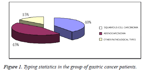
We did pathological typing statistics in the group of gastric cancer patients, of which we found that 6 (6/20) had squamous cell carcinoma, 12 (12/20) adenocarcinoma, and 2 (2/20) other pathological types, as shown in Table 3 and Figure 1.
| Total number of cases (n) | Squamous cell carcinoma (n) | Adenocarcinoma (n) | Other pathological types |
|---|---|---|---|
| 20 | 6 (6/20) | 12 (12/20) | 2 (2/20) |
Table 3: Clinical pathological type of patients.
Expression of MAP4K4 in normal tissue adjacent to cancer and cancer tissue
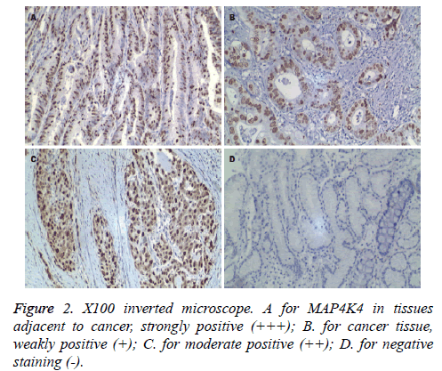
MAP4K4 gene can be expressed in both normal tissues and cancer tissues. As immunohistochemistry results showed, MAP4K4 gene increased in diseased tissue, the difference of which was statistically significant (P<0.05). We divided immunohistochemistry results according to (-)~(), as shown in Figures 2A-2D.
Regression analysis on kinds of influencing factors of prognosis
According to previous literature and reports, we pointedly selected pathological types relevant to gastric cancer expression. Moreover, we made univariate analysis on whether distant metastasis occurred, carcinoma tissue volume, recurrence situation, and patients’ age and weight, etc. discovering that level of MAP4K4 gene expression was correlated with lymph node metastasis and pathological staging of gastric cancer in cancer tissue (P<0.05). However, it had no relation to tumor size of gastric cancer and recurrence and metastasis, etc. (P>0.05), as shown in Table 4.
| Variable (n=20) | Cases number | 1 (>++) | P |
|---|---|---|---|
| Age (years) | 0.149 | ||
| ≥ 45 | 12 | 4 (4/12) | |
| <45 | 8 | 3 (3/8) | |
| Weight (kg) | 0.135 | ||
| ≥ 60 | 9 | 5 (5/9) | |
| <60 | 11 | 5 (5/11) | |
| Tumor diameter (cm) | 0.372 | ||
| ≥ 10 | 8 | 5 (5/8) | |
| 5~10 | 3 | 2 (2/3) | |
| ≤ 5 | 9 | 47 (4/9) | |
| Degree of differentiation | 0.451 | ||
| Poor differentiation | 3 | 1 (1/3) | |
| Middle differentiation | 9 | 6 (6/9) | |
| High differentiation | 8 | 8 (8/9) | |
| Tumor types | 0.563 | ||
| Squamous cell carcinoma | 4 | 4 (4/4) | |
| Adenocarcinoma | 12 | 5 (5/12) | |
| Other pathological types | 4 | 2 (2/4) | |
| TNM stage | <0.001 | ||
| I | 5 | 4 (4/5) | |
| II | 7 | 4 (4/7) | |
| III | 4 | 1 (1/5) | |
| IV | 4 | 2 (2/4) | |
| Regional lymph node staging | <0.001 | ||
| Nx | 2 | 0 (0/2) | |
| N0 | 5 | 1 (1/5) | |
| N1 | 7 | 4 (4/7) | |
| N2 | 4 | 1 (1/4) | |
| N3 | 2 | 0 (0/2) | |
| Distant metastasis | 0.783 | ||
| Mx | 8 | 2 (2/8) | |
| M0 | 3 | 3 (0/3) | |
| M1 | 9 | 3 (3/9) | |
| Helicobacter pylori infection history | 0.144 | ||
| Yes | 18 | 16 (16/18) | |
| No | 2 | 0 (0/2) | |
| Radiation exposure history | 0.579 | ||
| Yes | 9 | 7 (7/9) | |
| No | 11 | 4 (4/11) | |
| Family history | 0.483 | ||
| Yes | 13 | 8 (8/13) | |
| No | 7 | 3 (3/7) | |
Table 4: Univariate analysis on kinds of influencing factors of prognosis.
Comparison of progression free survival rates
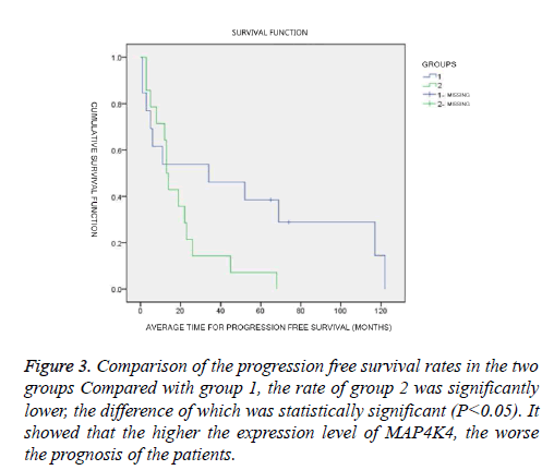
We divided patients into groups according to the expression level of MAP4K4, and -~+ was for group 1, ++~+++ for group 2. The analysis found that, in group -~+, average time for progression free survival was (49.80 ± 14.42) months; median time (34.00 ± 27.56) months; and in group ++~+++, average time for progression free survival (19.5 ± 74.76) months, median time (13.00 ± 1.25) months. Comparing the progression free survival of the two groups, the difference had statistical significance (χ2=13.167; P=0.005), as shown in Table 5 and Figure 3.
Figure 3: Comparison of the progression free survival rates in the two groups Compared with group 1, the rate of group 2 was significantly lower, the difference of which was statistically significant (P<0.05). It showed that the higher the expression level of MAP4K4, the worse the prognosis of the patients.
| Groups | Average time for progression free survival (Months, ± s) | Median time for progression free survival (Months ± s) | χ2 | P |
|---|---|---|---|---|
| Group -~+ | 49.80 ± 14.42 | 34.00 ± 27.56 | 13.167 | 0.005 |
| Group++~+++ | 19.57 ± 4.76 | 13.00 ± 1.25 |
Table 5: Comparison of progression free survival rates in two groups by Kaplan-Meier method.
Comparison of total survival time
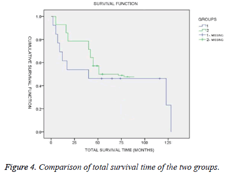
The average and median total survival time of MAP4K4 in patients in group -~+ and ++~+++-~ were compared, and in group -~+, the average total survival time was (65.00 ± 116.70months), the median (50.00 ± 39.90) months, the difference of which was statistically significant (χ2=10.084; P=0.003). We considered there was some relation between the expression level of MAP4K4 and total survival time of patients, as shown in Table 6 and Figure 4.
| Groups | Average total survival time (Months, ± s) | Median total survival time (Months, ± s) | χ2 | P |
|---|---|---|---|---|
| Group -~+ | 65.00 ± 116.70 | 50.00 ± 39.90 | 10.084 | 0.003 |
| Group++~+++ | 51.03 ± 6.79 | 51.00 ± 4.44 |
Table 6: Comparison of total survival time.
Compared with group 1, the time of group 2 was significantly lower, the difference of which was statistically significant (P<0.05). It showed that the higher the expression level of MAP4K4, the worse the prognosis of the patients.
Discussion
Gastric cancer is ranked the top five in common cancers, and at present, in China's elderly patients over 60 years old, the morbidity of patients with gastric cancer has a increasing trend year by year. Due to symptoms of early gastric cancer are similar to that of gastritis, usually presenting gastralgia, stomach discomfort, acid reflux and other non-specific symptoms mainly. Therefore, the diagnosis of it is difficult, and it seriously affects the patient's quality of life. Thus, it has very important significance for understanding the clinical prognosis of patients and rationally using medical resources to conduct the diagnosis of gastric cancer and clinical prognostic analysis for patients.
MAP4K4 is abbreviation of MAPK kinase isoform 4, belongs to the Mitogen Activated Protein Kinase (MAPK) family. MAPK family is a conserved serine/threonine protein kinase system and plays a very important role in the process of extracellular signal delivering to cytoplasm [3]. The main function of MAPKs includes the regulation of cell growth, migration, differentiation, and apoptosis and stress related reactions [3]. Liu et al. used biological information analysis and quantitative polymerase chain reaction for detecting expression level of MAP4K4 in gastric carcinoma, and eventually found in gastric cancer, the expression level of MAP4K4 was significantly increased, the conclusion of which were basically similar to ours [20]. Using small interfering RNA to knock MAP4K4 genes in cancer cells, we found that the proliferation of gastric cancer cells received significant inhibition, and most cells stalled in the G1 period. The results showed that, after the silence of MAP4K4, the apoptosis of cancer cells could be induced and increased by raising the rate of Bax/Bcl-2. And previous studies on Bax/Bcl-2 showed that it played a main role in regulating cell proliferation and apoptosis [20].
In our study, the average height of 20 cases gastric cancer patients was (168.4 ± 29.8) cm, and weight (66.9 ± 18.7) kg, (56.7 ± 12.3) years of age, BMI (21.4 ± 4.8) kg/m2, blood oxygen saturation (93.5 ± 9.8), red blood cell count (3.29 ± 1.27) × 109; haemoglobin (56.5 ± 12.7) g/L; average heart rate (102.4 ± 9.7) times/min.
The average ages of the patients were close to 60 years old, which was consistent with the age of high incidence in gastric cancer in China. Blood oxygen saturation of patients was (93.5 ± 9.8%), and red blood cell count (3.29 ± 1.27) × 109; haemoglobin (56.5 ± 12.7) g/L, which were lower than the normal reference values, which we thought was related to the cancer tissue invasion and anaemia caused by chronic consumption, and anaemia caused secondary decreasing of patients’ blood oxygen saturation. After screening, no collected patients with gastric cancer were found heart disease and lung disease, whose pulmonary functions were normal. The symptoms of anaemia may have relationship with patients’ primary gastric cancer diseases.
In addition, as also found in our study, in 20 patients, 9 (9/20) patients had a history of radiation exposure; 18 (18/20) patients Helicobacter pylori infection; 13 (13/20) cases family history; 14 (14/20) cases family history of gastric cancer; 7 (7/20) patients a history of smoking; 11 (11/20) cases a history of drinking; 8 (8/20) cases reflux esophagitis. 6 cases were squamous cell carcinoma, and 12 (12/20) cases were adenocarcinoma; 2 (2/20) cases other pathological types of. We further understood the patients’ medical history by questionnaires about them; COX regression analysis showed that the expression level of MAP4K4 gene in cancer tissues had something to do with lymph node metastasis and stage of gastric carcinoma (P<0.05), and nothing to do with tumor size and recurrence, metastasis, etc. factors of gastric cancer (P>0.05). This showed that the detection of MAP4K4 level in cancer tissues had certain implications for indicating the pathological types of patients and the risk degree of lymph node metastasis. The higher the malignancy degree of gastric cancer, the higher the expression level of MAP4K4. This was inseparable from the regulation function of MAP4K4, and after silence, the expression levels of Notch signalling pathway could be reduced, which activated Bax/Bcl-2 and started cell apoptosis.
Though further analysis by Kaplan-Meier on the relationship between expression level of MAP4K4 and median survival time as well as total survival time, we finally found that in group -~+, average time for progression free survival was (49.80 ± 14.42) months; median time (34.00 ± 27.56) months; and in group ++~+++, average time for progression free survival (19.57 ± 4.76) months, median time (13.00 ± 1.25) months. Comparing the progression free survival of the two groups, the difference had statistical significance (χ2=13.167; P=0.005). And in group -~+, the average total survival time was (65.00 ± 116.70 months), the median (50.00 ± 39.90) months, the difference of which was statistically significant (χ2=10.084; P=0.003). We believed that in the gastric cancer tissues, high expression levels of MAP4K4 usually indicated strong proliferation state and usually the higher the expression level of MAP4K4, the worse the prognosis or outcomes of patient, which were consistent with previous studies of gastric cancer. By detecting the expression level of MAP4K4 in cancer tissues of patients, it has a certain indicating function on the prognosis and survival situation of patients.
In summary, we believe that it has important theoretical and clinical significance to detect expression level of MAP4K4 in patients with gastric cancer for indicating patients’ survival and prognosis as well as severity of disease, which is also of some economic significance for tips on mortality risk in patients with gastric cancer, rational use of medical resources and reduction of the burden on the family.
References
- Vogelaar IP, van der Post RS, Bisseling TM, van Krieken JH, Ligtenberg MJ, Hoogerbrugge N. Familial gastric cancer: Detection of a hereditary cause helps to understand its etiology. Hered Cancer Clin Pract 2012; 10: 18.
- Jemal A, Clegg LX, Ward E, Ries LA, Wu X, Jamison PM, Wingo PA, Howe HL, Anderson RN, Edwards BK. Annual report to the nation on the status of cancer, 1975–2001, with a special feature regarding survival. Cancer 2004; 101: 3-27.
- Turjanski AG, Vaque JP, Gutkind JS. MAP kinases and the control of nuclear events. Oncogene 2007; 26: 3240-3253.
- Collins CS, Hong J, Sapinoso L, Zhou Y, Liu Z, Micklash K, Schultz PG, Hampton GM. A small interfering RNA screen for modulators of tumor cell motility identifies MAP4K4 as a promigratory kinase. Proc Natl Acad Sci USA 2006; 103: 3775-3780.
- Qiu MH, Qian YM, Zhao XL, Wang SM, Feng XJ. Expression and prognostic significance of MAP4K4 in lung adenocarcinoma. Pathol Res Pract 2012; 208: 541-548.
- Vitorino P, Yeung S, Crow A, Bakke J, Smyczek T, West K, McNamara E, Eastham-Anderson J, Gould S, Harris SF. MAP4K4 regulates integrin-FERM binding to control endothelial cell motility. Nature 2015; 519: 425-430.
- Danai LV, Flach RJ, Virbasius JV, Menendez LG, Jung DY, Kim JH, Kim JK, Czech MP. Inducible deletion of protein kinase Map4k4 in obese mice improves insulin sensitivity in liver and adipose tissues. Mol Cell Biol 2015; 35: 2356-2365.
- Yue J, Xie M, Gou X, Lee P, Schneider MD, Wu X. Microtubules regulate focal adhesion dynamics through MAP4K4. Dev Cell 2014; 31: 572-585.
- Wright JH, Wang X, Manning G, Lamere BJ, Le P, Zhu S, Khatry D, Flanagan PM, Buckley SD, Whyte DB, Howlett AR, Bischoff JR, Lipson KE, Jallal B. The STE20 kinase HGK is broadly expressed in human tumor cells and can modulate cellular transformation, invasion, and adhesion. Mol Cell Biol 2003; 23: 2068-2082.
- Feng XJ, Pan Q, Wang SM, Pan YC, Wang Q, Zhang HH, Zhu MH, Zhang SH. MAP4K4 promotes epithelial-mesenchymal transition and metastasis in hepatocellular carcinoma. Tumour Biol 2016.
- Haas DA, Bala K, Busche G, Weidner-Glunde M, Santag S, Kati S. The inflammatory kinase MAP4K4 promotes reactivation of Kaposis sarcoma herpesvirus and enhances the invasiveness of infected endothelial cells. PLoS Pathog 2013; 9: e1003737.
- Machida N. Mitogen-activated protein kinase kinase kinase kinase 4 as a putative effector of Rap2 to activate the c-Jun N-terminal kinase. J Biol Chem 2004; 279: 15711-15714.
- Yao Z. A novel human STE20-related protein kinase, HGK, that specifically activates the c-Jun N-terminal kinase signalling pathway. J Biol Chem 1999; 274: 2118-2125.
- Read MA. Tumor necrosis factor alpha-induced E-selectin expression is activated by the nuclear factor-kappa B and c-JUN N-terminal kinase/p38 mitogen-activated protein kinase pathways. J Biol Chem 1997; 272: 2753-2761.
- Zhao Y, Li F, Zhang X, Liu A, Qi J. MicroRNA-194 acts as a prognostic marker and inhibits proliferation in hepatocellular carcinoma by targeting MAP4K4. Int J Clin Exp Pathol 2015; 8: 12446-12454.
- Roth Flach RJ, Skoura A, Matevossian A, Danai LV, Zheng W, Cortes C, Bhattacharya SK, Aouadi M, Hagan N, Yawe JC, Vangala P, Menendez LG, Cooper MP, Fitzgibbons TP, Buckbinder L, Czech MP. Endothelial protein kinase MAP4K4 promotes vascular inflammation and atherosclerosis. Nat Commun 2015; 21: 8995.
- Liu AW, Cai J, Zhao XL, Jiang TH, He TF. ShRNA-targeted MAP4K4 inhibits hepatocellular carcinoma growth. Clin Cancer Res 2011; 17: 710-720.
- Meng Z, Moroishi T, Mottier-Pavie V, Plouffe SW, Hansen CG, Hong AW, Park HW, Mo JS, Lu W, Lu S, Flores F, Yu FX, Halder G, Guan KL. MAP4K family kinases act in parallel to MST1/2 to activate LATS1/2 in the Hippo pathway. Nat Commun 2015; 6: 8357.
- Schwaid AG, Su C, Loos P, Wu J, Nguyen C, Stone KL, Kanyo J, Geoghegan KF, Bhattacharya SK, Dow RL, Buckbinder L, Carpino PA. MAP4K4 is a threonine kinase that phosphorylates FARP1. ACS Chem Biol 2015; 10: 2667-2671.
- Yuan FL, Guo QQ, Yun ML, Wu MK, Yuan L, Wei XC, Xiao HL. Silencing of MAP4K4 by short hairpin RNA suppresses proliferation, induces G1 cell cycle arrest and induces apoptosis in gastric cancer cells. Mol Med Rep 2016; 13: 41-48.


