Research Article - Biomedical Research (2017) Health Science and Bio Convergence Technology: Edition-II
Study on the injury of shoulder joint and should rotator cuff of tennis players in receiving ball
Haolin Fang1, Jian Li2, Ru Gong3, Yaping Liu4*1Department of Emergency Trauma Surgery, Shandong Jining No.1 People’s Hospital, PR China
2Department of Bone and Joint Surgery, Shandong Jining No.1 People’s Hospital, PR China
3Department of Hand and Foot Surgery, Shandong Jining No.1 People’s Hospital, PR China
4Department of Endocrinology, Shandong Jining No.1 People’s Hospital, PR China
- *Corresponding Author:
- Yaping Liu
Department of Endocrinology
Shandong Jining No.1 People’s Hospital, PR China.
Accepted date: April 30, 2017
Abstract
Background: The most common sports injury of the tennis players in the receiving ball is the injury of the shoulder joint and the shoulder rotator cuff. This research was aimed to find the diagnosis of shoulder joint injury and exploration of injury causes when receiving ball.
Methods: 31 patients with shoulder joint injury caused by the receiving ball in three hospitals were selected from 2010-2015. MRI was used if X-ray cannot confirm the result. The diagnosis was performed by arthroscopy for serious rotator cuff injury. The Zlatkin classification of MRI was used as the diagnostic criterion.
Results: In the rotator cuff injury, the first level was the largest, the second level was less, and the third level only had 3 cases. Further analysis of the injury showed that the receiving injury of tennis players was mainly caused by inadequate preparation activities.
Conclusion: The MRI imaging technology can clearly reflect the rotator cuff injury, which can be widely used in the clinical diagnosis of tennis players receiving injury.
Keywords
Tennis ball, Receiving, Shoulder joint, Rotator cuff, Injury.
Introduction
In 2014, during the Australian Open women's singles final, Li Na beat her opponent to win the championship, obtaining the second Grand Slam champions of her career, with the world ranking rising to the second. This reflects China's rapid development of tennis to a certain extent in recent years, such as Zheng Jie, Yan Zi, Peng Shuai and a number of excellent athletes have made a number of world class championships in international competition [1]. Although the tennis sport entered in China at the end of 1980s, it has had rapid development in the beginning of twenty-first century, and now it has become a national movement [2]. The popularity of tennis in China, to a certain extent, has improved people's physical quality, and enriched people's leisure activities. However, it is hard for athletes to avoid being injured in sports because the sport belongs to competitive sports with high speed and high strength. It is helpful for people to understand tennis sports injury, so as to avoid and reduce the tennis sports injury, and improve the level of tennis in our country, to research the cause of injury and damage in the process of tennis.
The key link in the tennis movement is the receiving stage, which is one of the key points to win the game [3]. Because of the fast running and high strength swing ball in the receiving ball, the tennis player may have the shoulder injury, and even fall in the field due to the body instability. In 2009, at the Wimbledon tournament, the famous tennis player Federer accidentally slipped in the receiving ball. Although there was no serious damage, he lost in the game. The receiving injury of tennis players is often shoulder accompanied by shoulder rotator cuff injury, so the use of MRI imaging technology can be more clear understanding of the situation, to facilitate the treatment. The data of tennis players with shoulder joint injury caused by the receiving at three municipal hospitals of one province are analysed to understand the causes of injuries and medical imaging diagnosis, which has very important significance for the diagnosis of shoulder and rotator cuff injury and for tennis players to avoid injury. Since entering into our country, tennis sports have been deeply loved by people. In recent years, tennis has become popular, and it has become a national fitness campaign [4]. In international competitions, the level of China's outstanding tennis players has also been greatly improved, and they have achieved good results. In order to improve tennis sports level, our country also vigorously develops the occupation of tennis, and there is a large number of professional tennis players trained active in the sports world [5]. The tennis has the characteristics of high antagonism. In order to get a point, athletes often make many rounds during the competition. It not only requires a very high skill, but also requires a strong explosive force, very ornamental in the game. A very important part in the tennis competition is receiving, and due to the presence of the receiver and the server, it requires the receiver to have better receive technology [6]. A player with good access to serve technology, on the one hand, can avoid the other side to get the opportunity to use the ball, thus interrupting the opponent's offensive rhythm. And on the other hand, he will make a psychological victory over each other. In contrast, poor reception may cause the other side to attack and score, and there will be more tension and fear psychologically. It is shown that receiving technology has a very important position in tennis, which is a very important link to improve the level of tennis [7].
Because of the athletic rules of tennis, when serving, the server must serve in the diagonal direction. On the basis of this, the technical movement of the tennis ball was studied, including the grip, stance and position, stroke and follow up [8]. Among them, the preparation stage is the initial stage of the action, such as Federer turns the racket, and no matter what kind of action is in order to let him in a relatively stable stage. At this time, the body has remained relatively stable in the vertical direction, and the main body of the body does not have a greater range of angular variations [9]. In order to take reasonable strategy of receiving, the receiver needs to observe the server’s details of movements, to predict the direction and rotating velocity of the ball, and through the advance preparation to make him adjust to the best catch. In this stage, the tennis players often use small steps or the body slightly shaking for the anticipation, and his hip angle has a certain change. Before stroke, athletes begin to split step, and start the racket, and racket moves relatively quickly, and with the body smaller begins to turn back with small extent [10]. The athlete swinging forward and hitting the ball to the other court process is the stroke stage, which is the most important stage of receiving, until the last single leg landing support. In this process, the body erects, takes-off and swings to hit the ball, thus completing the whole process of receiving. To better complete the receiving action, the receiver needs to have a keen observation, strong body conditioning and explosive power. For the whole body coordination movement, the role of the shoulder is very important [11].
The tennis serve needs strong power, which makes the players very easily injured because of improper movement, and it also brings a serious threat to the health of athletes [12]. In particular, for many amateur tennis players, the body joints or muscle damage is often caused by inadequate preparation as well as the excessive movement, and they are even subject to a long time of the disease. The research on tennis sports injury is helpful for people to make scientific tennis, as far as possible to avoid the damage caused by improper movement.
Shoulder joint and rotator cuff injury are very common in sports. Because, with higher frequency of use of shoulder joint in sports, shoulder joint is also the guarantee of human power and rotation, and the damage in the movement is mainly fracture or soft tissue injury [13]. The rotator cuff is a tendon complex around the humeral head, the upper and lower, respectively the supraspinatus tendon and the infraspinatus tendon, and behind is the infraspinatus tendon and small muscle tendon. Rotator cuff tendon is the support of the rotation of the shoulder joint and the lifting activity. When the rotator cuff is injured, it will show pain and pressure pain. During the shoulder activities, the exacerbation of pain will appear, with shoulder joint function limitation and sound. When the rotator cuff injury is severe, it may even lead to the atrophy of the rotator cuff muscles. According to the different damage conditions, the rotator cuff injury is divided into three types, which are bruise, incomplete fracture and complete fracture [14]. The most serious is the complete fracture. All the tendons are broken and the shoulder function is almost completely lost. Incomplete fracture is partial fracture of the rotator cuff tendon, which is divided into I, II, and III according to the different tearing depths. Contusion is relatively mild damage, which is generally the congestion and edema of the rotator cuff tendon caused by the impact of the shoulder, the backlog or the pull and so on [15]. In addition, for the shoulder joint movement injury, more cases are dislocation of the shoulder with greater external force, which is divided into anterior dislocation and posterior dislocation. For the dislocation of the shoulder joint injury, there will be pain and activity is limited. The head and trunk will tilt to the injured side, and the hand cannot be put on the side of the shoulder [16].
The diagnosis of shoulder joint injury is made by medical imaging data with clinical palpation method. The clinical manifestations of patients are used for the comprehensive analysis and judgment, and to choose the appropriate treatment plan. The commonly used medical imaging techniques in the diagnosis of shoulder joint sports injuries contain X-ray film, MRI, CT, and arthroscopy, of which the X-ray film and MRI are the main methods [17]. The X-ray film is often used for the first to do a preliminary diagnosis for the shoulder injury. For the presence of a fracture or dislocation, it can be judged according to the X-ray film. If there is damage to the soft tissue of the shoulder joint, we can choose whether to carry on the MRI and arthroscopic examination according to the situation. The X-ray film can clearly show the skeleton of the shoulder joint. When there is a fracture, the injury degree of the shoulder joint can be analysed according to the X-ray film. Although the X-ray film has the characteristics of convenient, fast, and low cost, it is difficult to accurately image the damage of soft tissue, and the image is not clear enough. Therefore, if necessary, MRI imaging technology is required. Despite the high cost, long time, MRI is relatively sensitive, strong discrimination [18]. For rotator cuff injury, the empty pot test, the test of the muscle strength of the lower muscle and the Neer positive sign also can be used. But no matter what medical imaging technology is used for diagnosis, it is necessary to combine the clinical manifestation of the patients to make a comprehensive judgment. Compared with MRI imaging technology, shoulder joint radiography is the most accurate diagnosis of rotator cuff tears, especially when the rotator cuff tear is less than 1 cm, the shoulder joint noise can be accurately diagnosed.
For the shoulder joint sports injury gradually popular with tennis movement, the X-ray film and MRI imaging technology can basically meet the diagnosis of the patients [19]. There are two main causes of injury when the tennis players receive the service. One is the injury caused by excessive damage. And on the other hand, it is fall caused by improper action of foot during receiving. So it is generally common to be a shoulder joint dislocation or a rotator cuff injury. The analysis of the injury of shoulder joint based on medical image technology is helpful to improve the accuracy of clinical diagnosis. At the same time, it can also be combined with injury cause to enhance the professional tennis players, and reduce the damage caused during the receiving stage [20].
Materials and Methods
Research objects
The objects of this study are the tennis players injured, and through the investigation, the injury caused by the relevant reasons of the receiving action is screened out. In the actual cases, the number of patients is relatively small. In order to increase the number of samples collected, the time and scope are expanded, and the data of patients at three hospitals in 2010-2015 are collected. The selection criteria include: (1) the clinical symptom is shoulder injury, obvious. (2) All of the patients are performed with X-ray film or further MRI and other medical imaging examinations. (3) The patients are willing to receive the appropriate examination of the shoulder joint. (4) The direct cause of injury is the result of the tennis ball receiving. (5) The patients have personal information records and case data, and they are willing to cooperate with the study after searching. After screening, there are a total of 31 patients, of which 18 cases are male, 13 cases of female, and male and female ratio is 1.38:1. The average age of the patients is 30.5 y old, the youngest is 14 y old, and the oldest is 39 y old. According to the relevant information and view statistics, the main reasons for the injured patients include too large receiving swing, fall, hitting mistakes, and insufficient preparation. Most of the patients are not hospitalized, and there are 3 cases of hospitalized patients with severe shoulder injury. Patient's personal information is shown in Table 1.
| Variable | Numerical value | |
|---|---|---|
| Age | Average age | 30.5 |
| Max/min | 39/14 | |
| Gender | Male/female | 18/13 |
| Male to female ratio | 1.38:1 | |
| Cause of injury | Excessive movement | 5 |
| Fall | 4 | |
| Action error | 13 | |
| Inadequate preparation | 9 |
Table 1: Clinical data of patients with shoulder joint injury.
It is also because the cause of injury patients is too large swing and insufficient preparation; the damage is more of the rotator cuff injury. There is a fracture or bone dislocation in some patients companied with a soft tissue injury of the rotator cuff. Therefore, the follow-up study should focus on the research of the rotator cuff tissue injury.
Research methods
In the hospital, all patients are performed with X-ray camera after simple palpation. Through the view of the X-ray film, further determination should be made whether do the MRI inspection or not to confirm the extent of soft tissue damage. When the complexity of the disease is difficult to accurately judge, the shoulder joint radiography technique will be used. All imaging findings are examined by more than 2 experienced physicians. When larger differences appear or it is difficult to diagnose, the department will do the consultation. When they read the film, they should carefully observe the change of the size and structure of the humeral head on the glenohumeral joint, and understand the changes of soft tissue, rotator cuff tendon, acromion inferior.
X-ray photography uses front and rear position, rear and front position, and front and back lying position, focusing on the front and rear position. At this time, according to the specific circumstances of the patient, stand or lie can be chosen. But in the center line of shooting, it must be aligned perpendicular above the scapula glenoid. If the acromioclavicular joint is subluxation, the gap width of the shoulder lock joint is more than 0.5 cm. In the complete dislocation, the distal clavicle is upturned, and the joint gap is very obvious. When using MRI examination, the head of patient takes the first supine, to ensure that the shoulder joint is as far as possible with the bed line alignment. The shoulder is in the horizontal direction, the forearm with flexion extending in the abdomen. The upper arm is close to the chest wall, and the two ends of the upper arm are fixed. And a neutral position scanning is generally taken. MRI scanning of the shoulder joint is generally divided into oblique coronal site and oblique sagittal site and axial position. The imaging results obtained should be able to clearly show the shape of the tendon, contour and the muscle, with no artifact.
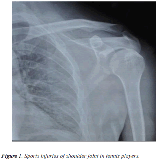
Take a case for example. The receiver’s running rate is too large and he does not adjust the pace, resulting in the fall and the shoulder joint injury. In the hospital, the patient has 10 degrees of front lift, 45 degrees of flexion abduction, not limited to basic level, and subacromial tubercle area has apparent tenderness. After the initial reading of the X-ray film, it is judged as the left humeral large nodule avulsion fracture, and with a certain degree of soft tissue injury. And the MRI check is made to confirm the left shoulder sleeve is not completely torn. The corresponding imaging results are shown in Figure 1.
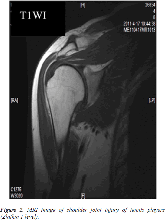
The tennis players receive injury is more of the injured rotator cuff muscle group. Therefore, imaging examination based on the MRI is the main basis for diagnosis. MRI diagnosis is used to observe whether the shape and signal change of the rotator cuff interruption, atrophy, thinning, thickening and other exists. Diagnostic criteria for reference Zlatkin are classified into four types. (1) Zero level: the signal and morphology of the shoulder tendon is normal, and the tendon is in good condition. (2) The first level: there is a local signal abnormality, but the tendon continuity still maintains a good shape. (3) The second level: although the continuity of the tendon is still present, but its shape is irregular, the signal on T1WI and the proton image has the limited increase. (4) The third level: tendon continuity is obviously interrupted, showing irregular shape, often accompanied by the subacromial bursa effusion, increasing signal on the T1WI and proton.
Results
After medical imaging examination, according to MRI classification statistics, there is 0 cases of zero level, 18 cases of the first level, 10 cases of the second level, and 3 cases of the third level. Among them, 11 cases show full-thickness supraspinatus tear, 13 cases of partial tear of the supraspinatus, 9 cases of infraspinatus tear, 6 cases of subscapularis injury, and 18 cases of trees minor injury, 4 cases of avulsion fracture, 2 cases of tendon calcification, and 20 cases of articular cavity effusion. There are 3 cases of the tear of the shoulder lower tendon with a tear of supraspinatus, and the image is presented as a high signal of the consolidated anterior tendon of the small nodule. There are 2 cases with calcification of the supraspinatus; and 1 case is accompanied with the infraspinatus tear. In the tear of teres minor, its edge is blurred and patchy high signal appears in nodules, and T2WI shows high signal in the tendon. There are 4 cases of the avulsion fracture of the large nodule on the humerus, long T1 and T2 signal displaying on MRI image. The specific imaging results are shown in Table 2.
| Name | The number of cases |
|---|---|
| Full thickness tear of the muscle of the upper | 11 |
| Partial tear of the upper part of the | 13 |
| Tear of the lower | 9 |
| Injury of the muscle of the shoulder | 6 |
| Teres minor injury | 18 |
| Avulsion fracture | 4 |
| Calcification of tendon | 2 |
| Effusion of joint cavity | 20 |
Table 2: Image detection results of shoulder joint injury.
According to the Zlatkin classification, 3 cases of specific description are made, the first level, the second level and the third level of Zlatkin. The Figure 2 is the first level of Zlatkin; normal morphology of the supraspinatus tendon, slightly higher signal, continuous running, and T2WI shows a slightly higher signal. The patients have inadequate preparation, and feel discomfort of shoulder when swinging. Although there is no obvious press pain, the activity is accompanied with local restrictions. This is also a kind of more common injury when a tennis player receives the ball. This injury can be treated with a simple treatment.
Take a tennis player injured when receiving as an example. He fails to grasp the physical center of gravity, and the body falls down to the ground, resulting in damage to the shoulder joint. In addition to the obvious visible trauma on the surface, there is a certain degree of redness and swelling, pain in the shoulder joint, difficult to go to sleep. The patient is admitted to hospital after imaging diagnosis. The diagnosis results are as follows: the third grade of Zlatkin, complete rupture of supraspinatus tendon, total rupture of the tendon on the large nodules of the humerus, interrupted continuity. There is a certain degree of retraction at the end of the break. In order to further examine the patient's shoulder soft tissue damage, arthroscopy is used for in-depth examination.
Discussion
Through the research and analysis, we can see that for tennis players in the course of receiving, the rotator cuff muscles is more easy to be injured, resulting in rotator cuff tear, to a certain extent. Because the rotator cuff tissue is the organization to maintain the normal activities of the shoulder joint, when the rotator cuff muscle group is injured, it will affect the tennis player's shoulder joint activities, which is more difficult to carry out sports activities. Rotator cuff injury in shoulder joint injury is mainly the first level and the second level of Zlatkin. Under normal circumstances, unless it is a tennis player in the case of a faster speed and a larger force to hit the ground or the surrounding wall, it may cause the third level of Zlatkin damage. It is also the case, the main causes of damage of tennis player when receiving are inadequate preparation and too large swing force. In order to reduce the damage, the players should be fully prepared before the sports activities. In particular, at the beginning of movement, the players should gradually increase their strength as well as their intensity step by step.
Although the X-ray film is a common medical imaging diagnosis technology, for the damage of tennis players to receive the ball, the MRI effect is better, which can clearly reflect the rotator cuff muscle damage. Through the Zlatkin grading standard, we can accurately carry on the shoulder sleeve damage grading diagnosis judge, thus formulating the corresponding treatment plan. When the shoulder joint injury is more complex, it is necessary to use arthroscopy to further check, using the joint inspection to accurately determine the extent of damage. According to the results of the diagnosis, the treatments of patients generally include nonsurgical treatment, surgical treatment, and open treatment. Because of the relatively low degree of injury of the shoulder joint, the tennis player can use the nonsurgical treatment, and use surgical treatment for serious injury.
In order to make the study on the shoulder and rotator cuff injury of tennis players caused in the receiving stage, patients with shoulder joint injury of three hospitals in one province during 2010-2015 are retrospectively analysed. According to the inclusion criteria, 31 patients are selected, the average age is 30.5 years old, and the ratio of male to female is 1.38:1. By the reason analysis of injury, it can be seen that insufficient preparation and too large swing are the main reasons, and the rotator cuff muscle damage is the main form in the shoulder joint damage. All patients are first carried out with X-ray radiography, and then MRI is used to confirm the degree of damage to the soft tissue. In the case of complex injury, arthroscopy is combined. It can be seen through the image inspection that 11 cases show full-thickness supraspinatus tear, 13 cases of partial tear of the supraspinatus, 9 cases of infraspinatus tear, 6 cases of subscapularis injury, and 18 cases of teres minor injury, 4 cases of avulsion fracture, 2 cases of tendon calcification, and 20 cases of articular cavity effusion. According to MRI classification statistics, there is 0 case of zero level, 18 cases of the first level, 10 cases of the second level, and 3 cases of the third level. Though the further analysis, it is shown that the main causes of damage of tennis player when receiving are inadequate preparation and too large swing force. In order to reduce the damage, the players should be fully prepared before the sports activities and gradually increase their strength. Because the number of samples is only 31 cases, it is difficult to reflect the general situation within a larger range. Therefore, the follow-up study needs to continue to expand the number of research samples, in order to obtain a more accurate conclusion.
References
- Zhang Y, University AT. Comparative study on development chinese tennis and tennis in Czech. Contemp Sport Technol 2015.
- Chen X, Sun CL. On the development of professional quality of Chinese tennis players exemplified by LI Na. J Phys Edu 2014.
- Zhou W, Zhenmin Z, Shang X. Research on the neurobiological features of the Chinese elite woman table tennis players. J Sci Med Sport 2015; 19: 55.
- Wei-Guo LI, University CS. Promotion strategy research on chinese mens tennis competitive level. J Chengdu Sport Univ 2014.
- Zhao LX, Liu BH. Chinese tennis professionalization: research progress and problem analysis. J Hebei Inst Phys Edu 2014.
- Whiteside D, Elliott BC, Lay B, Reid M. Coordination and variability in the elite female tennis serve. J Sports Sci 2015; 33: 675-686.
- Chiang WWF, Wang YT, Liang LC. The relationships between tennis serve and neuromuscular control in young athletes. Northeast Asia Conference on Kinesiology 2015.
- Murata M, Fujii N, Suzuki Y. Mechanical energy flow of the racquet holding arm in the tennis serve, focusing on the energy form. Jap J Phys Edu Health Sport Sci 2015; 60: 177-195.
- Mendes P, Couceiro M, Rocha R. Effects of an extrinsic constraint on the tennis serve. Int J Sport Sci Coach 2015; 10: 1-14.
- Cosac G, Ionescu DB. Research approach for outlining the biomechanical parameters of the tennis serve. Palestrica of the Third Millennium Civilization Sport 2015.
- Sam KL, Smith AW. Tennis serves performance in rested and exhaustion conditions of college-level players. Int J Develop Res 2015; 5: 4418-4412.
- Ma Y. Investigation on the sport injuries of Chinese excellent tennis athletes. Sports Forum 2011.
- Citak M, Grasmucke D, Cruciger O, Konigshausen M, Meindl R. Heterotopic ossification of the shoulder joint following spinal cord injury: an analysis of 21 cases after single-dose radiation therapy. Spinal Cord 2016; 54: 303-305.
- Thakrar GN, Patel DA, Sejpal JJ. Effect of physiotherapy rehabilitation in acute burn injury around shoulder joint. Ind J Physiother Occup Ther 2015; 9.
- Seminati E, Marzari A, Vacondio O. Shoulder 3D range of motion and humerus rotation in two volleyball spike techniques: injury prevention and performance. Sports Biomech 2015; 14.
- Yi Y, Kim JW. Coronal plane radiographic evaluation of the single Tight rope technique in the treatment of acute acromioclavicular joint injury. J Am Shoulder Elbow Surg 2015; 24:1582-1587.
- Rogowski I, Creveaux T, Genevois C. Upper limb joint muscle/tendon injury and anthropometric adaptations in French competitive tennis players. Eur J Sport Sci 2015; 16: 1-7.
- Madi S, Pandey V, Khanna V. A dual injury of the shoulder: acromioclavicular joint dislocation (type IV) coupled with ipsilateral mid-shaft clavicle fracture. BMJ Case Rep 2015; 2015.
- Castelein B, Cools A, Parlevliet T. Modifying the shoulder joint position during shrugging and retraction exercises alters the activation of the medial scapular muscles. Manual Ther 2015; 21: 250-255.
- Fyhr C, Gustavsson L, Wassinger C, Sole G. The effects of shoulder injury on kinaesthesia: a systematic review and meta-analysis. Man Ther 2015; 20: 28-37.

