Research Article - Biomedical Research (2017) Volume 28, Issue 6
Sexual dimorphism in permanent canine teeth and formulas for sex determination
Zeinab Davoudmanesh1,2, Mahsa Shariati3,4*, Nasim Azizi1, Saeed Yekaninejad5, Hamed Hozhabr2 and Fatemeh Kadkhodaei-Oliadarani61Member of Craniomaxillofacial Research Center, Tehran University of Medical Sciences, Iran
2Member of Craniomaxillofacial Research Center, Islamic Azad University, Dental Branch, Tehran, Iran
3Department of Orthodontics and Dentofacial Orthopaedics, Tehran University of Medical Sciences, Tehran, Iran
4Executive Editor of Journal of Craniomaxillofacial Research CMFRC, Tehran University of Medical Sciences, Tehran, Iran
5Department of Epidemiology and Biostatistics, School of Public Health, Tehran University of Medical Sciences, Tehran, Iran
6Post-Graduate Student, Department of Paediatric Dentistry, Dental School, Shahed University, Tehran, Iran
- *Corresponding Author:
- Mahsa Shariati
Craniomaxillofacial Research Center
North Kargar Ave, Shariati Hospital, Tehran, Iran
University of Tehran, 16 Azar ST, Tehran, Iran
Email: mahsa.shariati@gmail.com
Accepted date: November 12, 2016
Abstract
Background and Aims: Investigation of sexual dimorphism and sex determination has important implications in forensic sciences, anthropology and archaeology. As the strongest human teeth, canines are excellent for this purpose. This study investigated sexual dimorphism in maxillary and mandibular canines. In addition, cut-off points for sex determination were measured.
Method: The sample comprised 220 dental casts, taken from dental students of Azad University in Tehran, aged from 18 to 22 years. The mesiodistal and buccolingual dimensions of all 4 canines were measured using callipers. Data were compared using independent samples and paired t-tests. A formula was drawn to identify gender based on canine measurements.
Results: The mean values of mesiodistal dimensions of four canines and buccolingual dimension of maxillary left and right canines were statistically greater in males compared to females (P<0.05). The first equation can be written as follows: Logit p=-13.53+1.48 (MD of canine #13)+1.27 (MD of canine #43)-0.84 (BL of canine #33). The regression equation was computed as: Logit p=-12.67+2.08 (MD canine 13)-1.30 (BL canine 13)+0.9 (BL canine 23). For the mandible the equation was: Logit p=-5.52+1.68 (MD of right canine #43)-0.78 (BL of left #canine 33). In these equations if the value is greater than zero, the individual will be classified as male otherwise as female.
Conclusion: Dimorphism of the canines could be used as a reliable device to identify gender in forensic sciences.
Keywords
Canine teeth, Sexual dimorphism, Permanent dentition, Gender identification.
Introduction
Gender determination is an important aspect of forensic sciences and archaeological examinations. Anthropometric measurements of the skeleton and its comparison with standards may assist in differentiating between male and female [1]. When bones are fragmented or burned, a proper choice is usually the teeth [2,3]. Teeth are known as the most durable body components and can stand at high temperatures, air disasters, hurricanes, and decay for a duration much longer than other organs [4,5].
Among teeth, canines might be the key tooth, because they are rather large, are less likely to decay and periodontal diseases, usually remain in the mouth when most or all other teeth are missing or extracted due to caries, and due to stronger structures (greater bulks and root lengths), might endure severe post-mortem conditions such as explosions and air disasters [1,6-8].
Studies show that dental traits depend on genetics and environmental factors such as the area of residence [6-17]. Studies have attempted to relate tooth dimensions with factors such as sex [6-17]. To our knowledge, no study has been done so far in Iran regarding this issue. Therefore, the current study was conducted to evaluate the gender differences in maxillary and mandibular canines of Iranian people and possibly to draw a proper formula for identifying gender based on dental measurements.
Materials and Methods
The study sample comprised 220 dental casts (110 males, 110 females) all students of Islamic Azad University Dental Branch of Tehran aged between of 18-22 years. The inclusion criteria were lack of any caries, filling, attrition, abrasion, erosion, abfraction, bruxism, crowding, dental/occlusal abnormalities, dental diseases, proximal stripping and history of orthodontic treatment. Dental impressions were made using irreversible hydrocolloid (alginate) impression material. Impressions were poured immediately with dental stone. Ethics had been approved by the university committee, and written consents had been taken from participants.
The mesiodistal and buccolingual diameters of all four canines were measured by a digital Vernier calliper with an accuracy of 0.01 mm. All the measurements were carried out by a single general hospital dentist in order to eliminate inter-observer error. The Buccolingual (BL) measurement was defined as the greatest distance between the proximal surfaces of the crown on a line parallel to the occlusal plane of the canine crown. In a similar way, the Mesiodistal (MD) measurement was defined as the greatest distance between the heights of contour of labial and lingual surfaces of the crown parallel to the occlusal plane.
Data analysis
Normality of all measurements was confirmed using Shapiro- Wilk test. Independent sample t-test was used for comparing the mesiodistal and buccolingual size in males and females. Paired sample t-test was applied to compare measurements in left and right sides. Multiple logistic regression models were used to assess sexual dimorphism and develop statistical equation models to determine sex. For this purpose, stepwise forward technique was employed for generating several equations by considering preservation or the availability of dentition. A predicted probability of 0.5 (corresponding to Logit P (or Log P/(1-P)) or a logistic regression equation of zero was considered as the cut-off for determination of gender. The used dimensions were a combination of (1) mesiodistal and buccolingual dimensions of all canine teeth, (2) MD and BL of maxillary canines, and (3) MD and BL of mandibular canines. A p value less than 0.05 was considered as statistically significant. All analyses were performed with SPSS v20.0 software (IBM, USA).
Results
Men vs. women
The mean mesiodistal dimensions were greater in males compared to females, in terms of all of the four canines. The buccolingual dimensions were greater in men compared with women only in terms of the maxillary canines and not the mandibular ones (Table 1).
| Variables | Maxilla | Mandible | ||||||||
|---|---|---|---|---|---|---|---|---|---|---|
| Males | Females | P value | Males | Females | P value | |||||
| Mean | SD | Mean | SD | Mean | SD | Mean | SD | |||
| I1 MD | 8.51 | 0.66 | 8.41 | 0.6 | 0.274 | 5.54 | 0.58 | 5.65 | 0.57 | 0.19 |
| I2 MD | 6.48 | 0.72 | 6.5 | 0.57 | 0.778 | 6.04 | 0.5 | 6.11 | 0.61 | 0.355 |
| C MD | 7.84 | 0.54 | 7.26 | 0.65 | <0.001 | 7.03 | 0.49 | 6.68 | 0.61 | <0.001 |
| P1 MD | 6.98 | 0.49 | 6.93 | 0.86 | 0.622 | 6.98 | 0.64 | 7.13 | 0.66 | 0.099 |
| P2 MD | 6.78 | 0.62 | 6.85 | 0.85 | 0.51 | 7.34 | 0.59 | 7.48 | 1.09 | 0.234 |
| M1 MD | 10.33 | 0.61 | 10.01 | 0.72 | <0.001 | 10.78 | 0.81 | 10.32 | 0.61 | <0.001 |
| M2 MD | 9.67 | 1.04 | 9.62 | 0.77 | 0.733 | 10.11 | 0.85 | 9.97 | 0.83 | 0.236 |
| I1 BL | 7.28 | 0.65 | 7.3 | 0.67 | 0.844 | 6.17 | 0.42 | 6.17 | 0.51 | 0.951 |
| I2 BL | 6.5 | 0.61 | 6.52 | 0.59 | 0.833 | 6.46 | 0.54 | 6.59 | 0.66 | 0.132 |
| C BL | 8.31 | 0.78 | 7.96 | 0.69 | <0.001 | 7.6 | 0.76 | 7.6 | 0.79 | 0.986 |
| P1 BL | 9.28 | 0.67 | 8.75 | 0.87 | <0.001 | 8.1 | 0.7 | 8.08 | 0.67 | 0.853 |
| P2 BL | 9.49 | 0.72 | 8.91 | 1.06 | <0.001 | 8.71 | 0.73 | 8.55 | 1.11 | 0.202 |
| M1 BL | 11.19 | 0.73 | 10.85 | 0.85 | 0.002 | 10.75 | 0.71 | 10.53 | 0.57 | 0.012 |
| M2 BL | 11.18 | 0.77 | 12.57 | 8.38 | 0.089 | 10.59 | 0.82 | 10.49 | 0.6 | 0.317 |
Table 1. Descriptive statistics of Mesiodistal (MD) and Buccolingual (BL) dimensions in an Iranian sample (N=109 males, 111 females) and t test analysis results.
Right vs. left
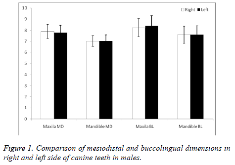
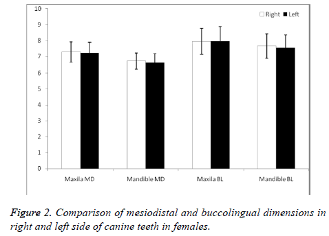
In males, BL dimension of canine teeth on the left side of the upper jaw was significantly greater than that on the right side (p=0.009). However, no other significant difference was observed in BL and MD dimensions between canine teeth on the right versus left sides (p>0.10, Figure 1). In females, BL and MD dimensions of lower jaw in right side were significantly higher than left side (p=0.007 and p=0.031 respectively). However, no significant difference in BL and MD dimensions observed in upper jaw of subjects in our study (Figure 2).
Sex determination
Several forward stepwise multiple logistic regression models were used to develop formulae to determine sex and the accuracy of estimation. The first equation contained all MD and BL dimensions of canine teeth (Table 2). This model selected the variables MD of maxillary right canine (#13), MD of mandibular right canine (#43) and BL of mandibular left canine (#33) as the most contributory for classification purposes (Table 2). The first equation can be written as follows: Logit p=-13.53+1.48 (MD of canine #13)+1.27 (MD of canine #43)-0.84 (BL of canine#33). In this equation if the value is greater than zero, the individual will be classified as male otherwise as female.
| Equations | Coefficient | Standard error | Sensitivity (correctness of the model for males and females) | |
|---|---|---|---|---|
| Male | Female | |||
| Equation 1: All variables | 69.8 | 74.3 | ||
| MD canine 13 | 1.48 | 0.29 | ||
| MD canine 43 | 1.27 | 0.34 | ||
| BL canine 33 | -0.84 | 0.25 | ||
| Constant | -13.53 | 2.68 | ||
| Equation 2: All maxilla | 70.1 | 73.4 | ||
| MD canine 13 | 2.08 | 0.36 | ||
| BL canine 13 | -1.3 | 0.39 | ||
| BL canine 23 | 0.9 | 0.31 | ||
| Constant | -12.67 | 2.29 | ||
| Equation 3: All mandibles | 70.1 | 75.7 | ||
| MD canine 43 | 1.68 | 0.32 | ||
| BL canine 33 | -0.78 | 0.23 | ||
| Constant | -5.52 | 1.84 | ||
Table 2. Logistic regression models for determining sex.
The second and third equations were computed based on each jaw. For the maxilla, MD of right canine (#13), BL of right canine (#13), and BL of left canine (#23) were selected by the stepwise algorithm. The regression equation was computed as: Logit p=-12.67+2.08 (MD canine 13)-1.30 (BL canine 13)+0.9 (BL canine 23).
For the mandible the equation was: Logit p=-5.52+1.68 (MD of right canine #43)-0.78 (BL of left #canine 33). Again, if the equation value is more than zero, the individual will be classified as male otherwise as female.
Discussion
In this study the mean values of all mesiodistal dimensions and maxillary buccolingual dimensions were statistically greater in males than in females. The findings of this study agree with other odontometric studies that same result not only for humans but also anthropoid apes and some monkeys [18]. Canine teeth of both jaws are more dimorphic than others and male have significantly greater buccolingual dimensions in all upper teeth [18]. Also, a research in 2013 found that sizes of male teeth were larger than female and canines were the greatest sexually dimorphic teeth [19,20]. Sex dimorphism is not limited to canines, as it is shown that in Iranians all permanent teeth except maxillary central and lateral incisors and mandibular lateral incisors might show sexual dimorphism at least in one of their mesiobuccal or labiolingual dimensions [21].
Different factors can affect human tooth size and dimorphism [6-17,22]. Current gender differences in tooth size relate to sex-dependent differences in body bulk that exist in any given human population. These are attributed to adaptive reasons and development of food processing techniques which led to the reduction of both male and female dental dimensions [23]. Canines in both jaws might show sexual difference while other teeth are less likely showing such differences [24,25], and dimensions are larger in males [25]. In another study, it was observed that the mean values for right and left maxillary and mandibular canine widths were greater for males than females but not statistically significant [4]. In a longitudinal study on mesiodistal diameters of the primary and permanent teeth in southern Chinese, it was found that the most dimorphic permanent teeth were canines; mesiodistal width of male teeth were more than those of female except for mandibular lateral and central incisors in both dentition [26]. The present study also indicated that the mandibular and maxillary canine teeth were larger in males than in females. The teeth with highest sex dimorphism were canines also in Nepalese [26]. Nevertheless, the study on Nepalese dentition showed that the mean values of mesiodistal and buccolingual widths of the mandibular left canine and the mean values of mesiodistal determinations of the mandibular right canine were larger in females than males [26]. It may be due to different genetic and environmental factors [26].
In this study, a formula was established to determine gender based on a given dental measurements. This formula might be reliable mostly to its ethnicity of origin, as it is known that many dentoskeletal features might vary between ethnicities [27-31]. Future studies should evaluate its accuracy in other populations. Also similar formulas should be found in other ethnicities. It is better to assess reliability of measurements in future studies. Additionally, all human teeth are under constant wearing throughout life time, which can narrow down the validity of our equations to sound young tooth.
Conclusions
Canine teeth can be used for gender determination, according to the formula established for the first time, in the present study. Future research is warranted to evaluate other teeth as well as other ethnicities.
References
- Kakkar T, Sandhu JS, Sandhu SV, Sekhon AK, Singla K, Bector K. Study of mandibular canine index as a sex predictor in a Punjabi population. Ind J Oral Sci 2013; 4: 23.
- Rastogi P, Jain A, Kotian S, Rastogi S. Sexual diamorphism-An odontometric approach. Anthropol 2013; 1: 2332-0915.
- Gray H. Grays anatomy: with original illustrations by Henry Carter. Arcturus Publ 2009.
- Al-Rifaiy MQ, Abdullah MA. Dimorphism of mandibular and maxillary canine teeth in establishing sex identity. Saudi Dent J 1997; 9: 17-20.
- Petersen KB, Kogon SL. Dental identification in the Woodbridge disaster. J Can Dent Assoc (Tor) 1971; 37: 275-279.
- Acharya AB, Angadi PV, Prabhu S, Nagnur S. Validity of the mandibular canine index (MCI) in sex prediction: Reassessment in an Indian sample. Forensic Sci Int 2011; 204: 1-4.
- Rao NG, Rao NN, Pai ML, Kotian MS. Mandibular canine index--a clue for establishing sex identity. Forensic Sci Int 1989; 42: 249-254.
- Saunders SR, Chan AHW, Kahlon B, Kluge HF, FitzGerald CM. Sexual dimorphism of the dental tissues in human permanent mandibular canines and third premolars. Am J Phys Anthropol 2007; 133: 735-740.
- Fernandes TM, Sathler R, Natalicio GL, Henriques JF, Pinzan A. Comparison of mesiodistal tooth widths in Caucasian, African and Japanese individuals with Brazilian ancestry and normal occlusion. Dental Press J Orthod 2013; 18: 130-135.
- Macko DJ, Ferguson FS, Sonnenberg EM. Mesiodistal crown dimensions of permanent teeth of black Americans. ASDC J Dent Child 1979; 46: 314-318.
- Richardson ER, Malhotra SK. Mesiodistal crown dimension of the permanent dentition of American Negroes. Am J Orthod 1975; 68: 157-164.
- Anderson DL, Thompson GW. Interrelationships and sex differences of dental and skeletal measurements. J Dent Res 1973; 52: 431-438.
- Garn SM, Lewis AB, Kerewsky RS. Sexual dimorphism in the buccolingual tooth diameter. J Dent Res 1966; 45: 1819.
- Garn SM, Lewis AB, Swindler DR, Kerewsky RS. Genetic control of sexual dimorphism in tooth size. J Dent Res 1967; 46: 963-972.
- Lladeres E, Saliba-Serre B, Sastre J, Foti B, Tardivo D. Odontological approach to sexual dimorphism in southeastern France. J Forensic Sci 2013; 58: 163-169.
- Muller M, Lupi-Pegurier L, Quatrehomme G, Bolla M. Odontometrical method useful in determining gender and dental alignment. Forensic Sci Int 2001; 121: 194-197.
- Schwartz GT, Dean MC. Sexual dimorphism in modern human permanent teeth. Am J Phys Anthropol 2005; 128: 312-317.
- Zingeser M. Sexual dimorphism in monkey canine teeth. Proceedings of the VIIIth International Congress of Anthropological and Ethnological Sciences. Sci Council Japan 1968.
- Acharya AB, Mainali S. Univariate sex dimorphism in the Nepalese dentition and the use of discriminant functions in gender assessment. Forensic Sci Int 2007; 173: 47-56.
- Angadi PV, Hemani S, Prabhu S, Acharya AB. Analyses of odontometric sexual dimorphism and sex assessment accuracy on a large sample. J Forensic Leg Med 2013; 20: 673-677.
- Mackinejad SA, Kaviani R, Rakhshan V, Khabir F. Assessment of the cut-off point of mesiodistal and buccolingual widths of permanent teeth for determination of sex. J Isfahan Dent Sch 2015; 11: 151-160.
- Plavcan JM. Sexual size dimorphism, canine dimorphism, and male-male competition in primates: where do humans fit in? Hum Nat 2012; 23: 45-67.
- Brace CL, Ryan AS. Sexual dimorphism and human tooth size differences. J Human Evol 1980; 9: 417-435.
- Hashim H, Murshid Z. Mesiodistal tooth width. A comparison between Saudi males and females. Part 1. Egypt Dent J 1993; 39: 343-346.
- Kaushal S, Patnaik V, Agnihotri G. Mandibular canines in sex determination. J Anat Soc India 2003; 52: 119-124.
- Yuen KK, So LL, Tang EL. Mesiodistal crown diameters of the primary and permanent teeth in southern Chinese-a longitudinal study. Eur J Orthod 1997; 19: 721-731.
- Rakhshan V. Meta-analysis of observational studies on the most commonly missing permanent dentition (Excluding the third molars) in non-syndromic dental patients or randomly-selected subjects, and the factors affecting the observed rates. J Clin Pediatr Dent 2015; 39: 199-207.
- Rakhshan V, Rakhshan A. Systematic review and meta-analysis of congenitally missing permanent dentition: Sex dimorphism, occurrence patterns, associated factors and biasing factors. Int Orthod 2016; 14: 273-294.
- Rakhshan V, Rakhshan H. Meta-analysis of congenitally missing teeth in the permanent dentition: Prevalence, variations across ethnicities, regions and time. Int Orthod 2015; 13: 261-273.
- Rakhshan V, Rakhshan H. Meta-analysis and systematic review of the number of non-syndromic congenitally missing permanent teeth per affected individual and its influencing factors. Eur J Orthod 2016; 38: 170-177.
- Amini F, Rakhshan V, Jamalzadeh S. Prevalence and pattern of accessory teeth (hyperdontia) in permanent dentition of Iranian orthodontic patients. Iran J Public Health 2013; 42: 1259-1265.

