Research Article - Journal of Trauma and Critical Care (2017) Volume 1, Issue 2
Role of mini C-arm in orthopedic emergency department, Karachi, Pakistan "save time, money and radiation exposure".
Ranjeet Kumar*, Muhammad Muzzammil, Kiran Maqsood, Anisuddin Bhatti
Department of Orthopedics, Jinnah Post Graduate Medical Centre, Karachi, Pakistan
- *Corresponding Author:
- Ranjeet Kumar
Department of Orthopedics
Jinnah Post Graduate Medical Centre
Karachi
Pakistan
Tel: +92333-7263417
E-mail: ranjeetkumartalib@yahoo.com
Accepted Date: September 07, 2017
Citation: Kumar R, Muzzammil M, Maqsood K, et al. Role of mini C-arm in orthopedic emergency department, Karachi, Pakistan “save time, money and radiation exposure”. J Trauma Crit Care. 2017;1(2):34-37
Abstract
Objectives: To determine role of mini C-Arm in orthopedic Emergency department of tertiary care hospital in Karachi, Pakistan.
Method: A retrospective cohort study was conducted in accident &emergency department of tertiary care hospital in Karachi, JPMC from 15 Nov 2015 to 15 May 2016. 500 forearm and wrist displaced fractures were treated under sedation with closed manipulation and casting in accident & emergency Department and the data was compared with historical controls. In 250 patients fractures were reduced and assessed with a mini-c-arm device while in 250 patients manipulation results were evaluated with radiographs. Patients aged between 2yrs to 60yrs with uncomplicated, isolated fractures were included in the study after informed written consent. .Subjects eligible for the study were randomly divided in two groups. Group 1 includes patients in which mini-c-arm fluoroscopy was used to check manipulation results (case group) and in Group 2 patients we used post reduction x-rays (control group).Pre- and post-reduction radiographs were obtained in each patient.
Results: Significant improvement in reduction quality seen in fractures undergoing closed reduction with mini c-arm assistance, had a (average angulation [and standard deviation], 7° ± 3° vs. 8 ± 5°; p = 0.02), significant decrease in repeated manipulations and need for subsequent operative intervention. (2% of 250 fractures of control group vs. 8.4% of 250 fractures of historical group; p ≤ 0.0003), and a decrease in hazardous radiation exposure to the patients (mean, 13.0 ± 11.3 mrem vs. 48.0 ± 11.7 mrem).The average decrease in orthopedic consultation time with the use of a mini c-arm (25 ± 12 min vs. 45 ± 20 min, p = 0.003) was observed. Conclusion: With the use of the mini c-arm in closed reduction of forearm and wrist fractures in the accident & emergency department can improve the quality of fracture alignment, decrease in radiation exposure to the patient and decrease in need for repeat fracture reduction or additional procedures. Decrease in average orthopedic consultation time for fracture reduction can also be achieved by Mini-c-arm imaging.
Keywords
mini c-arm, trauma, fractures, emergency department, fracture reduction.
Introduction
Improving patient throughput is one of the biggest challenge Accident and Emergency Department's faces today and highvolume of orthopedic cases makes a major bottleneck to patient flow in emergency departments. In USA approximately 2 million emergency departmental visits are documented on high account in adults due to Upper extremity fractures annually [1]. Visits for humeral fractures 8 percent; radial or ulnar fractures are 31 percent; and carpal, metacarpal, or phalangeal fractures are 51 percent. Interestingly, leading cause of upper extremity fractures has been acknowledged as falls [2]. Despite of aggressive campaigns for injury prevention, there has been increasing overall rate of fractures [3,4], and approximately during childhood half of all children would fracture a bone [5].
In Pakistan the situation is worse because number of trauma (road traffic accidents) victims and violence is increasing day by day and pre hospital or hospital based medical care is lacking [6]. Yearly overall incidences of traumatic injuries were found to be 41 injuries for every 1000 persons revealed in the first national injury survey Pakistan [7]. Study conducted in Karachi shows high incidence of road traffic accidents mostly by motor bikes. Incidence of road traffic accident causes approximately minor injuries 65/day, serious 15/day and fatal 3/day [8]. Road Traffic Injuries (RTA) as one of the major causal factors for injury presenting to Emergency department also identified by survey. Yearly incidence of 15 injuries for every 1000 persons by Road traffic injuries are reported [7].
About 2 million accidents occurred in Pakistan in year 2006 according to National road safety secretariat and 0.418 million were seriously injured nature [8]. The economic cost estimated to be over Rs100 billion by road crashes and injuries in Pakistan [9]. The costs of prolonged medical care, the loss of the family breadwinner, the loss of income due to disability and the cost of a funeral can 10 push families into poverty in many developing countries [10].
Fractures closed manipulation and reduction in the emergency department is a common procedure performed by orthopaedic residents/surgeons. Most Emergency Departments use radiographic images as diagnostic and post relocation/reduction conformation tool.
In pediatric patients, reduction in radiation exposure is of been paramount importance because their cells are more sensitive to radiation. For visualization of the skeletal anatomy requiring during surgical procedure, modern uses of the mobile fluoroscope have been vastly expanded. With the admittance of smaller c-arms, fluoroscopic imaging is now frequently used for fracture treatment in the accident and emergency department and for inn and outpatient orthopedic procedures because of the portability, convenience, and ease of use of the equipment [11].
By using the mini C-arm in the Emergency Departments we can reduce patients as well as attendants sufferings in roaming to radiology suit and wheeling back in orthopedic bay with post manipulation x-rays after waiting long in queue which usually backs up patient flow and provide real time access. Often sedation or regional anesthetic applied to these patients, so they cannot be moved while they are under sedation or their regional anesthetic is in place.
The use of the mini C-arm decreases the patients Emergency Department stays and helps to get treatment done in a timely fashion. That has really helped in early management of patients in Emergency Department. Having the mini C-arm substantially improves patient’s outcome as well as saves time and money.
Because of short view field and low power X-ray generator there are some disadvantages as well; due to low power and small field of view the image quality of mini C-arm is not better than conventional C- arm [12].
Method
A retrospective cohort study was conducted in accident and emergency department of tertiary care hospital in Karachi, JPMC from15 Nov 2015 to 15 May 2016. 500 displaced forearm and wrist fractures were treated with closed reduction and casting under sedation in the emergency department and the data was compared with historical controls. 250 fractures were reduced and assessed with a mini-c-arm device, and 250 fracture reductions were evaluated with radiographs. 2 years to 60 years of age patients were eligible for participation with inform consent.
A study inclusion criterion was isolated and uncomplicated fractures. Associated ipsilateral upper extremity injury or polytrauma patients were excluded from the study.
Eligible subjects for the study were randomly assigned to fracture reductions using the mini-c arm fluoroscopy (case group), or fracture reductions without the aid of mini-c-arm fluoroscopy (control group). On call junior orthopedic surgery resident carried out fracture reductions. Pre- and post-reduction radiographs were obtained for each patient.
Result
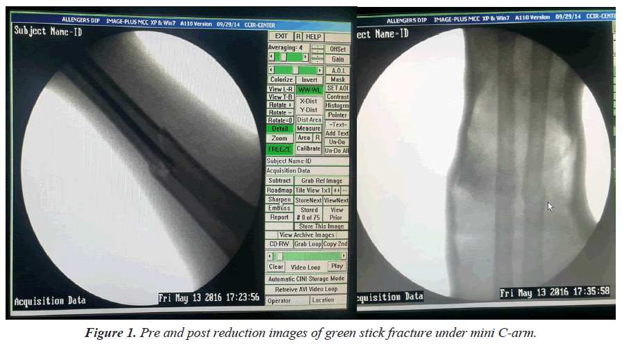
Significant improvement in bone alignment was seen in fractures undergoing closed reduction with assistance of the mini c-arm and also improves residents manipulation and reduction skills (Tables 1 and 2), had a (average angulation (and standard deviation), 7° ± 3° vs. 8° ± 5°; p=0.02) (Figure 1), significant decrease in repeat fracture reduction and need for subsequent operative treatment (2% of 250 fractures of mini c-arm group vs. 8.4% of 250 fractures of historical group; p ≤ 0.0003), and a decrease in radiation exposure to the patient (mean, 13.0 ± 11.3 mrem vs. 48.0 ± 11.7 mrem). Decreased average orthopedic consultations with use of a mini c-arm were (25 ± 12 min vs. 45 ± 20 min, p ≤ 0.003) (Tables 1 and 2).
| Control Group | Mini-C-Arm Group | |
|---|---|---|
| No. of patients | 250 | 250 |
| Average age (yrs) | 30 ± 39.59 (2-58) | 31 ± 41.01 (2-60) |
| Gender: Male | 211/250 (84.4%) | 178/250 (71.2%) |
| Gender: Female | 38/250 (15.2%) | 71/250 (28.4%) |
Table 1. Patient demographics.
| Control Group | Mini-C-Arm Group | |
|---|---|---|
| Number of repeat reductions | 77/250 (30.8%) | 18/250 (7.2%) |
| Consultation Time | 45 ± 20 min | 25 ± 12 min |
| Future Surgical Fixation | 8.40% | -2% |
| radiation exposure to the patient | Mean, 48.0 ± 11.7 mrem | mean, 13.0 ± 11.3 mrem |
Table 2. Study outcomes.
Discussion
Mini C-arm as compared to conventional X-ray causes less radiation exposure to the orthopedic surgeons, patients and theatre personals. Fluoroscopic imaging systems such as mini C-arm use X-rays to provide images, but by the mini C-arm there are many advantages as compared to the conventional X-ray machine. Due to smaller size and extensive Mini C-arm range of movements allows better and multiple desired views in minutes. It’s simple use makes it possible to be used without radiographer, as mini C-arm can be operated by surgeon itself, which will helps to reduce delays, decrease the cost of surgery and great reduction in the radiology department [12,13,14] work load.
It significantly reduces screening time and scattered radiation dose with the use of device it can be controlled by surgeon with foot pedal [13]. The significant benefit of mini C-arm as compared to conventional X-ray machine is reduce dose of scattered radiation to patient, orthopedic surgeon and theatre personnel [15,16].
Most countries of the world are experiencing an epidemic of trauma, but in the developing countries there is most serious increase. Due to rapid motorization and other factors the problem in developing countries is increasing at a fast rate. It is helpful in screening, diagnosis and post manipulation fracture reduction assessment. The burden of musculoskeletal trauma could be considerably decreased by implementing affordable and sustainable strategies to reinforce orthopedic trauma care, C-arm used for decreasing such burden of trauma by early identification and reduction of fractures.
The purpose of this study is to compare the results of fracture reductions when using mini C-arm fluoroscopy completed by orthopedic residents versus traditional closed reduction with use of conventional X-ray machine. In the view of these benefits, usefulness of fluoroscopy guided closed reductions in the emergency room can be understand. Studies have been shown usefulness of mini C-arm over the conventional X-ray machine for extremity surgery, mainly hand and foot surgery [12,13].
If it is used in an injudicious manner, It may cause considerable radiation exposure. Further, the radiation induce complications can be reduced and adjusted by radiation reducing recommendations. It is essential to prevent the useless shots by optimum positioning of C-arm prior to imaging. It requires devotion and practice [12]
From X-ray source/generator every attempt should be made to keep as far away as possible (30cm minimum requirement by British law) [12]. Appropriate positioning of limb in a way that the intensifier should be closer to extremity being examined and the surgeons and operation theater personals should make an adequate distance [17] .It will reduce the direct exposure to the surgeon from scattered exposure [18]. Moreover, minimal radiation subjected to the surgical team. Even with mini C-arm, the protective measures in the form of lead covers should be taken as per recommendations [19]. Lead cover usually used for thyroid, chest and abdomen and can also be used to cover patient’s abdomen and chest. However, with the mini C-arm 0.25 mm lead equivalence is sufficient for protective measures [14,15].
Conclusion
With usage of the mini c-arm in the closed reduction of forearm and wrist fractures in the emergency department can improve the quality of the reduction of fractures, decrease in radiation exposure to the patient, and decrease the need for repeat fracture reduction or additional procedures. Decrease in average orthopedic consultation time for fracture reduction can also achieved by Mini-c-arm imaging.
References
- American Academy of Orthopaedic Surgeons. Joint motion: method of measuring and recording. 2011.
- Chung KC, Spilson SV. The frequency and epidemiology of hand and forearm fractures in the United States. J Hand Surg Am. 2001;26(5):908-15.
- Khosla S, Melton LJ, Dekutoski MB, et al. Incidence of childhood distal forearm fractures over 30 years: A population based study. JAMA. 2003;290(11):1479-85.
- Jónsson B, Bengnér U, Redlund-Johnell I, et al. Forearm fractures in Malmo, Sweden. Changes in the incidence occurring during the 1950s, 1980s and 1990s. Acta Orthop Scand. 1990;13(12):129-32.
- Jones IE, Williams SM, Dow N, et al. How many children remain fracture-free during growth? A longitudinal study of children and adolescents participating in the Dunedin Multidisciplinary Health and Development Study. Osteoporos Int. 2002;13(12):990-5.
- Jamali AR. Trauma care in Pakistan. J Pak Med Assoc. 2008;58(3):102-3.
- Gaffer A, Hyder AA, Masud TI. The burden of road traffic injuries in developing countries: The 1st national injury survey of Pakistan. Public Health. 2004;118(3):211-7.
- Kumar R, Muzzammil M, Mahmood K, et al. Frequency of motor bike injuries, helmet vs. non helmet wearing in Karachi Pakistan. Trauma Int. 2015;1(2):12-6.
- Ahmed A. Road safety in Pakistan. National Road Safety Secretariat, Islamabad. 2007
- Hijar, M., Vazquez-Vela, E, Arreola-Risa. Pedestrian traffic injuries in Mexico: a country update. Injury Control and Safety Promotion. 2003;10(1-2):37-43.
- Badman BL, Rill L, Butkovich B, et al. Radiation exposure with use of the mini-C-arm for routine orthopaedic imaging procedures. JBJS. 2005;87(1):13-7.
- Giordano BD, Ryder S, Baumhauer JF, et al. Exposure to direct and scatter radiation with use of mini-C arm fluoroscopy. JBJS. 2007;89(5):948-52.
- Gangopadhyay S, Scammell BE. Optimising use of the mini C-arm in foot and ankle surgery. Foot and Ankle Surgery. 2009;15(3):139-43.
- White SP. Effect of introduction of mini C-arm image intensifier in orthopaedic theatre. The Annals of the Royal College of Surgeons of England. 2007;89(3):268-71.
- Giordano BD, Baumhauer JF, Morgan TL, et al. Patient and surgeon radiation exposure: Comparison of standard and mini-C-arm fluoroscopy. JBJS. 2009;91(2):297-304.
- Shoaib A, Rethnam U, Bansal R, et al. A comparison of radiation exposure with the conventional versus mini C arm in orthopedic extremity surgery. Foot Ankle Int. 2008;29(1):58-61.
- Mehlman CT, DiPasquale TG. Radiation exposure to the orthopaedic surgical team during fluoroscopy:" how far away is far enough?”. J Orthop Trauma. 1997;11(6):392-8.
- Singer G. Occupational radiation exposure to the surgeon. Journal of the American Academy of Orthopaedic Surgeons. 2005;13(1):69-76.
- Balter S. An overview of radiation safety regulatory recommendations and requirements. Catheterization and Cardiovascular Interventions. 1999;47(4):469-74.
