Case Report - Case Reports in Surgery and Invasive Procedures (2021) Volume 5, Issue 1
RIEDEL thyroiditis: A case report from Nigeria.
Martin Nzegwu*
Department of Pathology, University of Nigeria, Nsukka, Nigeria
Corresponding Author:
- Martin Nzegwu
Department of Pathology, University of Nigeria, Nsukka, Nigeria
E-mail: martin_nze@yahoo.com
Accepted date: 23 August, 2021
Citation: Nzegwu M. RIEDEL thyroiditis: A case report from Nigeria. Case Rep Surg Invas Proced 2021;5(2):1-3.
Abstract
It is common to have a patient present with a thyroid gland mass in clinical practice. Often the clinical diagnosis will be logically correct from the signs and symptoms elicited. The main concern is to distinguish benign from malignant lesions. Diagnosing Riedel thyroiditis correctly happens only when one has a high index of suspicion for it, and this is not probable because of rarity of the variant in clinical practice and consequent lack of exposure to its management. The significance of our reporting this case was to share our experience treating a patient with this rare unreported tumor at least in Africa, the lessons learnt, its contributions to the body of knowledge on the subject matter and to show that such disease can occur in Africans
Keywords:
Riedel thyroiditis, Riedel struma, Thyroid mass, Histopathology
Case Representation
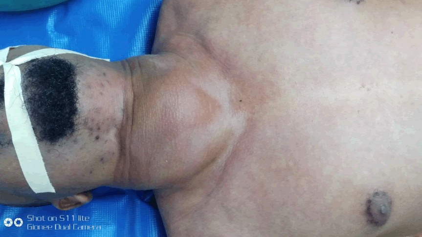
We present a 36-year-old male, self-employed business man of Igbo extraction, South East Nigeria, seen in our facility on referral from a General Surgeon. The General Surgeon attended to the patient 7 months prior to his referral. He noted a rapid rate of growth of a euthyroid goiter with tracheal deviation and suspected a malignancy of the thyroid gland. The presentation was as a painful anterior neck swelling of one year, 5 months duration. It started as a small sized painless mass with no other associated symptoms. However, 3 months prior to presentation, patient observed high a rapid increase in the size of the swelling and correspondingly increasing pains over the swelling. Occasionally he will have difficulty breathing while asleep depending on the position he lay in bed. There was neither difficulty in swallowing nor change in voice. Past medical history was non-contributory [1]. He had lived an active healthy lifestyle. No family member had a similar illness in the present nor past. Physical examination showed right anterior lower neck swelling that is multinodular with woody hard consistency. This mass extended into the superior mediastinum and displacing the trachea to the left. The lower end of the mass was not palpable even when the mass moved cephalad with swallowing action. Cervical lymph nodes were not palpably enlarged in the left side but on the right could neither be ascertained to be enlarge nor separate from the neck swelling. There was no sign of thyroid toxicity in the neither eyes nor vital signs (Figure 1).
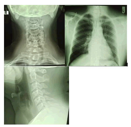
Plain radiographs of the neck confirmed tracheal deviation to the left and normal lung fields; See Figures 2a, b and c below. Ultrasonographic scan of the neck reported a right lobe multinodular mass 5.1 by 4.5 by 6.1 cm with tracheal deviation to the left (Figure 2).
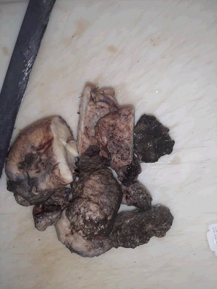
Thyroid hormones profile were normal values with hThyroid stimulating hormone (hTSH) value of 3.5 μU/ml (reference normal value range:0.5–4.8) He was non-reactive to human immunodeficiency virus I and II antigens. Hematological counts were normal. He had no significant systemic disorder The scheduled total thyroidectomy and functional neck dissection ended up as a right lobectomy and isthmectomy under general anesthesia [2]. Operative findings were a woody hard pale mass of the right lobe of thyroid gland that extended slightly across the midline on the isthmus and retrosternal into superior mediastinum displacing the trachea to the left. The cervical lymph nodes along the right carotid vasculature were frozen with and unidentifiable from the thyroid gland mass and had same gross appearance. No identifiable cervical lymph nodes in proximity with the right thyroid lobe. The left thyroid gland lobe grossly was palpably normal as well as the lymph nodes of the left. These were spared during the thyroidectomy because of unexpected challenges encountered that made the surgical team to limit extent of excision to play safe especially when no gross tumor was being left behind. Surgical excision was challenging because of the degree of hardness of the mass and obliteration of tissue planes, such that the mass had to be excised piece meals fashion from the isthmus towards the whole of the right thyroid gland lobe (Figure 3) [3].
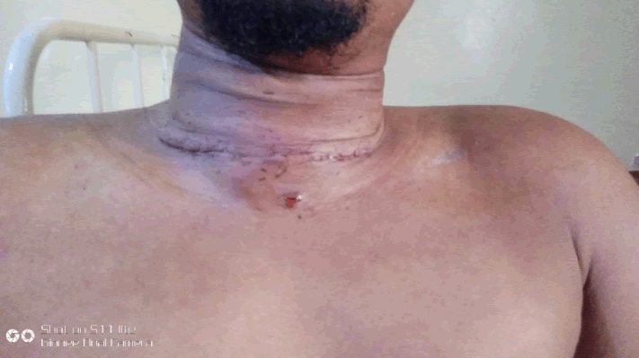
Peri-operative period was uneventful, and patient was discharged home on medications on the fifth post-operative day (Figure 4).
The initial small sized swelling in the neck was of little concern to the patient because it was considered insignificant. However, when the rate of increase in size became rapid with its attendant pains, the patient went back to the general surgeon he consulted months earlier, who in turn immediately referred because of suspicion of malignancy of the thyroid gland. This patient had pain as a main presenting symptom but not any compressive symptom. Other writers reported compressive symptom, voice changes, swallowing disorders among the reasons for seeking medical care. We believe that if our patient had delayed seeking medical care further, that these symptoms will appear. Already the trachea is deviated to the left [4]. Another factor is that he was euthyroid, that is neither hyper- nor hypo- thyroid.
There is still uncertainty of the aetilogy of RT. The aetiology of Riedel thyroiditis had been linked to autoimmune disorder and IgA4 related disorders 4 Knowledge of RT is being hindered by the rarity of the disease that many people will not have the benefit of treating many such patients.
The standard thyroid functions tests are not specific in the diagnosis of RT. The patiebt could be hyperthyroid hypothyroid or euthyroid These would depend on the stage of the disease and the involvement of the lobes of the thyroid gland.
The diagnosis of RT is still confirmed on histopathology findings. This was what we relied upon in making the diagnosis in this case. The dense fibrosing arrangement that replaced glandular elements in the slides [5]. The imaging and clinical features findings can still be present in thyroid gland malignancy.
References
- Shafi AA, Saad NB, AlHarthi B. Riedel's thyroiditis as a diagnostic dilemma: A case report and review of the literature. Ann Med Surg. 2020;52:5-9.
- James VH. Riedel’s Thyroiditis: A Clinical Review J Clin Endocrinol Metab. 2011;96:3031-41.
- Darouichi M, Constanthin PE. Riedel's thyroiditis. Radiology Case Report. 2016;11:175-77.
- Stone John H, Zen Yoh, Deshpande Vikram. Mechanisms of Disease: IgG4-Related Disease. N Engl J Med. 2012;366:539-51.
- Rajkovaca Z, Gajanin R, Pavkovic I, et al. A case of riedel’s thyroiditis Acta Endocrinologica (Buc). 2016;12:339-43.


