Research Article - Journal of Clinical Dentistry and Oral Health (2022) Volume 6, Issue 2
Retrospective study on type of instrument separated during root canal treatment-an institutional study
Yuvashree CS, Mahalakshmi J*
Department of Conservative Dentistry and Endodontics, Saveetha Dental College and Hospitals, Saveetha Institute of Medical and Technical Sciences, Saveetha University, Chennai, India
- *Corresponding Author:
- Mahalakshmi J
Department of Conservative Dentistry and Endodontics
Saveetha Dental College and Hospitals
Saveetha Institute of Medical and Technical Sciences
Saveetha University, Chennai, India
E-mail: mahalakshmij.sdc@saveetha.com
Received: 25-Feb-2022, Manuscript No. AACDOH-22-52398; Editor assigned: 28-Feb-2022, PreQC No. AACDOH-22-52398(PQ); Reviewed: 14-Mar-2022, QC No. AACDOH-22-52398; Revised: 17-Mar-2022, Manuscript No. AACDOH-22-52398(R); Published: 24-Mar-2022, DOI:10.35841/aacdoh-6.2.107
Citation: Yuvashree CS, Mahalakshmi J. Retrospective study on type of instrument separated during root canal treatment- an institutional study. J Clin Dentistry Oral Health. 2022;6(2):107
Abstract
Endodontic instruments becoming separated within the root canal is an undesirable occurrence. A different device prevents the root canal system from being completely debrided and sealed. As a result, every effort must be taken to recover the shattered instrument. When an instrument separates from the canal, the clinician is left in a state of despair, worry, and, finally, optimism that nonsurgical retreatment approaches will aid in the removal of the device. Aim: The aim of the current study is to find the most common type of instrument separated during root canal treatment among 10 -70 years of age visiting private Dental College and Hospitals. Materials and Methods: The current study is a descriptive study which is performed under university settings where the instrument retrieved data was collected from the Hospital Records management system at a Private Dental College. All the instruments fractured / separated during root canal treatment data’s were collected.The sample size of the study was found to be n=40. The data was obtained and tabulated in excel, imported to SPSS software by IBM, a statistical software with variables defined. The significance of this study was set at p<0.05, obtained using Chi square test and the results were interpreted. Results: From the statistical analysis, it is observed that the type of instrument separated has equal gender predilection and the most commonly involved age group is around 30-40 years. The most commonly involved tooth for instrument separation in both the arches was found to be canines and the most commonly involved type of instrument separation was found to be rotary files Chi square statistical test was done and the p value was found to be 0.492 (Chi square -p value >0.05, not significant. Conclusion: Within the limitations of the current study, it is found that instrument separation is more common among 30- 40 years of age with equal gender predilection, and higher incidence with canines.
Keywords
Instrument separation, Type of instrument, Fracture.
Introduction
Instrument fracture in endodontics is a common and troublesome occurrence that can hinder sufficient root canal cleaning and shaping and have a negative impact on the endodontic treatment prognosis [1]. Tooth, separated instrument, operator, and patient are just a few of the factors that can cause an instrument fracture [2]. Excessive torque causes most stainless steel instruments to fail, and torsional stress and cyclic loads cause Niti rotary files to fracture [3].
Although Niti instruments are believed to be more flexible, the introduction of Niti alloys has not resulted in a lower incidence of instrument fracture, with stainless steel separation rates ranging from 0.25 percent to 6% [4]. This difficulty can arise even in the most seasoned hands, frustrating both the clinician and the patient [5]. The success rates for removing separated instruments may vary depending on the devices, procedures, methodologies, and protocols used [6]. One of the various procedures for removing the shattered fragment is using a Masserman kit [7]. Before a clinician makes the decision to remove a separated fragment, they should ensure the availability of and successful manipulation of the required materials, instruments and devices [8].
Each case has its own distinct qualities that will determine how the case is handled [9]. However, a clinician may be fortunate enough to remove the detached instrument while attempting to bypass it, dislodging it coronally with other hand files, or irrigating the root canal [10]. A loose piece, on the other hand, may be difficult to remove despite the use of many methods and technologies. Many technologies, procedures, and strategies for removing separated instruments have been described over the last few decades. Some are still in use, while others are just of historical significance.
Our team has extensive knowledge and research experience that has translated into high quality publications [11-30]. As a result, the study's goal is to analyse the different methods used for the management of retrieval of broken instruments.
Materials and Methods
This was a descriptive study which was performed under a University setting where all the patients between 10-70 years of age reported to a private dental hospital, Chennai, India. The data was collected by reviewing the patient’s records and analysed the data of 86000 patients who underwent Root Canal treatment between June 2019 to February 2021. The ethical approval was obtained from the Institutional Ethical Committee. The population size of the study who underwent instrument separation during root canal treatment was found to be n=38. The data was cross verified with photographs and was compiled for statistical analysis on SPSS (version 23.0) software. The minimising sampling bias was done by collecting data within the University and by using the simple random sampling method. There was a high internal validity and low external validity in our study. The patients between 10-70 years of age and the patients who underwent instrument separation during root canal treatment were included in the study. Improper and incomplete data, repeated data, were excluded. Chi square test was used to compare the groups (p<0.05) was considered significant and the results were interpreted.
Results
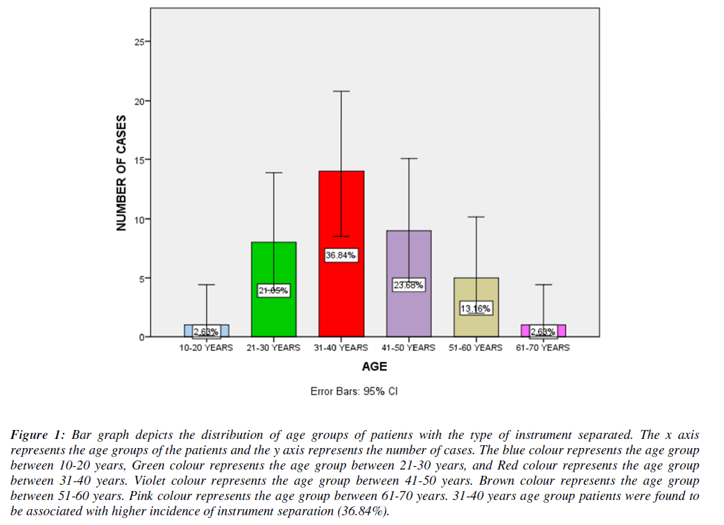
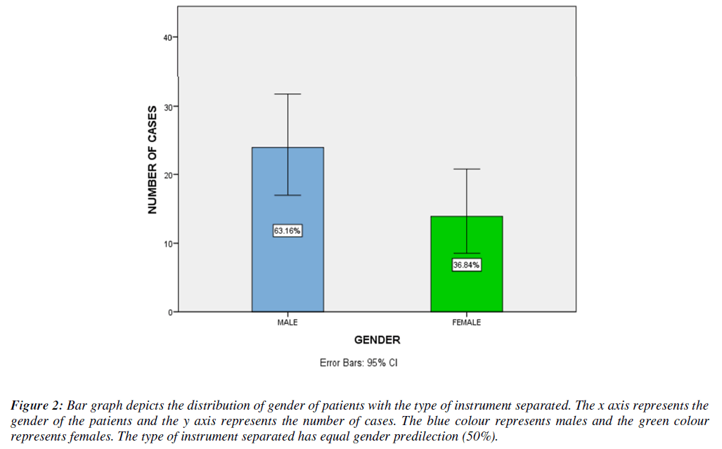
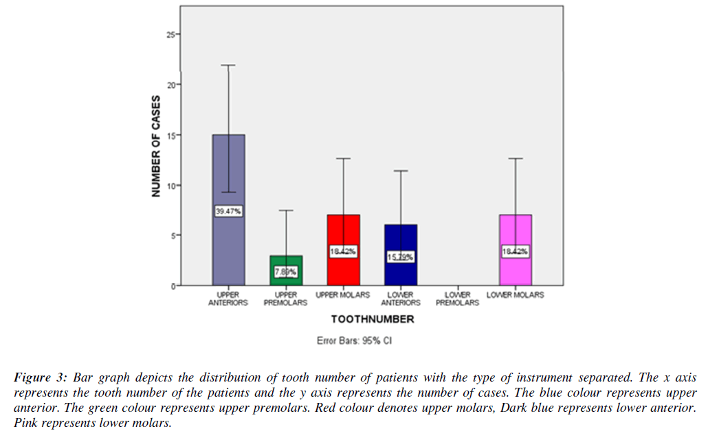
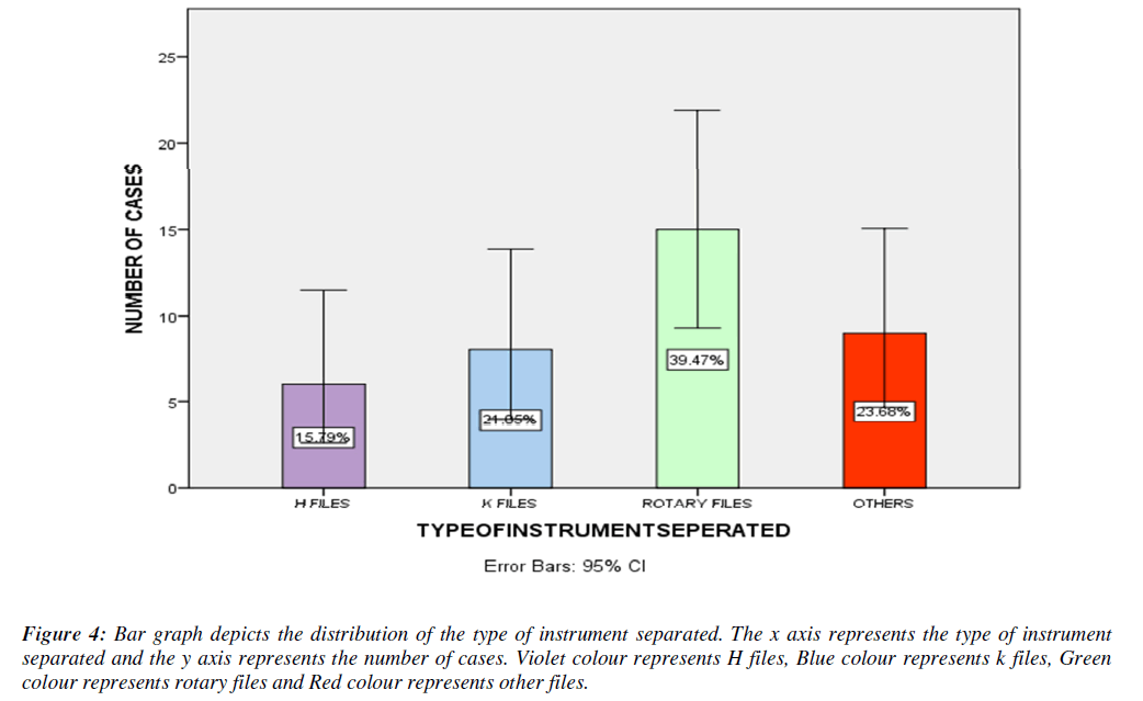
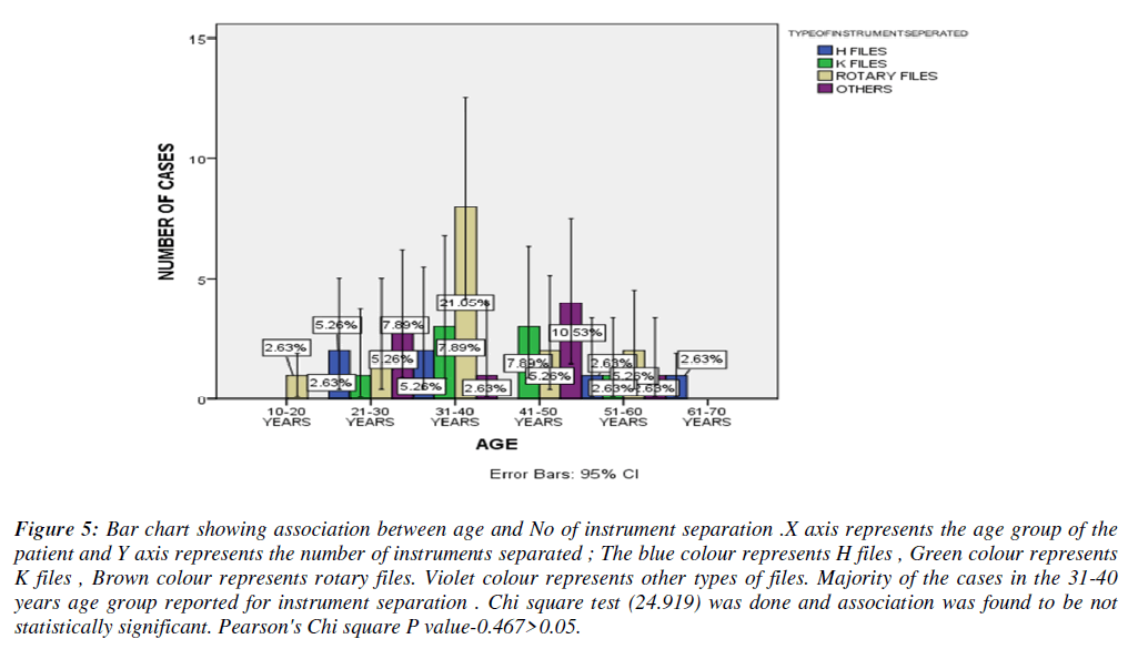
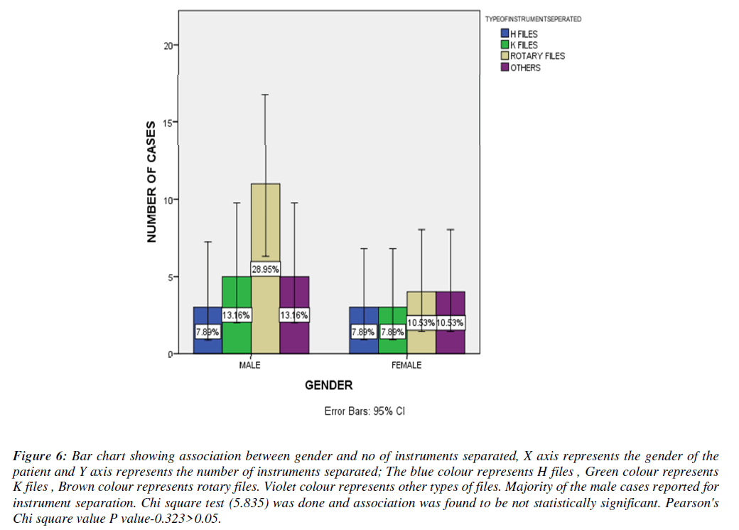
From the statistical analysis, it is observed that the type of instrument separated has equal gender predilection and the most commonly involved age group is around 30 - 40 years. The most commonly involved tooth for instrument separation in both the arches was found to be canines and the most commonly involved type of instrument separation was found to be rotary files (Figures 1-7).
Figure 1: Bar graph depicts the distribution of age groups of patients with the type of instrument separated. The x axis represents the age groups of the patients and the y axis represents the number of cases. The blue colour represents the age group between 10-20 years, Green colour represents the age group between 21-30 years, and Red colour represents the age group between 31-40 years. Violet colour represents the age group between 41-50 years. Brown colour represents the age group between 51-60 years. Pink colour represents the age group between 61-70 years. 31-40 years age group patients were found to be associated with higher incidence of instrument separation (36.84%).
Figure 2: Bar graph depicts the distribution of gender of patients with the type of instrument separated. The x axis represents the gender of the patients and the y axis represents the number of cases. The blue colour represents males and the green colour represents females. The type of instrument separated has equal gender predilection (50%).
Figure 3: Bar graph depicts the distribution of tooth number of patients with the type of instrument separated. The x axis represents the tooth number of the patients and the y axis represents the number of cases. The blue colour represents upper anterior. The green colour represents upper premolars. Red colour denotes upper molars, Dark blue represents lower anterior. Pink represents lower molars.
Figure 4: Bar graph depicts the distribution of the type of instrument separated. The x axis represents the type of instrument separated and the y axis represents the number of cases. Violet colour represents H files, Blue colour represents k files, Green colour represents rotary files and Red colour represents other files.
Figure 5: Bar chart showing association between age and No of instrument separation .X axis represents the age group of the patient and Y axis represents the number of instruments separated ; The blue colour represents H files , Green colour represents K files , Brown colour represents rotary files. Violet colour represents other types of files. Majority of the cases in the 31-40 years age group reported for instrument separation . Chi square test (24.919) was done and association was found to be not statistically significant. Pearson's Chi square P value-0.467>0.05.
Figure 6: Bar chart showing association between gender and no of instruments separated, X axis represents the gender of the patient and Y axis represents the number of instruments separated; The blue colour represents H files , Green colour represents K files , Brown colour represents rotary files. Violet colour represents other types of files. Majority of the male cases reported for instrument separation. Chi square test (5.835) was done and association was found to be not statistically significant. Pearson's Chi square value P value-0.323>0.05.
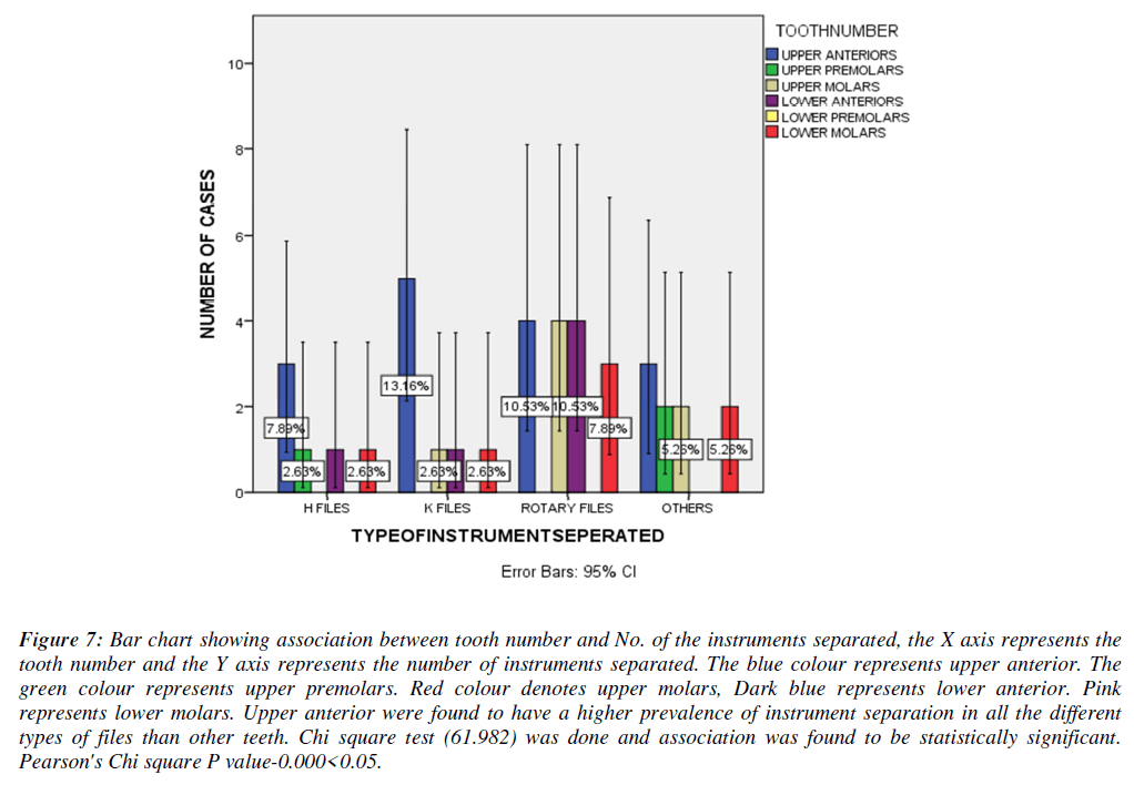
Figure 7: Bar chart showing association between tooth number and No. of the instruments separated, the X axis represents the tooth number and the Y axis represents the number of instruments separated. The blue colour represents upper anterior. The green colour represents upper premolars. Red colour denotes upper molars, Dark blue represents lower anterior. Pink represents lower molars. Upper anterior were found to have a higher prevalence of instrument separation in all the different types of files than other teeth. Chi square test (61.982) was done and association was found to be statistically significant. Pearson's Chi square P value-0.000<0.05.
Discussion
Two major issues must be addressed in order to improve longterm outcomes when the instrument separates in the root canal system [31]. The initial step is to remove the metal fragment from the tooth while also preventing it from corroding. In a study, Barbosa et al. found that stainless-steel fragments did not corrode after two years [10,32]. Strindberg et colleagues discovered that when separated instruments were present, the rate of apical tissue healing was lowered by 19% compared to control cases with no separated instruments [33]. Fox et al. conducted a follow-up study on 66 cases, with a two-year average follow-up period [34]. A favorable outcome was found in teeth with vital and necrotic pulp without periapical lesions [35]. In contrast, when a periapical lesion was present at the time of instrument separation the success rate reduced to 47%.
The type of instrument separated has an equal gender preference in our study, and the most usually involved age group is between 30 and 40 years. Rotary files were also the most prevalent kind of instrument separation [36]. Similarly, the study conducted by Amish et al. yielded the same results. NiTi devices prefer to thread to the canal walls, and they have a higher tendency to fracture frequently, according to her research [8] .Niti instruments are more difficult to remove compared to stainless steel instruments for the following reasons [37-41], Niti tends to thread to the canal walls; they have greater tendencies to fracture repeatedly particularly when ultarsonics are used. They usually remain against the walls, not in the centre. They fracture in shorter lengths making its retrieval difficult.
The upper anteriors were revealed to be the most typically implicated tooth with instrument separation in our investigation. The results were consistent with the findings of Crystal et al investigation. The cause could be linked to a number of factors, including tooth structure, operator expertise, and instrument typ on.
Conclusion
Regardless of little experience of the endodontic residents, they were successfully managed to remove or bypass most of the separated instruments. Ultrasonic device was very helpful in removing the separated instrument.
Acknowledgements
The authors would like to acknowledge the help and support rendered by the Department of Conservative Dentistry and Endodontics, Saveetha Dental college and Hospitals, Saveetha Institute of Medical and Technical Sciences, Saveetha University for their constant assistance with the research.
Funding
The present project is funded by
Saveetha Institute of Medical and Technical Sciences, Saveetha Dental College and Hospitals, Saveetha University, India
Jembu Printers Private Ltd., Chennai
Conflict of Interest
None declared.
References
- Ali MJ. Instrument Fracture. InAtlas of Lacrimal Drainage Disorders. 2018;643-646.
- Boutsioukis C, Lambrianidis T. Factors affecting intracanal instrument fracture. Manag Fract Endo Instru. 2018;31-60.
- Johnson WT. The Impact of Instrument Fracture on Outcome of Endodontic Treatment. Yearbook of Dentistry. 2007;2007:238–9.
- Cheung GS. Instrument fracture: mechanisms, removal of fragments, and clinical outcomes. Endo Topics. 2007;16(1):1-26.
- Parashos P, Messer HH. Rotary NiTi instrument fracture and its consequences. J Endo. 2006;32(11):1031-43.
- Spili P, Parashos P, Messer HH. The impact of instrument fracture on outcome of endodontic treatment. J Endo. 2005;31(12):845-50.
- Chopra V. Clinical Case 16–Management of root canal treatment with an instrument fracture in a mandibular molar. Clin Atlas Retrea Endo. 2021:121-7.
- Alfouzan K, Jamleh A. Fracture of nickel titanium rotary instrument during root canal treatment and re-treatment: a 5-year retrospective study. Int Endo J. 2018;51(2):157-63.
- Matelski J, Rendahl A, Goldschmidt S. Effect of alternative palatal root access technique on fracture resistance of root canal treated maxillary fourth premolar teeth in dogs. Fron Vet Sci. 2020:1078.
- Barbosa AF, Silva EJ, Coelho BP, et al. The influence of endodontic access cavity design on the efficacy of canal instrumentation, microbial reduction, root canal filling and fracture resistance in mandibular molars. Int Endo J. 2020;53(12):1666-79.
- Muthukrishnan L. Imminent antimicrobial bioink deploying cellulose, alginate, EPS and synthetic polymers for 3D bioprinting of tissue constructs. Carbo Poly. 2021;260:117774.
- PradeepKumar AR, Shemesh H, Nivedhitha MS, et al. Diagnosis of vertical root fractures by cone-beam computed tomography in root-filled teeth with confirmation by direct visualization: a systematic review and meta-analysis. J Endo. 2021;47(8):1198-214.
- Chakraborty T, Jamal RF, Battineni G, et al. A review of prolonged post-COVID-19 symptoms and their implications on dental management. Int J Enviro Res Public Health. 2021;18(10):5131.
- Muthukrishnan L. Nanotechnology for cleaner leather production: a review. Enviro Chem Lett. 2021;19(3):2527-49.
- Teja KV, Ramesh S. Is a filled lateral canal–A sign of superiority?. J Dental Sci. 2020;15(4):562.
- Narendran K, MS N, Sarvanan A. Synthesis, Characterization, Free Radical Scavenging and Cytotoxic Activities of Phenylvilangin, a Substituted Dimer of Embelin. Ind J Pharmac Sci. 2020;82(5):909-12.
- Reddy P, Krithikadatta J, Srinivasan V, et al. Dental caries profile and associated risk factors among adolescent school children in an urban South-Indian city. Oral Health Prev Dent. 2020;18(1):379-86.
- Sawant K, Pawar AM, Banga KS, et al. Dentinal Microcracks after Root Canal Instrumentation Using Instruments Manufactured with Different NiTi Alloys and the SAF System: A Systematic Review. App Sci. 2021;11(11):4984.
- Bhavikatti SK, Karobari MI, Zainuddin SL, et al. Investigating the Antioxidant and Cytocompatibility of Mimusops elengi Linn Extract over Human Gingival Fibroblast Cells. Int J Enviro Res Public Hea. 2021;18(13):7162.
- Karobari MI, Basheer SN, Sayed FR, et al. An In Vitro Stereomicroscopic Evaluation of Bioactivity between Neo MTA Plus, Pro Root MTA, BIODENTINE & Glass Ionomer Cement Using Dye Penetration Method. Mat. 2021;14(12):3159.
- Rohit Singh T, Ezhilarasan D. Ethanolic extract of Lagerstroemia Speciosa (L.) Pers., induces apoptosis and cell cycle arrest in HepG2 cells. Nutr Cancer. 2020;72(1):146-56.
- Ezhilarasan D. MicroRNA interplay between hepatic stellate cell quiescence and activation. Euro J Pharmacol. 2020;885:173507.
- Romera A, Peredpaya S, Shparyk Y, et al. Bevacizumab biosimilar BEVZ92 versus reference bevacizumab in combination with FOLFOX or FOLFIRI as first-line treatment for metastatic colorectal cancer: a multicentre, open-label, randomised controlled trial. Lancet Gastroenterol Hepatol. 2018;3(12):845-55.
- Raj R K. ß-Sitosterol-assisted silver nanoparticles activates Nrf2 and triggers mitochondrial apoptosis via oxidative stress in human hepatocellular cancer cell line. J Biomed Mat Res Part A. 2020;108(9):1899-908.
- Vijayashree Priyadharsini J. In silico validation of the non-antibiotic drugs acetaminophen and ibuprofen as antibacterial agents against red complex pathogens. J Periodontol. 2019;90(12):1441-8.
- Priyadharsini JV, Girija AS, Paramasivam A. In silico analysis of virulence genes in an emerging dental pathogen A. baumannii and related species. Archiv Oral Biol. 2018;94:93-8.
- Uma Maheswari TN, Nivedhitha MS, Ramani P. Expression profile of salivary micro RNA-21 and 31 in oral potentially malignant disorders. Braz Oral Res. 2020;34.
- Gudipaneni RK, Alam MK, Patil SR, et al. Measurement of the maximum occlusal bite force and its relation to the caries spectrum of first permanent molars in early permanent dentition. J Clini Pediatr Dent. 2020;44(6):423-8.
- Chaturvedula BB, Muthukrishnan A, Bhuvaraghan A, et al. Dens invaginatus: a review and orthodontic implications. Br Dent J. 2021;230(6):345-50.
- Kanniah P, Radhamani J, Chelliah P, et al. Green synthesis of multifaceted silver nanoparticles using the flower extract of Aerva lanata and evaluation of its biological and environmental applications. Chem Select. 2020;5(7):2322-31.
- Sathorn C, Parashos P. Monitoring the outcomes of root canal re-treatments. Endo Topics. 2008;19(1):153-62.
- Parashos P. Prognosis of root canal treatment with retained instrument fragment (s). Manag Fract Endo Instru. 2018:247-269.
- Cohenca N, González Amaro AM. Root canal infection and endodontic apical disease. Disinfec Root Canal Sys. 2014:1-4.
- Balto K. Tooth survival after root canal treatment. Evid-Based Denti. 2011;12(1):10-1.
- Kirkevang LL. Root canal treatment and apical periodontitis: what can be learned from observational studies?. Endo Topics. 2008;18(1):51-61.
- Malakpour M. Assessment of Fracture Resistance of Maxillary First Molars after Root Canal Preparation Using Three Different Rotary Instruments (V-Taper, ProTaper, Vortex Blue): Finite Element Analysis. West Virginia University; 2019.
- Miccoli G, Seracchiani M, Del Giudice A, et al. Fatigue resistance of two Nickel-Titanium rotary instruments before and after ex vivo root canal treatment. J Contemp Dent Pract. 2020;21:728-32.
- Tan SW. Factors affecting the length of survival of permanent teeth after first-time non-surgical root canal treatment. HKU Theses Online (HKUTO). 2003.
- de Carvalho Felippini AL. Introductory Chapter: Some Important Aspects of Root Canal Treatment. InRoot Canal 2019.
- Shiyakov KK, Vasileva RI. Success for removing or bypassing instruments fractured beyond the root canal curve–45 clinical cases. J IMAB. 2014;20(3):567-71.
- XU DM. Three kinds of root canal sealers in one-visit root canal therapy: A comparison of clinical outcomes. Acad J Second Mili Med Univ. 2013:1029-31.
Indexed at, Google Scholar, Cross Ref
Indexed at, Google Scholar, Cross Ref
Indexed at, Google Scholar, Cross Ref
Indexed at, Google Scholar, Cross Ref
Indexed at, Google Scholar, Cross Ref
Indexed at, Google Scholar, Cross Ref
Indexed at, Google Scholar, Cross Ref
Indexed at, Google Scholar, Cross Ref
Indexed at, Google Scholar, Cross Ref
Indexed at, Google Scholar, Cross Ref
Indexed at, Google Scholar, Cross Ref
Indexed at, Google Scholar, Cross Ref
Indexed at, Google Scholar, Cross Ref
Indexed at, Google Scholar, Cross Ref
Indexed at, Google Scholar, Cross Ref
Google Scholar, Cross Ref, Indexed at
Indexed at, Google Scholar, Cross Ref
Indexed at, Google Scholar, Cross Ref
Indexed at, Google Scholar, Cross Ref
Indexed at, Google Scholar, Cross Ref
Indexed at, Google Scholar, Cross Ref
Indexed at, Google Scholar, Cross Ref
