Research Article - Journal of Clinical Dentistry and Oral Health (2022) Volume 6, Issue 6
RETROSPECTIVE ANALYSIS ON FREQUENTLY DISPLACED DIRECT POSTERIOR CLASS II RESTORATIONS - A TIME DEPENDENT DATA ANALYSIS
Sharon Keziah V, Kavalipurapu Venkata Teja*
Department of Conservative Dentistry and Endodontics, Saveetha Dental College and Hospitals, Saveetha Institute of Medical and Technical Sciences, Saveetha University, Chennai, India
- Corresponding Author:
- Kavalipurapu Venkata Teja
Department of Conservative Dentistry and Endodontics
Saveetha Dental College and Hospitals
Saveetha Institute of Medical and Technical Sciences
Saveetha University, Chennai, India.
E-mail: venkatatejak.sdc@saveetha.com
Received: 01-November-2022, Manuscript No. AACDOH-22-80356; Editor assigned: 03-November-2022, PreQC No. AACDOH-22-80356(PQ); Reviewed: 17-November-2022, QC No. AACDOH-22-80356 (QC); Revised: 21-November-2022, Manuscript No. AACDOH-22-80356 (R); Published: 28-November-2022, DOI: 10.35841/aacdoh-6.6.129
Citation: Sharon Keziah V, Kavalipurapu Venkata Teja. Retrospective analysis on frequently displaced direct posterior class ii restorations: A time dependent data analysis. J Clinic Dent Oral Health. 2022;6(6):129
Abstract
Aim: This study analysed the frequently displaced class 2 restorations in the posterior teeth. Materials and method: A total of 1568 patients who have undergone class 2 composite restorations from June 2020- March 2021 were evaluated. Patient details like age, gender, teeth no, surfaces involved, cause for replacement were included. The prevalence of frequently displaced class 2 restorations was determined. The collected Data was analysed using SPSS. Results: Of the 1568 patients that were analysed 44.25% were in the age group 31–45 yrs. There were 53% men and 47% women included in the study The majority of the displaced restorations were class 2 mesio occlusal [MO] with 85% followed by 8.32% MOD and 5.81% Disto- occlusal. Over 50% of the study population had dislodged upper molar restorations followed by 28.93% lower molar restorations. The association between the type of restorations and survival rate was found to be highly significant, p=0.001. Conclusions: The present study reported a high incidence of displacement in the restoration in the mesio occlusal surfaces, especially on the molars. The most common cause of dislodgement was found to be fracture and secondary caries. This study demonstrated that a wide variation of risk factors on the practice, patient, and tooth levels influences the survival of class II restorations. To provide personalized dental care, it is important to identify and record potential risk factors. Therefore, we recommend further clinical studies to include these patient risk factors in data collection and analysis.
Keywords
Tooth, Dental composite, Restoration, Dental innovation.
Introduction
Direct restorations have a limited life span and the longevity depends on various factors. Displacement or dislodgement of the direct restorations is a frequently seen incidence in clinical practice [1]. The replacement of failed restorations due to secondary caries or dislodgement constitutes a major part of all operative work in dental practice [2]. However the replacement of the dislodged restoration or secondary caries leads to an increase in cavity size [3], more loss of the tooth structure and eventually to the destruction requiring an endodontic intervention or even extraction – a ‘death spiral’ [4]. Therefore it is better to reduce the failure rate of restorations than to increase the chances of destruction which is a vital part in dentistry [4,5].
Previous literature has reported a greater longevity of amalgam restorations than resin composite [6]. As the restorations with resin composite require a more sensitive technique than amalgam, it often leads to the accumulation of more plaque, which further leads to the development of secondary caries [7]. However, there is very limited use of amalgam in operative dentistry in recent times and in India and most of the world, the majority of the restorations are done with composite rather than amalgam [8]. Certain other studies have shown comparable longevity of both composite and amalgam with good skills in placing the restorations. Literature and clinical studies published in the 1990’s showed poorer amalgam and composite longevity compared with studies published recently [9]. This could be because of the introduction of better materials and new methods of cavity preparation and not merely the improved skills among dentists in handling composites [10]. Conventionally, Class II cavity preparations are made with mechanical retention based on Black’s principles [11]. After the recent introduction of adhesive materials, there is more focus [10] on minimal intervention and techniques to preserve tooth structure, such as saucer-shaped or box-shaped preparations [11,12].
Previous studies have observed a shorter longevity in restorations that have a larger cavity but however very little studies are there on the preparation techniques and its impact on the longevity of the restoration [13]. Multiple surface restorations have shown poorer longevity comparatively [13]. Although there are many studies done on the longevity of restorations, very few had separate results for class II restorations.
Therefore, there is still a need for long-time studies on Class II composites placed in operative dentistry [14]. More studies need to be done giving more attention to material used preparation techniques, cavity size, and to patient-related factors [14,15]. The current study aimed to analyze the frequently displaced direct posterior class II restorations.
Materials and Method
Study designs
The current study was designed as a retrospective cross clinical study conducted among patients who underwent class 2 composite restorations due to dislodgement.
Study setting
This study was conducted in Saveetha Dental College and Hospitals, Chennai, India. Therefore the availability of the data l is of patients from the same geographic location having similar ethnicity. The digital case records of the patients who reported to the hospital were analysed and studied. Ethical clearance was obtained from the scientific review board of the hospital.
Sampling
A total of 1568 patients were reviewed. All the patients who have undergone class 2 composite restorations from June 2020- March 2021 were evaluated. The records with Incomplete medical documentation, replication of results in different time periods with improper clinical photographs or diagnosis were excluded from the study. Simple random sampling method was carried out. Cross verification was done by additional reviewers and by photographic evaluation.
Data collection
Patient details like age, gender, teeth no, surfaces involved, cause for replacement were included. The prevalence of frequently displaced class 2 restorations was determined. The collected Data was described as frequency distribution and percentile. All obtained data were tabulated into Microsoft excel documents.
Statistical analysis
The collected Data was described as frequency distribution and percentile. Statistical analysis was performed using Statistical Package for the Social Sciences, version 22(SPSS).Descriptive analysis were based on quantitative variables and frequencies for categorical variables. A Chi square test was applied to determine the significance between groups. P value <0.05 was considered to be statistically significant with a confidence interval of 95%.
Results
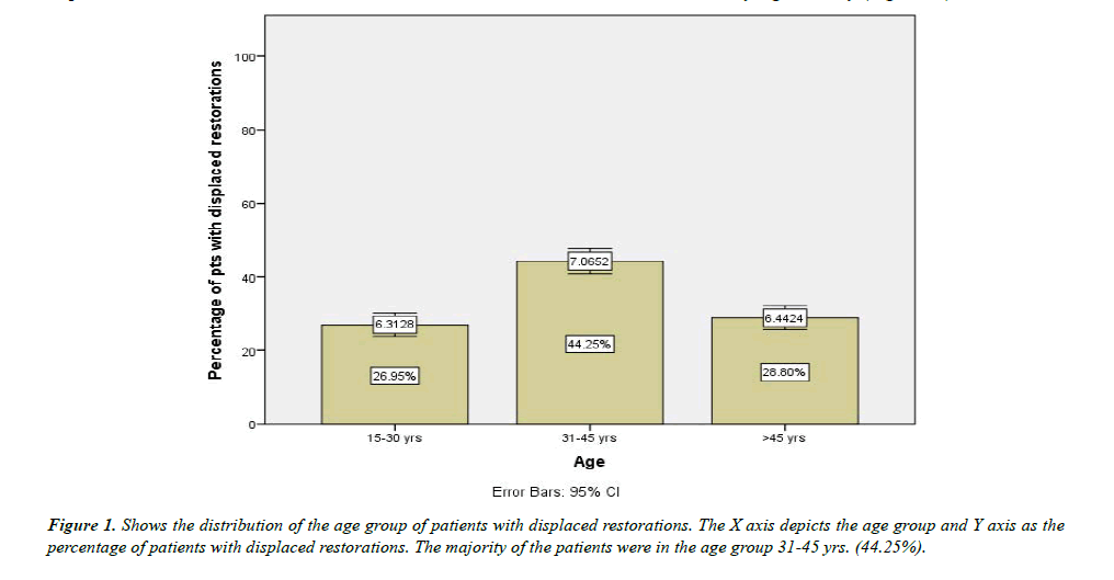
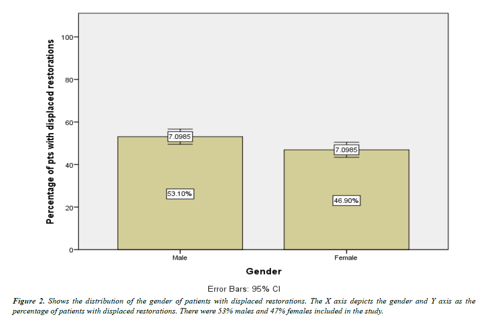
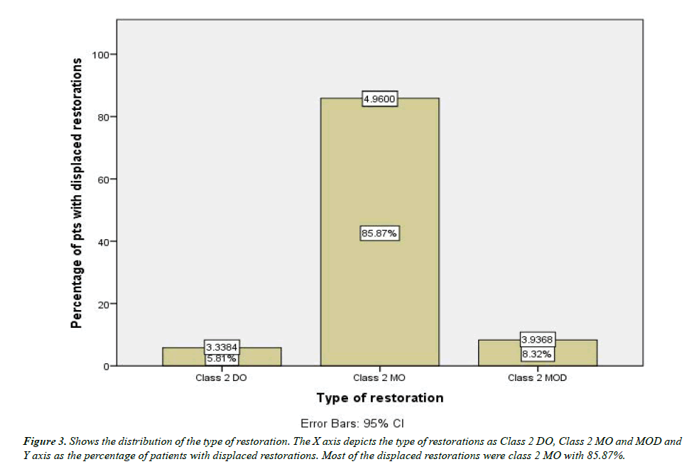
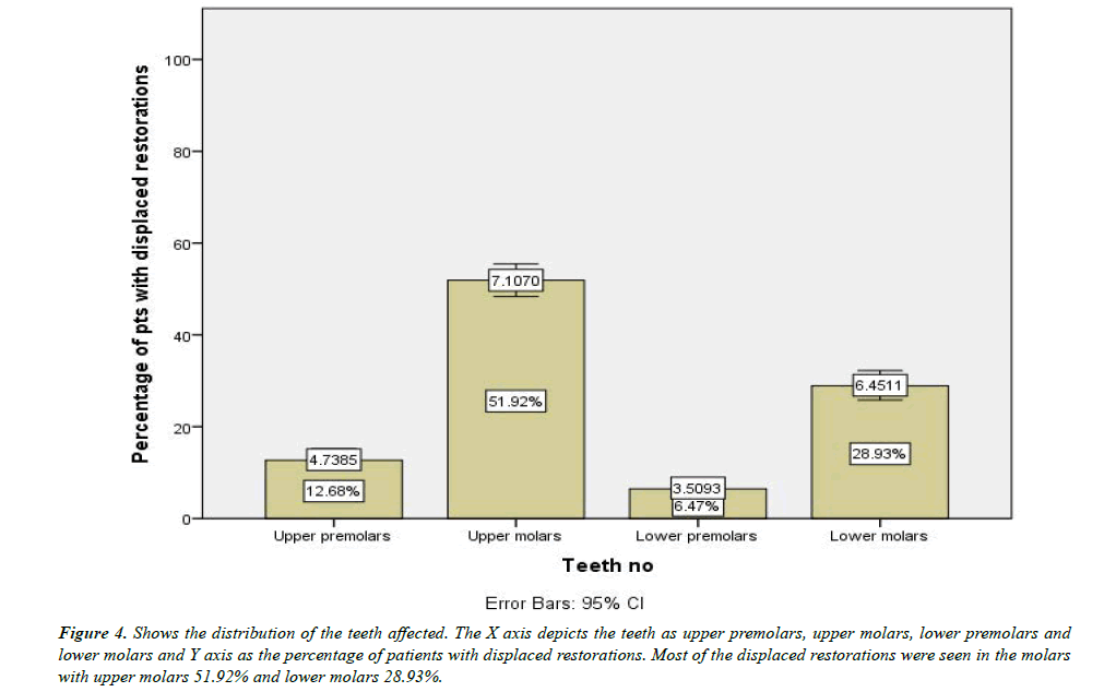
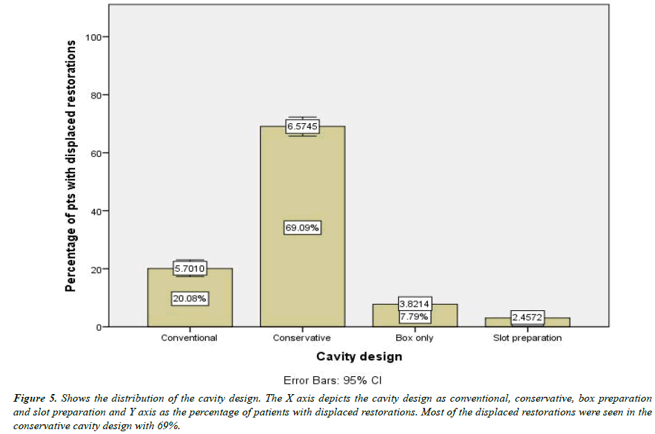
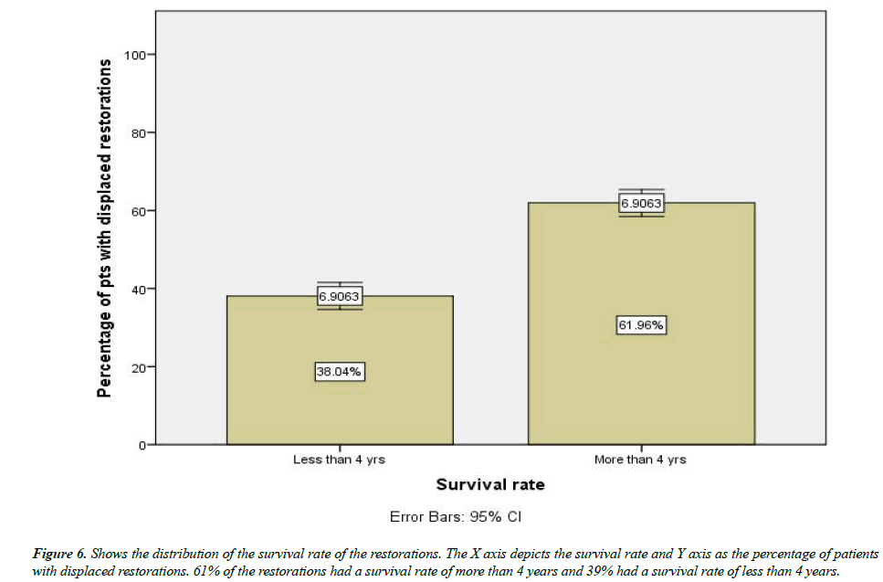
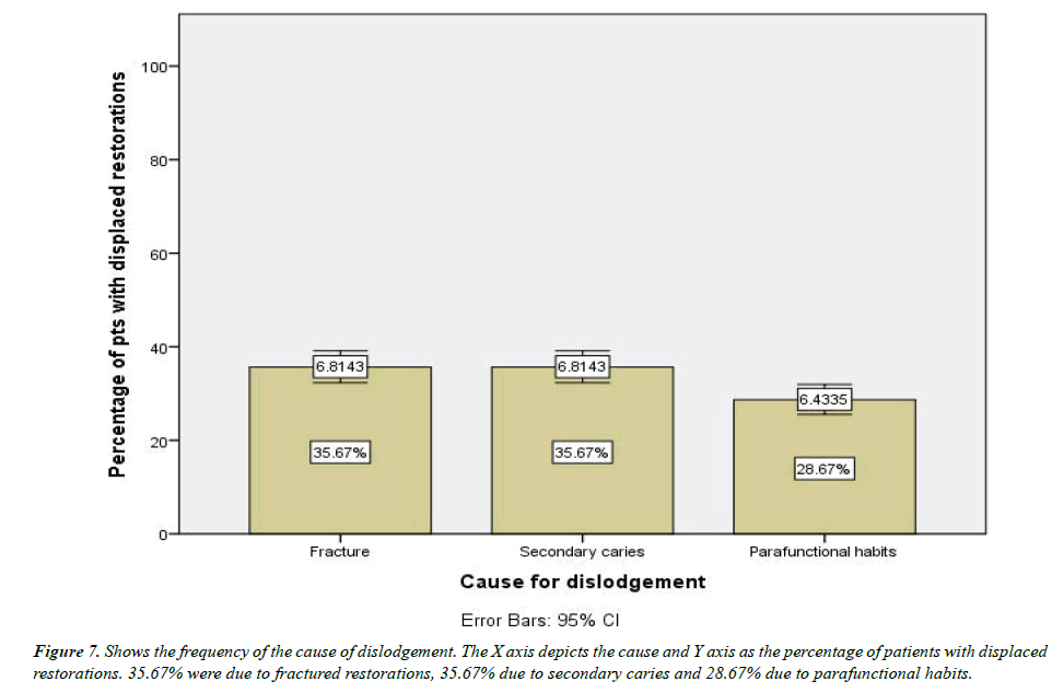
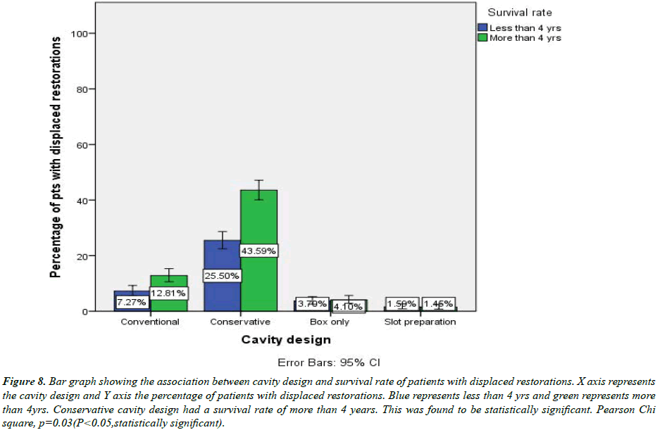
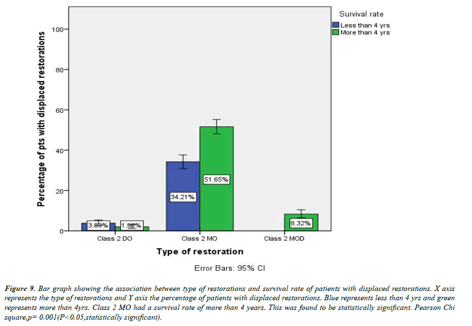
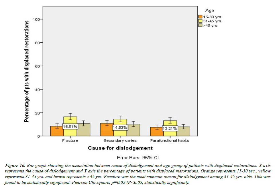
A total of 1568 patients were included in the present study among which 44.25% were in the age group 31–45 yrs. (Figure 1). There were 53% men and 47% women included in the study (Figure 2). The majority of the displaced restorations were class 2 mesio occlusal (MO) with 85% followed by 8.32% MOD and 5.81% Disto- occlusal (Figure 3). Over 50% of the study population had dislodged upper molar restorations followed by 28.93% lower molar restorations (Figure 4). Conservative cavity design was the frequently used cavity preparation with 69% followed by conventional with 20% and box preparation with 7.79% (Figure 5). 61% of the restorations had more than 4 yrs of survival rate (Figure 6). The most common cause for the displaced restorations was fracture and secondary caries with 35.67% each followed by para functional habits with 28.67% (Figure 7). The association between cavity design and survival rate was done and 43.59% of the conventional cavities had a survival rate of more than 4 years. The p value was 0.03 (p<0.05) which was statistically significant (Figure 8). The association between the type of restorations and survival rate was found to be highly significant, p=0.001(Figure 9). The cause of dislodgement and age group was associated and p value was found to be 0.02 which was statistically significantly (Figure 10).
Figure 4:Shows the distribution of the teeth affected. The X axis depicts the teeth as upper premolars, upper molars, lower premolars and lower molars and Y axis as the percentage of patients with displaced restorations. Most of the displaced restorations were seen in the molars with upper molars 51.92% and lower molars 28.93%.
Figure 5:Shows the distribution of the cavity design. The X axis depicts the cavity design as conventional, conservative, box preparation and slot preparation and Y axis as the percentage of patients with displaced restorations. Most of the displaced restorations were seen in the conservative cavity design with 69%.
Figure 8:Bar graph showing the association between cavity design and survival rate of patients with displaced restorations. X axis represents the cavity design and Y axis the percentage of patients with displaced restorations. Blue represents less than 4 yrs and green represents more than 4yrs. Conservative cavity design had a survival rate of more than 4 years. This was found to be statistically significant. Pearson Chi square, p=0.03(P<0.05,statistically significant).
Figure 9:Bar graph showing the association between type of restorations and survival rate of patients with displaced restorations. X axis represents the type of restorations and Y axis the percentage of patients with displaced restorations. Blue represents less than 4 yrs and green represents more than 4yrs. Class 2 MO had a survival rate of more than 4 years. This was found to be statistically significant. Pearson Chi square,p= 0.001(P<0.05,statistically significant).
Figure 10:Bar graph showing the association between cause of dislodgement and age group of patients with displaced restorations. X axis represents the cause of dislodgement and Y axis the percentage of patients with displaced restorations. Orange represents 15-30 yrs., yellow represents 31-45 yrs. and brown represents >45 yrs. Fracture was the most common reason for dislodgement among 31-45 yrs. olds. This was found to be statistically significant. Pearson Chi square, p=0.02 (P<0.05, statistically significant).
Discussion
In this present study, fracture of the restoration and Secondary caries was the most common reason for displaced restorations. Secondary caries are the presence of lesions at the margins of existing restoration [10]. The presence or recurrence of secondary caries is most commonly associated with the marginal areas of the restoration, and it has been stated in previous literature that 80% to 90% of secondary caries can be found at the gingival margin such as in the case of class II. The recurrence of these lesions is attributed to the accumulation of plaque and especially in susceptible individuals who have and to the overall difficulty in cleaning it, especially in the interproximal margin. Our study restorations were grouped by patients.
This study showed that conservative cavity preparation has a higher recurrence of secondary caries. In a previous literature, correlation between cavity preparation techniques like the traditional Class II Saucer-shaped restorations and their survival rates was observed. The Impact of cavity design on the longevity of composite restorations was recorded. Traditional class II preparations had a better longevity than saucer-shaped restorations.
A comparative study done previously observed that the majority of the patient’s choice of restorative material was composite. In the previous study, resin-composite restorations failed more than amalgam restorations. JOKSTAD et al in his study found the longevity of amalgam restoration to be 12–14 yrs. compared to 7–8 yrs. for composite-resin restorations. More studies observed better survival rate of amalgam restorations.
Studies conducted in the 1990’s on the longevity of dental restorations had similar results to the present study. It was observed that secondary caries was the reason for replacement in 33–65% of failed resin-composite restorations. Studies published later reported similar rates: 25%, 38%, 52%, 58%, and 88%. The lack of standardized diagnostic criteria for marginal failure could cause over-registration of secondary caries. The current study shows the association of age and survival rate of restorations to be significant. Lower survival rates were seen in the younger age group. Similar results were seen in the study conducted by Hawthorne and Smales indicated that lower survival rates occurred when the restorations were placed in patients younger than 20 yrs. of age compared with those 21–40 yrs. of age reported that approximal lesions progress through the enamel more slowly in young adults than in adolescents, and according to CARLOS & GITTELSOHN, caries-attack rates may reach a peak from 2 to 4 yrs. following eruption. Longevity of dental restorations.
Resin composite was and still is after the minute use of amalgam, the dominant material of choice for Class II restorations. For resin-composite restorations, secondary caries was the most common reason for replacement, and longevity was significantly greater in older patients. Resin composites performed better with traditional Class II preparations than with saucer-shaped restorations, and the risk of failure was higher in medium/deep cavities compared with shallow cavities. Limitations of the present study are assessing the shorter sample population, with no proper preoperative data [16-35]. Future studies can assess using a prospective standardized controlled trail.
Conclusion
The present study reported a high incidence of displacement in the restoration in the mesio occlusal surfaces, especially on the molars. The most common cause of dislodgement was found to be fracture and secondary caries. This study demonstrated that a wide variation of risk factors on the practice, patient, and tooth levels influences the survival of class II restorations. To provide personalized dental care, it is important to identify and record potential risk factors. Therefore, we recommend further clinical studies to include these patient risk factors in data collection and analysis.
Acknowledgements
This research was done under the supervision of the Department of Endodontics, Saveetha dental College and Hospital. We sincerely show gratitude to the corresponding guide who provided insight and expertise that greatly assisted the research.
Conflict Of Interest
There was no potential conflict of interest.
Source of Funding
Saveetha Dental College & Hospitals, Saveetha Institute of Medical and Technical Sciences, Saveetha University, India.
References
- Qvist J, Qvist V, Mjör IA. Placement and longevity of amalgam restorations in Denmark. Acta Odontol Scand. 1990;48(5):297-303.
- AINegrish AS. Reasons for placement and replacement of amalgam restorations in Jordan. Int Dent J. 2001;51(2):109-115.
- Qvist V, Qvist J, Mjör IA. Placement and longevity of tooth-colored restorations in Denmark. Acta Odontol Scand. 1990;48(5):305-311.
- Castillo J, Sasso L, Svendsen WE. Self-assembled peptide nanostructures: Advances and applications in nanobiotechnology. CRC Press. 2012;324.
- Banerjee A. Minimal intervention dentistry: Part 7. Minimally invasive operative caries management: Rationale and techniques. Br Dent J. 2013;214(3):107-111.
- Williams RC. Periodontal disease. N Engl J Med. 1990;322(6):373-382.
- Frencken JE, Holmgren CJ. Atraumatic restorative treatment (ART) for dental caries. STI book 1999.
- Jokstad A, Mjör IA, Qvist V. The age of restorations in situ. Acta Odontol Scand. 1994;52(4):234-242.
- Forss H, Widström E. Reasons for restorative therapy and the longevity of restorations in adults. Acta Odontol Scand. 2004;62(2):82-86.
- Björkman L, Musial F, Alræk T, et al. Removal of dental amalgam restorations in patients with health complaints attributed to amalgam: A prospective cohort study. J Oral Rehabil. 2020;47(11):1422-1434.
- Byrnes RR. Operative restoration of the teeth in relation to adjacent tissues. J Am Dent Assoc. 1932;19(7):1189-1196.
- Elderton RJ. Positive dental prevention: The prevention in childhood of dental disease in adult life. Butterworth-Heinemann. 1987.
- Vallittu P, Özcan M. Clinical guide to principles of fiber-reinforced composites in dentistry. Woodhead Publishing. 2017; 252.
- Craig RG. Craig’s restorative dental materials. Mosby. 2006;632.
- Youngson C. Summary of: The survival of Class V restorations in general dental practice. Part 2, early failure. Br Dent J. 2011;210(11):530.
- Li B, Moriarty TF, Webster T, et al. Racing for the surface: Antimicrobial and interface tissue engineering. Springer Nature. 2020;809.
- PradeepKumar AR, Shemesh H, Nivedhitha MS, et al. Diagnosis of vertical root fractures by cone-beam computed tomography in root-filled teeth with confirmation by direct visualization: A systematic review and meta-analysis. J Endodontics. 2021;47(8):1198–214.
- Lichtfouse E, Schwarzbauer J, Robert D. Environmental chemistry for a sustainable world. Remediation of air and water pollution. Springer Science and Business Media 2011;541.
- Bhavikatti SK, Karobari MI, Zainuddin SL, et al. Investigating the antioxidant and cytocompatibility of Mimusops elengi Linn extract over human gingival fibroblast cells. Int J Environ Res Public Health. 2021;18(13):7162.
- Karobari MI, Basheer SN, Sayed FR, et al. An in vitro stereomicroscopic evaluation of bioactivity between Neo MTA Plus, Pro Root MTA, BIODENTINE & glass ionomer cement using dye penetration method. Materials. 2021;14(12):3159.
- Rohit Singh T, Ezhilarasan D. Ethanolic extract of Lagerstroemia speciosa (L.) Pers., induces apoptosis and cell cycle arrest in HepG2 cells. Nutr Cancer. 2020;72(1):146-156.
- Shukla AK. Nanoparticles and their biomedical applications. Springer. 2021;286.
- Romera A, Peredpaya S, Shparyk Y, et al. Bevacizumab biosimilar BEVZ92 versus reference bevacizumab in combination with FOLFOX or FOLFIRI as first-line treatment for metastatic colorectal cancer: A multicentre, open-label, randomised controlled trial. Lancet Gastroenterol Hepatol. 2018;3(12):845–855.
- Raj R K. ß-Sitosterol-assisted silver nanoparticles activates Nrf2 and triggers mitochondrial apoptosis via oxidative stress in human hepatocellular cancer cell line. J Biomed Mater Res A. 2020;108(9):1899-1908.
- Vijayashree Priyadharsini J. In silico validation of the non-antibiotic drugs acetaminophen and ibuprofen as antibacterial agents against red complex pathogens. J Periodontol. 2019;90(12):1441-1448.
- Uma Maheswari TN, Nivedhitha MS, Ramani P. Expression profile of salivary micro RNA-21 and 31 in oral potentially malignant disorders. Br Oral Res. 2020;34.
- Chaturvedula BB, Muthukrishnan A, Bhuvaraghan A, et al. Dens invaginatus: a review and orthodontic implications. Br Dent J. 2021;230(6):345-350.
- Bhavikatti SK, Karobari MI, Zainuddin SL, et al. Investigating the antioxidant and cytocompatibility of Mimusops elengi Linn extract over human gingival fibroblast cells. Int J Environ Res Public Health. 2021;18(13):7162.
- Karobari MI, Basheer SN, Sayed FR, et al. An in vitro stereomicroscopic evaluation of bioactivity between Neo MTA Plus, Pro Root MTA, BIODENTINE and glass ionomer cement using dye penetration method. Materials. 2021;14(12):3159.
- Li FH, Zheng SJ, Zhao JC, et al. Phenolic extract of Morchella angusticeps peck inhibited the proliferation of HepG2 cells in vitro by inducing the signal transduction pathway of p38/MAPK. J Integr Agric. 2020;19(11):2829-2838.
- Ojo OA, Olayide II, Akalabu MC, et al. Nanoparticles and their biomedical applications. Biointerface Res Appl Chem. 2021;11(1):8431-8445.
- Romera A, Peredpaya S, Shparyk Y, et al. Bevacizumab biosimilar BEVZ92 versus reference bevacizumab in combination with FOLFOX or FOLFIRI as first-line treatment for metastatic colorectal cancer: A multicentre, open-label, randomised controlled trial. Lancet Gastroenterol Hepatol. 2018;3(12):845–855.
- Guo M, Zhang W, Niu S, et al. Adaptive regulations of Nrf2 alleviates silver nanoparticles-induced oxidative stress-related liver cells injury. Chem Biol Interact. 2022;110287.
- Ganesh PS, Girija AS, Priyadharshini JV, et al. Virtual screening to identify the protein network interaction of theophylline in red complex pathogens. J Pharm Res Int. 2021;374-81.
- Tu HF, Lin LH, Chang KW, et al. Exploiting salivary miR-375 as a clinical biomarker of oral potentially malignant disorder. J Dent Sci. 2022;17(2):659-665.
Indexed at, Google Scholar, Cross Ref
Indexed at, Google Scholar, Cross Ref
Indexed at, Google Scholar, Cross Ref
Indexed at, Google Scholar, Cross Ref
Indexed at, Google Scholar, Cross Ref
Indexed at, Google Scholar, Cross Ref
Indexed at, Google Scholar, Cross Ref
Indexed at, Google Scholar, Cross Ref
Indexed at, Google Scholar, Cross Ref
Indexed at, Google Scholar, Cross Ref
Indexed at, Google Scholar, Cross Ref
Indexed at, Google Scholar, Cross Ref
Indexed at, Google Scholar, Cross Ref
Indexed at, Google Scholar, Cross Ref
Indexed at, Google Scholar, Cross Ref
Indexed at, Google Scholar, Cross Ref
Indexed at, Google Scholar, Cross Ref
Indexed at, Google Scholar, Cross Ref
