Research Article - Journal of Clinical Dentistry and Oral Health (2022) Volume 6, Issue 5
RETROSPECTIVE ANALYSIS OF WIDELY USED BARRIERS AND ITS USES IN NON VITAL BLEACHING
Sunar S, Sharma S*
Department of Aesthetics, Saveetha Dental College and Hospitals, Saveetha University, Chennai, India
- Corresponding Author:
- Sharma S
Department of Aesthetics
Saveetha Dental College and Hospitals
Saveetha University, Chennai, India
E-mail: subash@saveetha.com
Received: 19-August-2022, Manuscript No. AACDOH-22-70339; Editor assigned: 20-August-2022, PreQC No. AACDOH-22-70339 (PQ); Reviewed: 05-September-2022, QC No. AACDOH-22-70339; Revised: 09-September-2022, Manuscript No. AACDOH-22-70339 (R); Published: 16-September-2022, DOI:10.35841/aacdoh-6.5.124
Citation: Sunar S, Sharma S. Retrospective analysis of widely used barriers and its uses in non-vital bleaching. J Clin Dentistry Oral Health.2022;6(5):124
Abstract
Background: Recently a visually pleasing smile has become a major concern for the patients; therefore, dental bleaching has gained importance due to its safety and great aesthetic results. Aesthetic dentistry has evolved, gained popularity and became one of the highly important factors in the dentistry field. Recently a visually pleasing smile has become a major concern for the patients, and a noticeable improvement in the perception of beauty concept was observed in the media.
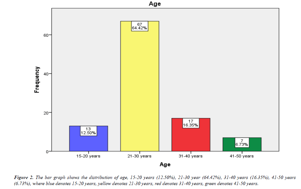
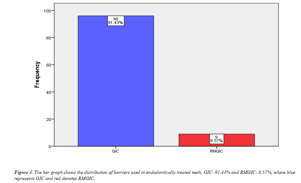
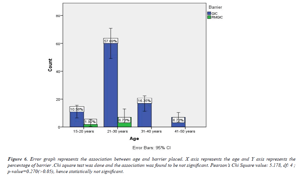
Aim: The aim of the study is to analyze various barriers and its uses in non-vital bleaching. Result: According to the study, the most commonly used barrier is GIC (91.43%) and RMGIC (8.57%). According to the study the most commonly used barrier is in the age group of 21-30 years- 57.69%, followed by 31-40 years - 16.35%
Conclusion: Within the limits of the study the most commonly used barrier is GIC and in the age group of 21-30 years.
Keywords
Innovative technique, Aesthetic dentistry, Dental bleaching, GIC, Barrier.
Introduction
Regenerative endodontic procedures (REPs) are a recently expanding field in endodontics. They are “biologically based procedures designed to physiologically replace damaged tooth structures''. Regeneration of damaged dentin and root structures, as well as the pulp-dentin complex, is fundamental goals of these procedures. REPs are increasingly applied in immature permanent teeth with pulpal necrosis (with or without apical periodontitis) as an alternative treatment option to apexification [1]. The discolouration of nonvital teeth could be a result of many factors such as dental trauma or the endodontic procedure itself [2,3]. Bacterial, mechanical, or chemical irritation to the pulp may result in tissue products that may penetrate tubules and discolor the surrounding dentin. Such discoloration can usually be bleached intracoronary. Teeth discoloration may negatively impact the quality of life in young patients and their families, especially if the problem concerns anterior teeth [4]. To minimize the risk of discoloration, placing of triple antibiotic paste (TAP) without containing minocycline (TAPM) [1,5]. Masking the discoloration with composite resin veneer or internal bleaching are the treatment options used to reduce or eliminate tooth discoloration after REPs. Simple, viable, and minimally invasive procedures should be considered as a treatment of choice. The ideal option would be dental bleaching [6,7]. Intrapulpal haemorrhage and lysis of erythrocytes are a common result of traumatic injury to a tooth. Blood disintegration products, presumably discolour the surrounding dentin. If the pulp becomes necrotic, the discoloration persists and usually becomes more severe with time [8,9]. The failure of the operator to remove blood or other organic material completely from the pulp chamber during the treatment appears to be the most important and common reason for this post endodontic discoloration [10]. Inadequate access to the cavity preparation results in the presence of shelves of dentin which the pulp horns and the lingual area of the pulp chamber [11]. Therefore, adequate access for the complete debridement of the pulp chamber is essential. Adequate obturation ensures a better overall prognosis of the treated tooth. It also provides an additional barrier against ligament and periapical tissues. This is essential to prevent leakage of bleaching agent into gutta percha and root canal walls, reaching the periodontal ligament via dentinal tubules, lateral canals, or the root apex. GIC plays an important role as barrier protection since GIC has chemical interlocking with dentin tubules and GIC has smaller micropores than zinc phosphate cements, it plays adequate sealed post endodontic treatment. Our team has extensive knowledge and research experience that has translate into high quality publications [12-31].
Materials and Methods
This is a retrospective clinical study, carried out at Saveetha Dental College. This study retrospective analysis of widely used barriers and its uses in non-vital bleaching in Saveetha Dental College that were taken over a period of 2 year, from June 2019 to March 2021. Ethical Approval was obtained from the Institutional Review Board. The data was cross verified by 2 examiners. The data were retrieved and examined to assess the micro esthetics in patients reporting for orthodontics treatment.
Inclusion criteria
? Age.
? Gender.
? Endodontic treated teeth.
? Non vital bleaching.
? Barrier used.
Exclusion criteria
Vital bleaching
A total of 105 patients were selected and data were collected and assessed for age, gender, endodontically treated teeth, barriers used. Collected data was tabulated in the excel sheet. The data was imported and transcribed in the statistical analyses package for 23 (SPSS) IBM Corporation. Chi square test was done. Analysis was based on quantitative variables and frequencies for categorical variables. P value less than 0.05 was considered to be statistically significant.
Results and Discussion
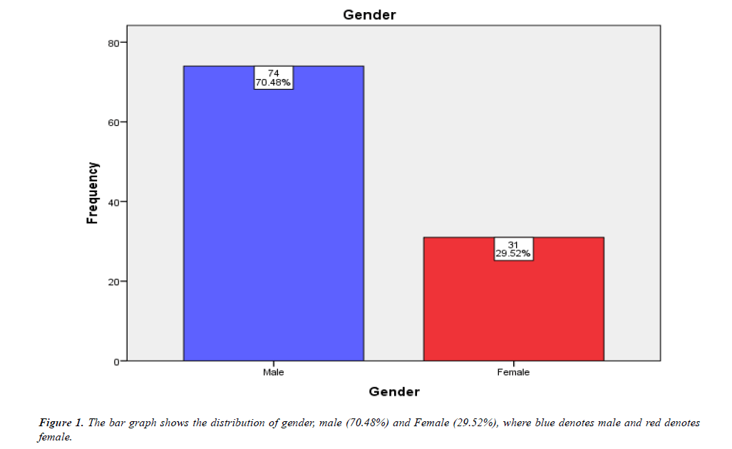
Adequate obturation ensures a better overall prognosis of the treated tooth. It also provides an additional barrier against ligament and periapical tissues. This is essential to prevent leakage of bleaching gutta percha and root canal walls, reaching the periodontal ligament via dentinal tubules, lateral canals, or the root apex. GIC plays an important role as barrier protection since GIC has chemical interlocking with dentin tubules and GIC has smaller micropores than zinc phosphate cements, it plays adequate sealed post endodontic treatment.According to the study most of the participants are male (70.48%) and female 29.52% (Figures 1 and Figure 2).
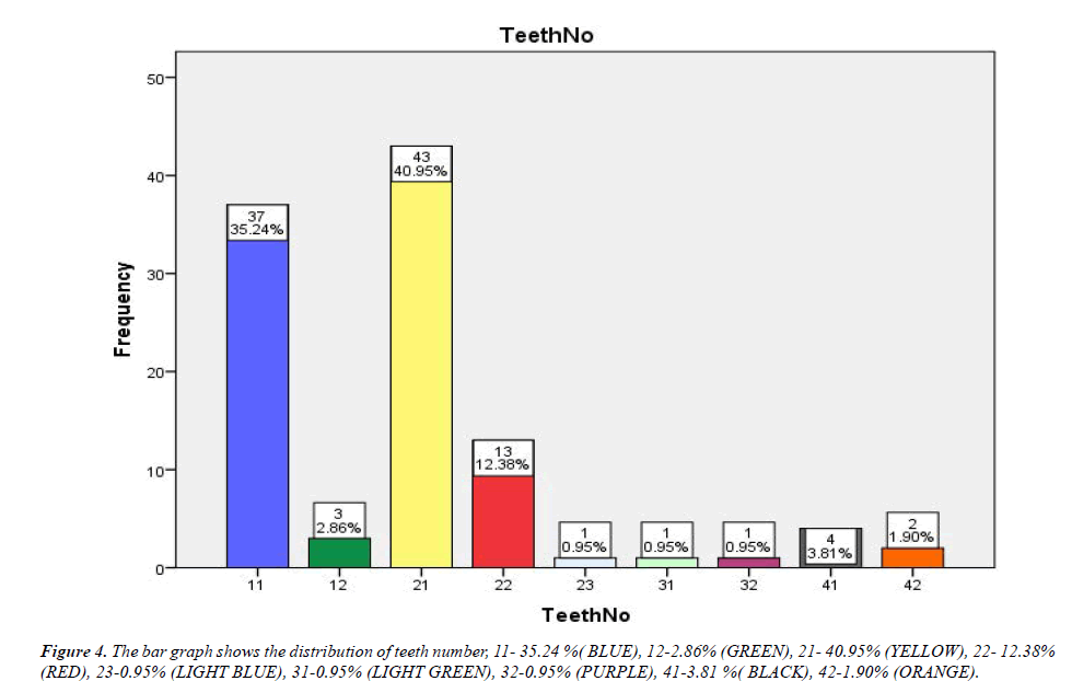
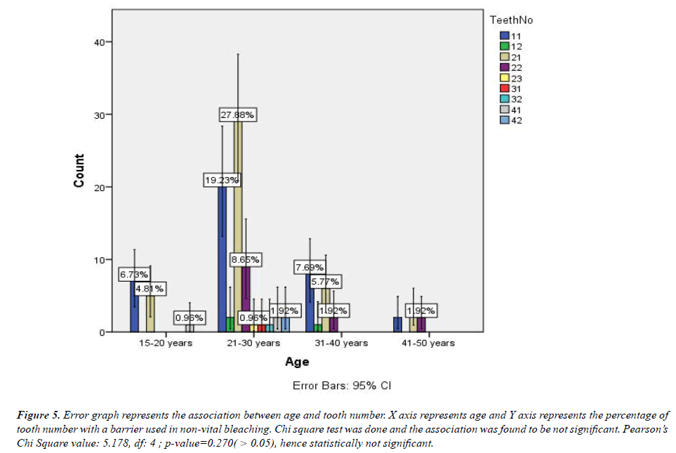
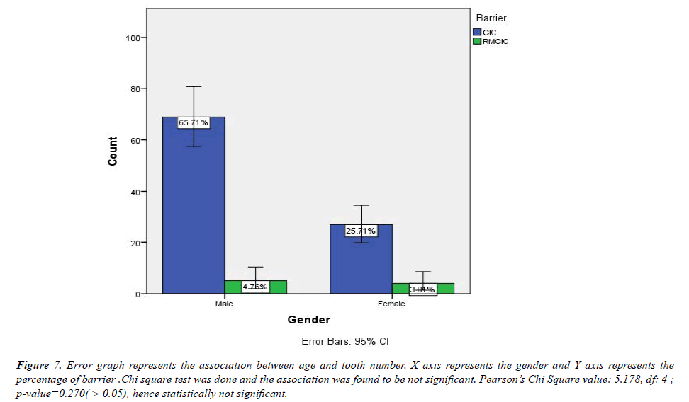
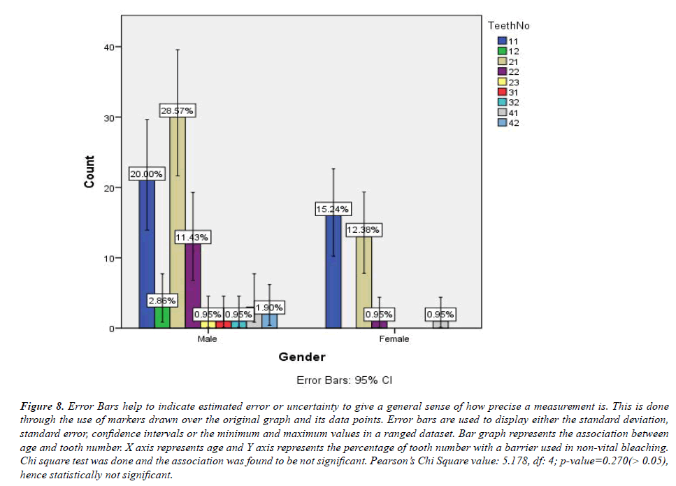
According to the study, the most commonly used barrier is GIC (91.43%) and RMGIC (8.57%). According to the study the most commonly used barrier is in the age group of 21- 30 years- 57.69%, followed by 31-40 years-16.35%, which is consistent with previous literature (Figure 3) [32]. The GIC restorative materials have many attractive features, such as chemical adhesion to enamel and dentin enabling minimal removal of sound tooth tissue, steady fluoride release with a potential cariostatic action, good biocompatibility and a colour similar to that of the tooth. The disadvantages are slow setting action with susceptibility to moisture contamination and dehydration during the early stages, low fracture toughness, and low resistance to wear and degradation. According to the study in tooth number 21 barriers are used for non-vital bleaching (40.95%), followed by 11 (35.24%), followed by 22 (12.38%) (Figure 4). According to the study, the age group 21-30 years has mostly used a barrier for non-vital bleaching on tooth number 21 (27.88%) (Figure 5). According to the study GIC barriers are mostly used on male (65.71%), which is consistent with previous literature [33,34].The resin-modified glass ionomers (RMGIC) were developed to improve the mechanical properties of GIC. These materials had a better fracture toughness and wear resistance compared to GIC as well as a higher moisture resistance and longer working-time. The RMGIC restoratives also had a continuous fluoride release and thus a potential cariostatic effect, although the long-term fluoride release might be somewhat reduced compared to GIC. According to the study, the most commonly endodontically treated tooth is at the age group 21-30 years (58.64%) and the most commonly treated teeth for non-vital bleaching is 21 (27.88%) (Figures 6-8).
Figure 5:Error graph represents the association between age and tooth number. X axis represents age and Y axis represents the percentage of tooth number with a barrier used in non-vital bleaching. Chi square test was done and the association was found to be not significant. Pearson’s Chi Square value: 5.178, df: 4 ; p-value=0.270( > 0.05), hence statistically not significant.
Figure 6:Error graph represents the association between age and barrier placed. X axis represents the age and Y axis represents the percentage of barrier .Chi square test was done and the association was found to be not significant. Pearson’s Chi Square value: 5.178, df: 4 ; p-value=0.270(>0.05), hence statistically not significant.
Figure 7:Error graph represents the association between age and tooth number. X axis represents the gender and Y axis represents the percentage of barrier .Chi square test was done and the association was found to be not significant. Pearson’s Chi Square value: 5.178, df: 4 ; p-value=0.270( > 0.05), hence statistically not significant.
Figure 8:Error Bars help to indicate estimated error or uncertainty to give a general sense of how precise a measurement is. This is done through the use of markers drawn over the original graph and its data points. Error bars are used to display either the standard deviation, standard error, confidence intervals or the minimum and maximum values in a ranged dataset. Bar graph represents the association between age and tooth number. X axis represents age and Y axis represents the percentage of tooth number with a barrier used in non-vital bleaching. Chi square test was done and the association was found to be not significant. Pearson’s Chi Square value: 5.178, df: 4; p-value=0.270(> 0.05), hence statistically not significant.
Conclusion
Within the limits of the study, the most commonly used barrier is GIC (91.43%) and RMGIC (8.57%). However, due to heterogeneity of the research design, the clinical relevance of the included studies, and the lack of adequate comparable studies, the applications of the current study's results should be considered with caution. On the basis of this study, there is a need for more evidence-based research in the area of analysis of widely used barriers and its uses in non-vital bleaching.
Acknowledgement
The authors would like to acknowledge the help and support rendered by the department of Department of Aesthetic and Department of Information Technology of Saveetha Dental College and Hospital and the management for their constant assistance.
Conflict of Interest
All the authors declare that there was no conflict of interest in present study.
Source of Funding
The present project is supported and funded by Saveetha institute of Medical and Technical Sciences, Saveetha Dental College and Hospitals, Saveetha University, India.
References
- Fagogeni I, Falgowski T, Metlerska J, et al. Efficiency of teeth bleaching after regenerative endodontic treatment: A systematic review. J Clin Med Res. 2021;10(2):316.
- Zimmerli B, Jeger F, Lussi A. Bleaching of nonvital teeth. Schweiz Monatsschr Zahnmed. 2010;120(4):306-13.
- Coelho AS, Garrido L, Mota M, et al. Non-vital tooth bleaching techniques: A systematic review. Coatings. 2020;10(1):61.
- Noorul AN, Nivedhitha MS, Ramakrishnan M, et al. Non-Vital bleaching done in discoloured maxillary central incisors. Ann RSCB. 2021;15639–50.
- Greenwall-Cohen J, Greenwall LH. The single discoloured tooth: Vital and non-vital bleaching techniques. Br Dent J. 2019;226(11):839–49.
- Casado BGS, Moraes SLD, Souza GFM, et al. Efficacy of dental bleaching with whitening dentifrices: A systematic review. Int J Dent. 2018;2018:7868531.
- Pandey SH, Patni PM, Jain P, et al. Management of intrinsic discoloration using walking bleach technique in maxillary central incisors. Clujul Med. 2018;91(2):229–33.
- Abou-Rass M. The elimination of tetracycline discoloration by intentional endodontics and internal bleaching. J Endod. 1982;8(3):101–6.
- Abou-Rass M. Long-term prognosis of intentional endodontics and internal bleaching of tetracycline-stained teeth. Compend Contin Educ Dent. 1998;19(10):1034–8.
- Anjum AS, Ganapathy D, Kumar K. Knowledge of the awareness of dentists on the management of burn injuries on the face. Drug Inven Today. 2019;11(9).
- Ari H, Ungor M. In vitro comparison of different types of sodium perborate used for intracoronal bleaching of discoloured teeth. Int Endod J. 2002;35:433–6.
- Muthukrishnan L. Imminent antimicrobial bioink deploying cellulose, alginate, EPS and synthetic polymers for 3D bioprinting of tissue constructs. Carbo Poly. 2021;260:117774.
- PradeepKumar AR, Shemesh H, Nivedhitha MS, et al. Diagnosis of vertical root fractures by cone-beam computed tomography in root-filled teeth with confirmation by direct visualization: a systematic review and meta-analysis. J Endo. 2021;47(8):1198-214.
- Chakraborty T, Jamal RF, Battineni G, et al. A review of prolonged post-COVID-19 symptoms and their implications on dental management. Int J Environ Res Public Health. 2021;18(10):5131.
- Muthukrishnan L. Nanotechnology for cleaner leather production: A review. Environ Chem Lett. 2021;19(3):2527-49.
- Teja KV, Ramesh S. Is a filled lateral canal–A sign of superiority?. J Dent Sci. 2020;15(4):562.
- Narendran K, MS N, Sarvanan A. Synthesis, characterization, free radical scavenging and cytotoxic activities of phenylvilang in, a substituted dimer of embelin. Ind J Pharmac Sci. 2020;82(5):909-12.
- Reddy P, Krithikadatta J, Srinivasan V, et al. Dental caries profile and associated risk factors among adolescent school children in an urban South-Indian city. Oral Health Prev Dent. 2020;18(1):379-86.
- Sawant K, Pawar AM, Banga KS, et al. Dentinal microcracks after root canal instrumentation using instruments manufactured with different NiTi alloys and the SAF system: A systematic review. App Sci. 2021;11(11):4984.
- Bhavikatti SK, Karobari MI, Zainuddin SL, et al. Investigating the antioxidant and cytocompatibility of Mimusops elengi Linn extract over human gingival fibroblast cells. Int J Enviro Res Public Hea. 2021;18(13):7162.
- Karobari MI, Basheer SN, Sayed FR, et al. An in vitro stereomicroscopic evaluation of bioactivity between Neo MTA Plus, Pro Root MTA, BIODENTINE & glass ionomer cement using dye penetration method. Mat. 2021;14(12):3159.
- Rohit Singh T, Ezhilarasan D. Ethanolic extract of Lagerstroemia speciosa (L.) pers., induces apoptosis and cell cycle arrest in HepG2 cells. Nutr Cancer. 2020;72(1):146-56.
- Ezhilarasan D. MicroRNA interplay between hepatic stellate cell quiescence and activation. Euro J Pharmacol. 2020;885:173507.
- Romera A, Peredpaya S, Shparyk Y, et al. Bevacizumab biosimilar BEVZ92 versus reference bevacizumab in combination with FOLFOX or FOLFIRI as first-line treatment for metastatic colorectal cancer: a multicentre, open-label, randomised controlled trial. Lancet Gastroenterol Hepatol. 2018;3(12):845-55.
- Raj R K. β?Sitosterol?assisted silver nanoparticles activates Nrf2 and triggers mitochondrial apoptosis via oxidative stress in human hepatocellular cancer cell line. J Biomed Mat Res Part A. 2020;108(9):1899-908.
- Vijayashree Priyadharsini J. In silico validation of the non?antibiotic drugs acetaminophen and ibuprofen as antibacterial agents against red complex pathogens. J Periodontol. 2019;90(12):1441-8.
- Priyadharsini JV, Girija AS, Paramasivam A. In silico analysis of virulence genes in an emerging dental pathogen A. baumannii and related species. Archiv Oral Biol. 2018;94:93-8.
- Uma Maheswari TN, Nivedhitha MS, Ramani P. Expression profile of salivary micro RNA-21 and 31 in oral potentially malignant disorders. Braz Oral Res. 2020;34.
- Gudipaneni RK, Alam MK, Patil SR, et al. Measurement of the maximum occlusal bite force and its relation to the caries spectrum of first permanent molars in early permanent dentition. J Clini Pediatr Dent. 2020;44(6):423-8.
- Chaturvedula BB, Muthukrishnan A, Bhuvaraghan A, et al. Dens invaginatus: A review and orthodontic implications. Br Dent J. 2021;230(6):345-50.
- Kanniah P, Radhamani J, Chelliah P, et al. Green synthesis of multifaceted silver nanoparticles using the flower extract of Aerva lanata and evaluation of its biological and environmental applications. Chem Select. 2020;5(7):2322-31.
- Qvist V, Manscher E, Teglers PT. Resin-modified and conventional glass ionomer restorations in primary teeth: 8-year results. J Dent. 2004;32(4):285–94.
- Hatibovi?-Kofman S, Koch G. Fluoride release from glass ionomer cement in vivo and in vitro. Swed Dent J. 1991;15(6):253–8.
- Davidson CL, Mjör IA. Advances in glass-ionomer cements. Quint Publishing Comp. 1999;303.
Indexed at, Google Scholar, Cross Ref
Indexed at, Google Scholar, Cross Ref
Indexed at, Google Scholar, Cross Ref
Indexed at, Google Scholar, Cross Ref
Indexed at, Google Scholar, Cross Ref
Indexed at, Google Scholar, Cross Ref
Indexed at, Google Scholar, Cross Ref
Indexed at, Google Scholar, Cross Ref
Indexed at, Google Scholar Cross Ref
Indexed at, Google Scholar, Cross Ref
Indexed at, Google Scholar, Cross Ref
Indexed at, Google Scholar, Cross Ref
Indexed at, Google Scholar, Cross Ref
Indexed at, Google Scholar, Cross Ref
Indexed at, Google Scholar, Cross Ref
Indexed at, Google Scholar, Cross Ref
Indexed at, Google Scholar, Cross Ref
Indexed at, Google Scholar, Cross Ref
Indexed at, Google Scholar, Cross Ref
Indexed at, Google Scholar, Cross Ref
Indexed at, Google Scholar, Cross Ref
