Research Article - Journal of Physical Therapy and Sports Medicine (2017) Volume 1, Issue 1
Reconstruction of coracoclavicular and acromioclavicular ligaments under small incision for the treatment of old acromioclavicular joint dislocation.
Tao Jun*, Chen Pu, Guo Huimin, Li Wenfeng, Yang Yongpeng, Tao Wei
The Second Affiliated Hospital of Nanchang University, 190 Jiangling West 2nd Rd, Qingyunpu Qu, Nanchang Shi, Jiangxi Sheng, China
- *Corresponding Author:
- Tao Jun
The Second Affiliated Hospital of Nanchang University 190 Jiangling West 2nd Rd Qingyunpu Qu, Nanchang Shi, Jiangxi Sheng China
E-mail: 2431835455@qq.com
Accepted date: December 29, 2017
Citation: Jun T, Pu C, Huimin G, et al. Reconstruction of coracoclavicular and acromioclavicular ligaments under small incision for the treatment of old acromioclavicular joint dislocation. J Phys Ther Sports Med 2017;1(1):14-18.
DOI: 10.35841/physical-therapy.1.1.14-18
Visit for more related articles at Journal of Physical Therapy and Sports MedicineAbstract
Acromioclavicular joint dislocation is a common shoulder joint injury, reported in the literature. Its incidence in the population is of about 0.15%, shoulder injury accounts for about 12%. For Rockwood type, who belong to type I and type II injuries, the coracoclavicular ligament injury is not obvious and can be a conservative treatment; type III (in dispute) and above injury i.e., acromioclavicular ligament, joint capsule and coracoclavicular ligament tear, all complete dislocation of the distal and the proposed surgical treatment. Acromioclavicular joint dislocation treatment methods such as grams of needles, coracoid screws, clavicular hook plate, micro-plate and so on. However, there are few articles about the treatment of old acromioclavicular joint dislocation. Recently, our hospital admitted to the old acromioclavicular joint dislocation. In 1 case, the patient through the reconstruction of the oblique, conical ligaments, and at the same time reconstruction of the acromioclavicular ligament and the upper joint capsule. No. 5 cherish line as a protection line, and then use a No. 5 line of love as a tension zone with acromioclavicular joint to further maintain Acromioclavicular joint stability. The patient underwent a small incision reconstruction coracoid and acromioclavicular ligaments shoulder function recovered well. Now, reporting as follows.
Keywords
Old acromioclavicular joint dislocation, Coracoclavicular ligament, Acromioclavicular ligament.
Introduction
Medical record information
Male patient, 35 years old, due to "Car accident caused by left shoulder pain, limited mobility more than 1 month" admitted. A private car accident took place in August 2016. X-ray and CT showed concussion and dislocation of the left acromioclavicular joint. They were also treated at a local hospital (unspecified acromioclavicular joint dislocation was not treated). After leaving the hospital feel left shoulder pain, limited mobility, so in our hospital, to be "left shoulder acromioclavicular dislocation" income hospital. The patient's previous normal body [1]. Physical examination showed: abnormal swelling of the left shoulder joint, tenderness significantly, the keys sign (+), left shoulder joint activity is limited, the left shoulder joint skin is complete, the feeling can be, without rupture; the other limbs showed no significant deformity.
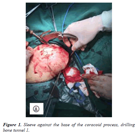
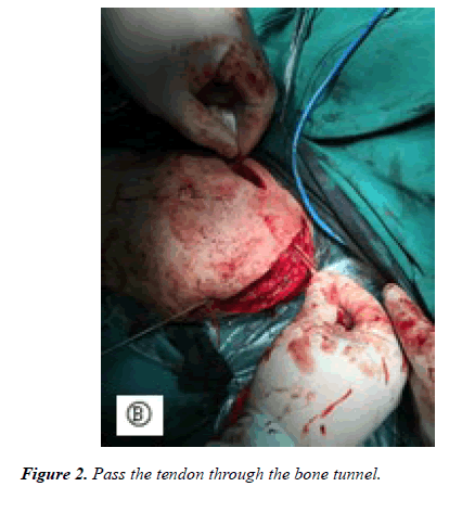
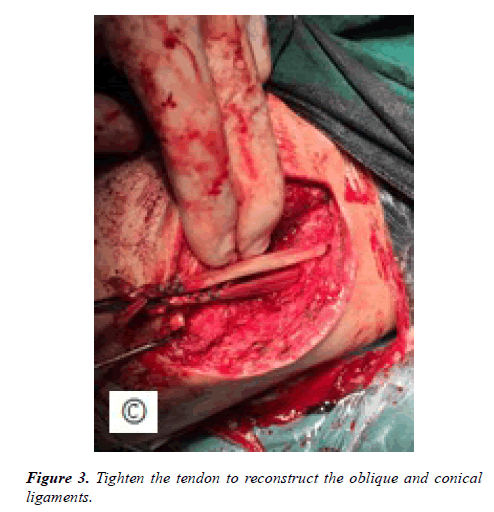
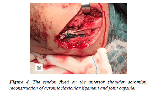
After admission to be routine electrocardiogram, chest radiograph, left shoulder dynamic image, Improve the preoperative routine examination, no obvious contraindications for surgery. It is under the acromioclavicular joint dislocation underwent open reduction and acromioclavicular ligament, coracoclavicular ligament reconstruction. The left distal clavicle incision about 7 cm, followed by incision skin, fascia, see the left acromioclavicular joint dislocation, acromioclavicular ligament and joint capsule rupture, acromioclavicular joint scar removal scar tissue, temporary reduction acromioclavicular joint [2]. Then, Across the shoulder to the distal clavicle, we drilled into two 2.0 Kirschner wire temporary fixation of acromioclavicular joint, perspective acromioclavicular joint reset well. Then the coracoid process as the center, cut a length of about 3 cm longitudinal incision, revealing the base of the coracoid process, with a cross-anchor hooked from the acromioclavicular joint 4.5 cm at the clavicle partial posterior (Figure 1). Locating the sleeve against the base of the coracoid process, drilling bone tunnel 1, and then at a distance of 2.5 cm from the acromioclavicular joint at the center of the clavicle to drill a bone tunnel 2, the tendon and a No. 5 care line from the bone tunnel 1 through the coracoid base bone tunnel, and then from the bone tunnel 2 piercing, the 5th line in the bone surface knot, as a protection line, to strengthen the fixed [3]. Tendon from the bone tunnel 1 radial coracoid base after the tunnel from the bone 2 piercing (Figure 2), Then the ligaments tightened (Figure 3), A 3.5 mm anchor was placed on the anterior superior margin of the acromion and the tendon was fixed on the acromion. A bundle was fixed in front of the acromioclavicular joint, a bundle was fixed above the acromioclavicular joint and the acromioclavicular joint was reconstructed (Figure 4). Using No. 5 to help wire tie Kirschner wire as a tension band to strengthen the fixed. Seeing it again in good position, then cleaning the wound and counting equipment gauze routinely closed wounds.
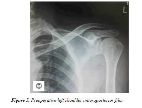
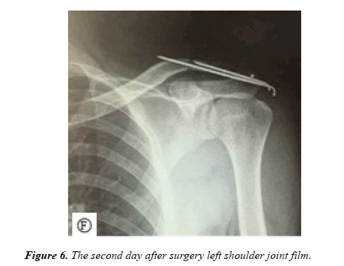
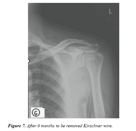
Postoperative limb scarf hanging 4-6 weeks (Figure 5). The day after the passive functional exercise, the premise is that functional exercise will not damage the ligament repair (Figure 6). For backward internal rotation, cross-chest position adduction, forward flexion of 90 degrees need to be cautious and controlled within the patient's pain tolerance range. 4 weeks after the start of active flexion, abduction and external rotation functional exercise. Flexion and outreach as much as possible 180°, 6 weeks after the beginning of a variety of resistance exercises on the shoulder joint, after 6 months in a variety of physical activity or exercise. And inform the patient 1, 2, 3, 6 months after the regular review, after 3 to 6 months to restore the situation to be removed Kirschner wire (Figure 7).
Discussion
Acromioclavicular joint anatomy and biomechanics
Acromioclavicular joint dislocation is a common clinical shoulder injury, mostly caused by direct violence, more common in men. The acromioclavicular joint belongs to a fretting joint, and activities between the scapula and the trunk have corresponding activities on acromioclavicular joint and sternoclavicular joint [4]. The study found that in the upper limb lift process, the clavicle will have an upward rotation of about 40-50°, while the scapula at this time there are downward rotation [5]. Urist et al. acromioclavicular joint anatomy according to the anatomy and orientation, the acromioclavicular joint is divided into 4 type, type : coronal plane joint space from the direction of the arrangement of the direction from the inside to the inside, that is, distal clavicular articular oblique Line covering the acromial joint surface, this type is more common, accounting for about 49%; Type II: the joint space was vertical arrangement, the two articular surfaces parallel to each other, this type accounted for about 27%; Type III: This type is also known as Matching acromioclavicular joint, acromioclavicular joint surface from the outside oblique up and down, the distal clavicular articular surface from the inside oblique down, this type accounts for about 21%; Type IV: joint space from the inside out, That is, acromioclavicular articular surface covering the distal clavicular articular surface, this type accounts for about 3% [6]. I believe that the type is unstable, type IV is stable. The main function of acromioclavicular joint is to coordinate the activity of shoulder joint. The stable structure of acromioclavicular joint includes acromioclavicular ligament and joint capsule structure, oblique ligament and conical ligament. In the study, Klassen et al. found that in the ligaments around the acromioclavicular joint, the strength of the condylar ligament is the largest, followed by the orthopedic ligaments and the acromioclavicular ligaments again [7]. However, when the acromioclavicular ligaments and the joint sacs are integrated, it’s strength is significantly higher than the conical ligament. Rios et al. studied the anatomy of the acromioclavicular ligaments and found that the distance from the distal part of the male clavicle to the midpoint of the oblique ligament was (25.4 ± 3.7) mm and that of the female (22.9 ± 3.7) mm; whereas the distal part of the male clavicle, the distance to the midpoint of the conical ligament was (47.2 ± 4.6) mm and in the female (42.8 ± 5.6) mm [8]. Chinese scholar Liu Yanjie et al. conducted an anatomical study of 26 adult fresh shoulder bodies (20 males and 6 females) in our country [9]. The distance from the distal end of the clavicle to the midpoint of the conical ligament and the oblique ligament was (43.67 ± 6.30) mm and (25.25 ± 3.06) mm. Renfree et al. showed that the distance between the proximal end of the conical ligament and the oblique ligament of the male to the distal clavicle was (33.5 ± 4.4) mm and (16.7 ± 2.4) mm, respectively, and (28.9 ± 2.5) mm and 16.1 ± 1.4) mm, suggesting that we should not exceed 10 mm in the distal clavicle resection surgery to avoid injury to the oblique ligament [10].
Acromioclavicular joint dislocation treatment
Up to 80 surgical treatments for acromioclavicular joint dislocation are still controversial for the best surgical modalities [11]. As early as 1861, some scholars proposed the use of wire cerclage fixation of acromioclavicular joint dislocation, creating a precedent for surgical treatment of acromioclavicular joint dislocation, and then as technology advances, followed by Kirschner wire, coracoclavicular screw, distal clavicle Resection, ligament repair, clavicular hook plate, tendon transposition and other surgical methods [12]. In 2007, Professor Strum first used double-Endobutton plate technique to reconstruct the coracoclavicular ligament for the treatment of acromioclavicular joint dislocation [13]. In 2007, acromioclavicular joint was considered as a juggle joint. Kirschner wire, hook plate and other rigid fixation were nonanatomical reconstruction. Good double Endobutton plate technology to reconstruct coracoclavicular ligament can still maintain the acromioclavicular joint fretting, closer to the anatomic reduction, and achieved good results. Lim on this basis, the use of three Endobutton plate in the treatment of acromioclavicular joint dislocation in 31 patients, while reconstruction of the oblique and conical ligaments, with more stable mechanical advantages, achieved good results [14]. However, because of drilling two bone tunnels at the base of the coracoid process, this approach increases the risk of coracoid fractures and the difficulty of the procedure. In 2013, Cook et al. studied the failure rate of acromioclavicular joint dislocation using coiled clavicular reconstruction and found that the rate of re-dislocation was as high as 29% [15]. It was also considered that the location of a good clavicular tunnel was again the key to dislocation.
Old acromioclavicular joint dislocation treatment characteristics
For the old acromioclavicular joint dislocation [16], most patients with type I, II can be achieved by conservative treatment of good therapeutic effect, recovery exercise. For patients with symptomatic symptoms, local closure of the acromioclavicular joint can be performed, and distal clavicle resection can be performed in patients with ineffective occlusion. For type III and type III or more of the old acromioclavicular joint dislocation need surgery.
For older acromioclavicular joint dislocations of type III and III, the acromioclavicular ligament and coracoclavicular ligament are completely torn, there is local contracture of the ligaments and scar adhesions, and the tension is large and there is no direct repair of the suture. Reconstruction of the coracoclavicular and acromioclavicular ligaments and even the upper joint capsule to maintain its stability.
Small incision under the coracoid reconstruction and acromioclavicular ligament treatment of acromioclavicular joint dislocation of the advantages and disadvantages and precautions
Advantages:
• belong to the soft fixation, in line with anatomical reconstruction, with mechanical advantage;
• reconstruction of oblique, conical ligaments, and reconstruction of acromioclavicular ligaments and joint capsule, Protect the line, and then use a No. 5 cherish state line as a tension zone with a fixed acromioclavicular joint, to further maintain the stability of acromioclavicular joint;
• the operation without destroying the rest of the shoulder joint structure, maintaining the original shoulder joint Anatomy of the beak shoulder arch, acromion space remain intact, conducive to early postoperative functional exercise.
Disadvantages:
• 3 to 6 months after surgery still need to be removed again Kirschner wire;
• need to take autologous tendon or purchase allogeneic tendon, there may be adverse reactions such as rejection, the higher the cost.
• intraoperative complete removal of broken cartilage disk, clean scar tissue;
• be careful of the coracoid drilling to protect the vascular nerves to avoid causing coracoid fracture;
• at the end of the screw into the anchor Pay attention to the direction to avoid twisting the lower surface of the acromion.
Conclusion
Acromioclavicular joint dislocation is one of the common clinical diseases, the old acromioclavicular joint dislocation has its special treatment, the current treatment of a variety of ways for the optimal surgical program is still meaningful. We need to select the appropriate treatment according to the patient's functional requirements. The patient underwent a small incision reconstruction coracoid and acromioclavicular ligaments shoulder function recovered well after June to be removed Kirschner wire, the last follow-up no dislocation, the need for re-dislocation still need long-term follow-up.
References
- Nordqvist A, Petersson CJ. Incidence and causes of shoulder girdle injuries in an urban population. J Shoulder Elbow Surg. 1995;4(2):107-12.
- Ming DZ, Wang PY. Development of acromioclavicular joint dislocation. Chin J Trauma Orthop. 2006;8(2):172-5.
- Rockwood Jr CA, Williams G, Young C. Injuries to the aeromioclavicular joint. In: Rockwood Jr CA, Green D, Bucholz R (eds.). Fractures in adults. Lippicott-Raven, Philadelphia, Pennsylvania, US. 1996;1341-414.
- Qiao HX, Jinzhong Z, Yaohua H. Arthroscopic coracoclavicular ligament augmentation for the treatment of acromioclavicular joint dislocation. Chin J Shoulder Elbow Surg. 2013;(1):40-5.
- Costic RS, Labriola JE, Rodosky MW. Biomechanical rationale for development of anatomical reconstructions of coracoclavicular ligaments after complete acromioclavicular joint dislocations. Am J Sports Med. 2004;32(8):1929.
- Urist MR. Complete dislocation of the acromioclavicular joint: The nature of the lesion and effective method of treatment with an analysis of 41 cases. J Bone Joint Surg Am Vol. 1946;28:813-37.
- Klassen JF, Morrey BF, An JE. Surgical anatomy and functionof the acromioclavicular and coracoclavicular ligaments. Oper Tech Sports Med. 1997;5(2):60-4.
- Rios CG, Arciero RA, Mazzocca AD. Anatomy of the clavicle and coracoid process for reconstruction of the coracoclavicular ligaments. Am J Sports Med. 2007;35(5):811.
- Yanjie L, Hongtao H, Yunfeng C, et al. Acromioclavicular joint anatomy and clinical significance. Pract Orthop J. 2012;18(2):139-42.
- Renfree KJ, Wright TW. Anatomy and biomechanics of the acromioclavicular and sternoclavicular joints. Clin Sports Med. 2003;22(2):219-37.
- You WG, Hua-Rui S, Sheng-Qiang Z. A total arthroscopic treatment of acromioclavicular joint dislocation. Zhonghua Shoulder and Elbow Surgery. 2014;(3):151-6.
- Qirong D, Ming C. Progress in the treatment of acromioclavicular joint dislocation. Chin J Shoulder Elbow Surg. 2013;8(1):13-7.
- Strum S. Double Endobutton Technique for Repair of Complete Acromioclavicular Joint Dislocations. Tech Shoulder Elbow Surg. 2007;8(4):175-9.
- Lim YW. Triple endobutton technique in acromioclavicular joint reduction and reconstruction. Ann Acad Med Singap. 2008;37(4):294.
- Cook JB, Shaha JS, Rowles DJ. Clavicular bone tunnel malposition leads to early failures in coracoclavicular ligament reconstructions. Am J Sports Med. 2013;41(1):142.
- Gao H, Jin HW, Jian ZK. Diagnosis and treatment of acromioclavicular joint dislocation. Trauma Surg Jour. 2012;14(4):369-72.
