Otolaryngology Online Journal

All submissions of the EM system will be redirected to Online Manuscript Submission System. Authors are requested to submit articles directly to Online Manuscript Submission System of respective journal.
Reach Us +44 151 808 1136

Case Report - Otolaryngology Online Journal (2016) Volume 6, Issue 3

Pyriform Sinus Fistula

Vijay Abraham*, Anjali Lepcha, Betty Simon, BS Chandran

Department of Surgery Unit 3, Christian Medical College and Hospital, Vellore, Tamilnadu, India

*Corresponding Author:
Vijay Abraham
Department of Surgery Unit 3, Christian Medical College and Hospital, Vellore, Tamilnadu, India
E-mail: vabraham@cmcvellore.ac.in

Received date: April 06, 2016; Accepted date: May 18, 2016; Published date: May 23, 2016

Visit for more related articles at Otolaryngology Online Journal

Abstract

Pyriform sinus fistula is the rarest of the cervical branchial anomalies. A recurrent left sided lower neck infection may be the only clue to this elusive entity. The ambiguity of the presentation, its diagnosis, delineation of its possible anatomical course and treatment options are highlighted in this review article.

Abstract

Traumatic injuries are very common in oral cavity and involve primarily the soft palate. Traumatic injuries involving the hard palate in adults are rare. Hard palate perforation secondary to animal trauma is a rare entity. This is an unusual case where a 50 yrs old male presented with midline hard palate injury by horn of a bull. The perforation was repaired using palatoplasty and patient had no complication postoperatively. This case report reviews other unusual causes of palatal perforation, mechanism of bull horn injuries and treatment options for such defects.

Keywords

Palate, Perforation, Traumatic

Introduction

Palatal perforation either acute or chronic is rarely caused by trauma. Penetrating trauma to the head and neck accounts for 1% to 2% of pediatric trauma admissions. Children have a propensity to place objects in their mouths, which leaves them at risk of trauma to the oral cavity. On the contrary palatal trauma in adults is rare [1].

Bull-horn wounds are incisive and contusive, they have special Characteristics [2]

• The entry opening is usually small and surrounded by an erosion zone. It also may not correlate with the gap in the aponeurosis.

• One or more in-depth tracts may be present, usually with important muscular destruction.

• These wounds are contaminated, and multiple foreign bodies may found at the bottom of the wound tract, including cloth fragments, dirt, and horn chips.

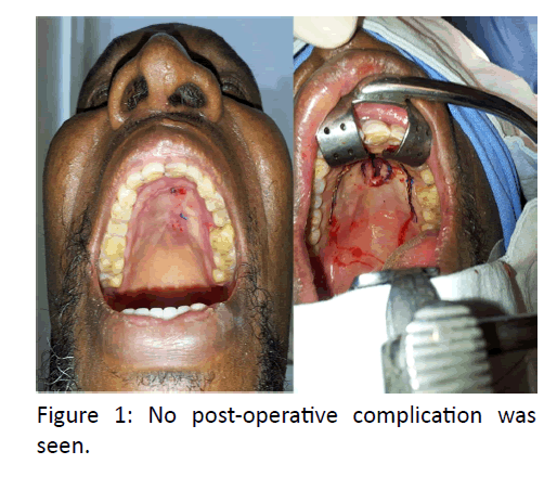
This contaminated wound has a high chance of failure in primary closure. Review of important causes of palatal fistula indicate Developmental causes being the most common cause, occur due to failure of fusion of palatal shelves by 6th week of prenatal life. Maternal alcohol consumption, cigarette smoking, deficiency of folic acid, teratogenic drugs and viruses are some of the known environmental factors leading to cleft palate [2] (Figure 1).

otolaryngology-online-journal-post-operative-complication

Figure 1: No post-operative complication was seen.

Various infectious and chronic granulomatous conditions are known to cause palatal perforation like leprosy, tertiary syphilis, tuberculosis, actinomycosis, histoplasmosis, and blastomycosis. Autoimmune conditions including sarcoidosis, Wegner’s and Crohn’s disease also lead to palatal perforation [3].

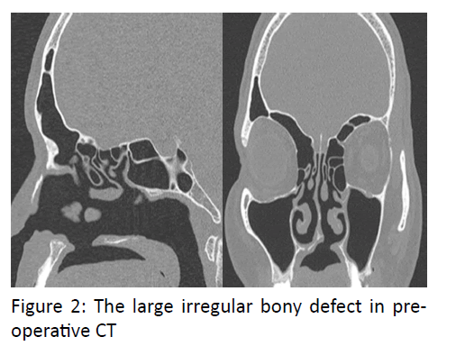
Malignancy of minor salivary gland tumor like adenoid cystic carcinoma also leads to palatal perforation [4]. Tumors’ from maxilla and nose also contributes to this either due to rapid spread leading to bulge in palatal area and later on perforation or sometimes perforation occur as a part of treatment5. Cocaine is a proven drug leading to perforation, other drugs involving the palate includes heroine and nicotine [6] (Figure 2).

otolaryngology-online-journal-large-irregular-bony

Figure 2: The large irregular bony defect in preoperative CT

Perforation might occur following a surgical procedure done for tumor removal or may be following intubation. Literature also describes role of rhinolith leading to perforation [7]. Aim of presenting this case is to emphasize the rare cause of palatal injury being animal.

Case Report

A 50 yrs old male from Nigeria presented to ENT OPD with a defect in hard palate since 6 months, which occurred owing to injury by the horns of bull. Patient also had laceration involving lips and right side ala of nose. Patient had difficulty while eating and change in speech. The patient was taking treatment from the primary health centre in Nigeria, where he underwent repair for the laceration but no intervention was done for the palatal wound. Examination revealed nasal speech, an oval palatal fistula with irregular margins measuring 3 x 1 cm; extending anteriorly up to the rughae area and posteriorly up to the canines. No signs of inflammation noted. CECT oral cavity revealed area of defect at the anterior part of hard palate.

Patient was taken up for V-Y Palatoplasty and intra operatively the bony defect was found to be 5 x 5 cm. The defect was then repaired in 3 layers using PDS suture for nasal layer and 4.0 vicryl for muscle and mucosa, and patient was allowed liquids orally next morning. Due to the large irregular bony defect in pre-operative CT, consent for bone grafting was also taken but intra operatively the closure was adequate.

Discussion

Palate contributes to an important part of oral cavity, the defect of which leads to nasal regurgitation of food, recurrent nose and ear infections and nasal speech.

It comprises of the palatine part of palatine process of maxilla covered by palatine muscles and mucosal layer.

Pathologies affecting palate can be congenital or acquired. Acquired causes include idiopathic or due to traumatic, infectious, malignancy related, granulomatous disease.

Case presented here is of interest as it is caused by trauma by horns of animal (bull). It is important to understand the complex mechanism causing the wound, as the result of the interaction of various distinct forces. The depth is dependent on the force of penetration of the bull´s horn into the body (which is the result of the animal´s weight and speed). There is an additional force because of the effect of the bull´s strong neck muscles when it raises its horns. This force causes upward tears at right angles to the ground. If the injured person is lifted, his body weight exerts an opposite force. Finally, when the patient´s body is lifted and suspended by the bull´s horns, it is in an unstable balance, which, depending on the location of the center of gravity relating to the horn, causes a rotational movement (with ensuing tears of arteries, veins, and nerves), combined with the animal´s efforts to disengage the person´s body [8].

Cases of traumatic perforation have been reported in literature. Hwang and Kim reported a case of submucosal cleft palate in 27 yr old women because of ingestion of hot food (Thermal injury). Perforation and midline notching at the posterior edge of the hard palate was seen noted [9]. A case of 69 years male patient was reported by Macleod in which perforation of the hard palate secondary to pressure atrophy was noted [10]. Ozul et al. [11] reported a case in which perforation of hard and soft palate is seen after a long intubation period. A case of palatal perforation in a 36-year-old female patient treated for empyema of maxillary sinus was reported by Pegler [12]. Other non-traumatic acquired causes presenting as palatal perforation should include infections (syphilis, leprosy, tuberculosis, diphtheria, mucormycosis, actinomycosis) tantrum oris, mechanical trauma, intranasal cocaine abuse, malignancies (especially nasal T cell lymphomas, carcinoma, melanoma), collagen vascular diseases (Wegener’s granulumatosis, systemic lupus erythematosus), sarcoidosis and idiopathic cause such as midline non-healing granuloma [13]. This patient was operated 6 months after the injury. This was sufficient time for optimal wound healing and contraction of fistula and union of adjacent maxilla facial bones. Pre-operative CT scan helps in determining the extent of surgery required and to consent the patient for possible bone grafting.

The treatment options comprise sealing of the defect, either surgically or with a removable obturator, once the lesion has been seen to remain stable. The surgical technique is chosen according to the location and dimension of the lesion, the presence of infections and the general patient conditions.

For reconstruction of the defect, the literature describes surgical techniques like primary closure, use of local pedicled flaps, lingual grafts, temporal muscle flaps, oral adipose tissue grafting, von Langenbeck technique, Furlow double opposing Z palatoplasty, Veau-Wardill-Kilner or VY pushback palatoplasty. In case of extensive lesions, free microvascular flap ie. radial forearm free flap 15 is indicated, with or without simultaneous bone transfer [14,15].

The literature also describes the resolution of small or medium-sized defects using a Le Fort I osteotomy and bilateral adipose flap of the Bichat bulla, in those cases where a temporal or microvascularized flap is contraindicated [16]. Anterior hard palate perforation are better dealt with von lagenbeck technique or its modifications , V Y pushback palatoplasty and posterior soft palate ones by furlow double opposing Z palatoplasty. Each of these techniques has their advantages and different failure rates depending on site of perforation and extent of perforation. This case was done by VY pushback palatoplasty without complications of VPI or fistula.

The use of prosthetic obturators as a solution to situations that pose social problems for the patient is often indicated in cases characterized by palatal communications secondary to highly mutilating surgery. These obturators may be offered as an alternative for patients with palatal perforations who do not wish to undergo surgery, in those cases where the cost/benefit ratio is not favorable, in patients who cannot or do not wish to abandon the habit, or as a temporary measure before surgical treatment [14,17].

Such obturators avoid nasal reflux, facilitating correct swallowing and sufficient speech performance. The only contraindication to such devices is patient tolerance of the obturator, since in some cases the obturator size required to fully seal the defect can cause nausea [17].

Conclusion

Traumatic injuries to maxillofacial region are quite common but this report presents a unique type of traumatic injury causing perforation of palate.

References

Introduction

Development of the neck takes place from the branchial apparatus which is an embryological complex. Abnormal development gives rise to branchial anomalies that most commonly present with neck masses and occasionally with external opening on the skin forming sinuses. Most often they are undiagnosed till multiple surgeries have been performed. Of the various branchial anomalies, the 2nd branchial anomaly is the most common. Third and 4th branchial anomalies account for only about 3 to 10% [1]. The latter are in close proximity or pass through the thyroid gland and originate in the pyriform fossa, hence they are called pyriform sinus fistula (PSF) [2]. Most reported cases are found on the left side with very few being reported on the right. This may be related to normal embryology of the branchial apparatus, where the fourth arch artery on the left side becomes part of aortic arch, whereas on the right side it becomes proximal part of right subclavian artery [2].

Case report

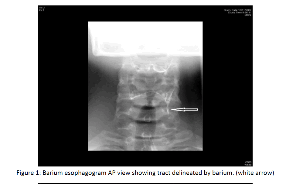
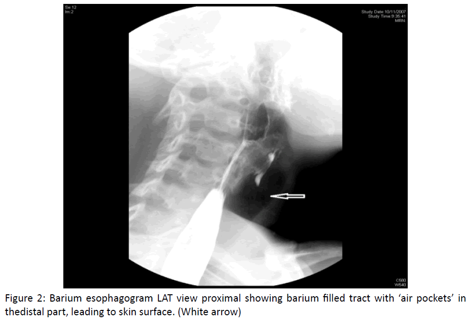
A 19 year old male presented with complaints of recurrent swelling in the anterior aspect of left lower neck. He had undergone multiple incision and drainage of the abscess and had received antituberculosis medications and various antibiotics empirically. However, this therapy only resulted in temporary relief. On presentation to us, he had tenderness and mild swelling in the left lower neck with scars of previous procedures. Barium esophagram (BO) suggested the presence of a pyriform sinus (Figures 1,2).

otolaryngology-online-journal-delineated-barium

Figure 1: Barium esophagogram AP view showing tract delineated by barium. (white arrow)

otolaryngology-online-journal-barium-filled

Figure 2: Barium esophagogram LAT view proximalshowingbarium filled tract with ‘air pockets’ in thedistal part, leading to skin surface. (White arrow)

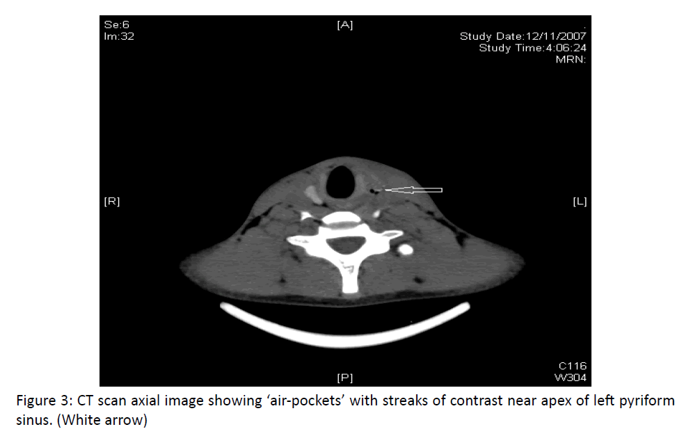
Computed tomogram (CT) showed a small tract containing air pockets and a streak of contrast, located anterior to the left carotid artery, leading to apex of the left pyriform sinus (Figure 3). He was reluctant to try non-invasive treatment and opted to undergo surgery. He successfully underwent excision of the tract along with a left hemithyroidectomy as the tract was going through the thyroid gland. He made an uneventful recovery and has not had any recurrence for the last 6 years.

otolaryngology-online-journal-contrast-near-apex

Figure 3: CT scan axial image showing ‘air-pockets’ with streaks of contrast near apex of left pyriform sinus. (White arrow)

References

PSFs occasionally present as a discharging sinus along the anterior part of sternomastoid, often with localised swelling and recurrent infection. The more typical presentation is with a recurrent neck abscess with a history of repeated surgical drainages, resulting in fistula formation. If a PSF courses through thyroid gland it may also present with acute suppuratives thyroiditis [3,4].

Knowledge of the embryological relationships of the branchial clefts to the adjoining structures, can predict a probable course of 3rd and 4th branchial fistulas. A typical course of the 3rd branchial fistula is to pierce the platysma, ascend along carotid sheath, pass over superior laryngeal nerve, deep to glossopharyngeal nerve (4th arch nerve), pass behind the internal carotid artery, pierce the thyrohyoid membrane and enter upper lateral pyriform sinus [5]. The expected course of a 4th branchial fistula would also pierce platysma; ascend along carotid sheath but pass under superior laryngeal nerve and over the recurrent laryngeal and hypoglossal nerve. Then it would dip back into mediastinum, passing the aortic arch on the left and the subclavian artery on the right side. Finally, the fistula would ascend to enter larynx near cricothyoid joint or lower part of thyroid cartilage, pass through inferior constrictor muscle and enter apex of pyriform sinus4. Other differentiating features may include presence of thymic tissue in the third sinus and thyroid tissue in the fourth sinus. Although the above description is of interest, there is considerable overlap between the two. None of the PSF has been reported to follow the above course entirety. Fibrosis developing after infection makes correct identification of anatomical relationships difficult.

An alternative embryological process is perhaps pertinent to the clinical presentation of branchial cleft fistulas. As the thymus descends during fetal development, a thymo-pharyngeal duct is formed. The thymus is derived from ventral portion of third pouch and it descends through fourth arch during 7th t o 8th week of intrauterine life. It fuses with its counterpart in the midline forming a single organ. The thymopharyngeal duct soon gets obliterated. Failure of this duct to close results in the formation of a branchial sinus lined by endodermal cells. These cells arise from the pyriform fossa and pass in close association to the thyroid gland as they head towards cervical inlet. This is analogous to the formation of a thyroglossal cyst between tongue base and thyroid gland [6,7].

Clinically a non-communicating cyst or communicating non infected cyst may present as a cold thyroid nodule or can be confused with a thyroglossal cyst [8]. The most common presentation is that of recurrent abscess with a repeated history of incision and drainage. A history of recurrent upper respiratory tract infections, neck or thyroid pain and tenderness as well as a neck mass is common. Other manifestations include cellulitis, hoarseness, odynophagia, thyroiditis, abscess and stridor.

Diagnosis of a PSF is based on demonstration of a sinus or a fistula, originating from pyriform sinus. BO has been widely used to demonstrate the sinus tract [9]. If there is acute inflammation, the chance of a false negative result increases due to the tract obliteration by inflammatory oedema [10]. Real time ultrasound can also be used to establish the connection of an abscess cavity to the pyriform sinus by performing the ‘trumpet, manoeuvre (exhale with pursed lips to distend the pyriform sinus) [11]. A CT scan is considered the investigation of choice, since it can delineate the location and extent of a PSF. If the scan is performed soon after BO, the sensitivity is considerably increased [12]. Carbonated beverages have also been used as alternative to barium to demonstrate air in the fistula’s tract [13]. In addition, the Trumpet manoeuvre can be used to facilitate demonstration of sinus tract during BO and CT. Indirect laryngoscopy often shows the internal opening of the tract. Catheterization of the internal opening with small Fogarty embolectomy catheters may facilitate dissection. Some authors have used methylene blue dye to delineate the entire tract but extravasation can hinder identification of important structures around the tract during surgery [14].

Meticulous dissection to identify superior and recurrent laryngeal nerve should be taken. The tract should be traced and dissected as high as possible towards pyriform fossa, ligated and excised. An oblique thyrotomy is performed above the cricothyroid joint to expose the apex of the pyriform sinus, thus, preventing injury to recurrent laryngeal nerve. Recurrence of the fistula can occur if the thyroid lobe is not removed with the tract, if the tract passes through the thyroid or if the resected specimen shows an absence of the epithelial lined tract. However, recurrent infection can destroy the epithelial lining. Chemo cauterization of the internal opening using trichloroacetic acid has been described with short term symptom free follow up [15]. Using a laser to obliterate the epithelium has also been advocated in the treatment of PSF. An endoscopic approach with use of a carbon dioxide laser has been reported to produce good outcomes [16,17]. These less invasive treatments are an attractive option, since they have a shorter surgical time and a low morbidity and can be performed as an outpatient procedure.

Conclusion

PSFs are uncommon developmental abnormalities of the branchial apparatus which are often misdiagnosed, resulting in multiple operations prior to making the correct. A barium esophagram often reveals the presence of a tract communicating with the pyriform sinus. However, a CT scan is considered as the investigation of choice. Complete excision of the tract, including its entry into the pyriform sinus with or without hemithyroidectomy, is essential to prevent recurrence. However, there have also been an increasing number of case reports managed by less invasive procedures, such as chemo or electrocauterization that have obtained good clinical outcome and reduced morbidity. More research in this area is needed.

References

Get the App