Research Article - Biomedical Research (2017) Volume 28, Issue 10
Prenatal ultrasound diagnosis of VACTERL syndrome and partial caudal regression syndrome
Qing Li1#, Na Liang2#, Qiu Zhang3 and Li Li3*
1Zaozhuang Maternity and Child Healthcare Hospital, No.25 Wenhuadong Road, Shizhong District, Zaozhuang, PR China
2Department of Ultrasound, Beijing Obstetrics and Gynecology Hospital, Capital Medical University, No.251 Yaojiayuan Road, Chaoyang District, Beijing, PR China
3Qilu Hospital of Shandong University (Qingdao), No.758 Hefei Road, Qingdao, PR China
#These authors contributed equally to this work
Accepted date: March 13, 2017
Abstract
Objective: To explore prenatal ultrasound diagnosis of VACTERL syndrome and partial caudal regression syndrome.
Methods: 60 patients with VACTERL syndrome and partial caudal regression syndrome treated in our hospital during September 2011 to December 2013 were retrospectively analysed. The prenatal sonographic of patient was analysed taking the postpartum result as a standard.
Results: Prenatal ultrasonic examination illustrated that there were 18 cases of anal atresia (30.00%), 26 cases of spine abnormalities (43.33%), 24 cases of malformation of upper limb (40.00%), 23 cases of malformation of lower limb (38.33%), 34 cases of renal malformation (56.67%), 46 cases of cardiac malformation (76.67%), 18 cases of tracheoesophageal fistula or esophageal atresia (30.00%), 9 cases of double umbilical arteries (15.00%). In the patients with VACTERL syndrome, there were 41 cases of 3 comprehensive deformities (68.33%), 15 cases of 4 comprehensive deformities (25.00%), 4 cases of 5 comprehensive deformities (6.67%), without 6 comprehensive deformities. There were 28 cases (46.67%) of patients with partial caudal regression syndrome, including 18 cases of 2 comprehensive deformities (30.00%), 8 cases of 3 comprehensive deformities (13.33%), 2 cases of 4 comprehensive deformities (3.33%). There were 31 cases of patients with other malformations (51.67%), with 23 cases of single umbilical artery (38.33%). All the ultrasonic examination results were in accordance with postpartum results, with no significant difference (p>0.05).
Conclusions: Prenatal ultrasonic diagnosis of VACTERL syndrome and partial caudal regression syndrome was of great value, which could guide obstetric treatment and was worthy of promotion clinically.
Keywords
VACTERL syndrome, Caudal regression syndrome, Ultrasonic diagnosis
Introduction
VACTERL syndrome includes 6 varieties of congenital malformations, they are spinal defects, anal atresia, cardiac malformation, tracheoesophageal abnormality, renal abnormality and limb abnormality, which are relatively rare in clinical with about 1/10000~1/40000 of incidence [1-4]. Caudal regression syndrome is also called sacrum or sacro-coccygeal dysplasia syndrome. The malformations include double umbilical arteries, malformation of lower limb, anal atresia and renal malformation. It is a rare congenital defect. Prenatal ultrasound diagnosis of VACTERL syndrome and partial caudal regression syndrome to fetus is rarely reported [5-8]. Missed diagnosis of one of the malformations will impact on postpartum of fetus. Early and accurate diagnosis can guide obstetric treatment and postpartum treatment on fetus [9]. 60 patients with VACTERL syndrome and partial caudal regression syndrome in our hospital were diagnosed prenatally. The details were reported below.
Materials and Methods
General information
60 patients with VACTERL syndrome and partial caudal regression syndrome treated in our hospital during September 2011 to December 2013 were retrospectively analysed. The entire study was conducted with the patient's informed consent and the approval ethics committee of our hospital. All the gravidae were in singleton pregnancy, at the age of 20~40, with average of 26.53 ± 4.68. The gestational weeks were 15~35 weeks, with average of 22.38 ± 4.29 weeks.
Methods
The inspection equipment were Siemens S2000 color Doppler ultrasonography instrument and Siemens Acuson Sequoia 512 color Doppler ultrasonography instrument. The probe was 2- dimentional, with the frequency of 4.0~6.0 MHz. Ultrasonic examination was conducted to fetus according to obstetric ultrasonic examination method. Specific method was as follows. 1. The gravida was in supine position to take abdominal ultrasonic examination. The processes were strictly in accordance with the method in Prenatal Ultrasound Guide (2012). 2. If 1 variety of the syndromes was discovered in examination, esophagus, trachea, anus, heart, spine, kidney and limbs were examined with pertinence. The spine of fetus was examined in coronal section and transverse section, and threedimensional reconstructed. The anus was examined in transverse section. Arch long axis view of ductus arteriosus and aorta, long axis view of superior vena cava and inferior vena cava, as well as coronary artery section examination under bilateral clavicle were implemented on the heart. Size, shape, location, bladder, renal artery and ureter of both kidneys were examined detailed. Continuous sequence tracking scanning was implemented on limbs. The existence of fingers and toes, shape, length, posture and number were observed detailed on the limbs. If the magenblase of the fetus was small and amniotic fluid amount large, dynamic observation needed to be conducted to magenblase and esophagus. The standard of ultrasonic diagnosis was listed as follows. If one type in the following abnormalities, which were spinal defects, anal atresia, cardiac malformation, tracheoesophageal abnormality, renal abnormality and limb abnormality, was examined, it was indicated as VACTERL syndrome. If one type of the following malformations, which were double umbilical arteries, malformation of lower limb, anal atresia and renal malformation, was examined, it was indicated as partial caudal regression syndrome. The 60 fetuses were followed up till postpartum after diagnosed with ultrasound. The postpartum results and examine results were compared.
Observational index
The malformation condition of the fetuses, as well as mal formation categories of VACTERL syndrome and partial caudal regression syndrome were counted and analysed.
Statistical analysis
SPSS 18.0 was employed for statistical analysis. The data were represented as mean ± standard deviation (x̄ ± s). T test was used for comparison of measurement data. χ2 test was used for comparison of enumeration data. Rank sum test (two-sample Wilcoxon test) was used for comparison of ranked data. p<0.05 was for significant difference.
Results
Examination results of malformations of fetuses
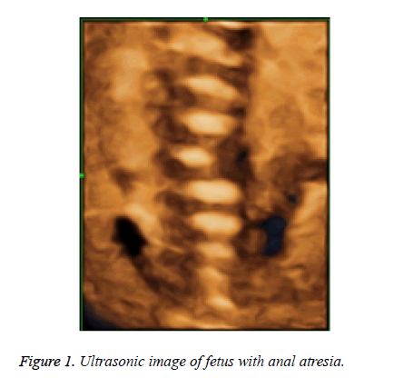
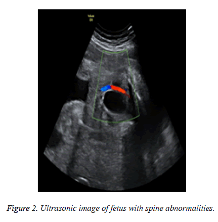
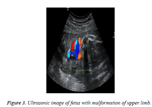
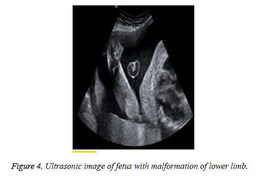
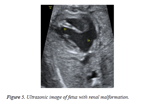
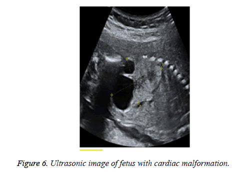
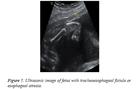
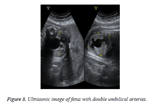
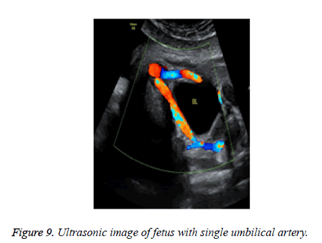
Prenatal ultrasonic examination illustrated that there were 18 cases of anal atresia (30.00%), 26 cases of spine abnormalities (43.33%), 24 cases of malformation of upper limb (40.00%), 23 cases of malformation of lower limb (38.33%), 34 cases of renal malformation (56.67%), 46 cases of cardiac malformation (76.67%), 18 cases of tracheoesophageal fistula or esophageal atresia (30.00%), 9 cases of double umbilical arteries (15.00%). There were 31 cases of patients with other malformations (51.67%), with 23 cases of single umbilical artery (38.33%). All the ultrasonic examination results were in accordance with postpartum results, with no significant difference (p>0.05). Figures 1-9 illustrated the results.
Malformation categories of VACTERL syndrome
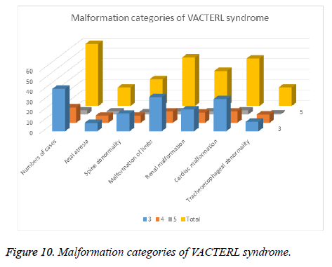
In the patients with VACTERL syndrome, there were 41 cases of 3 comprehensive deformities (68.33%), 15 cases of 4 comprehensive deformities (25.00%), 4 cases of 5 comprehensive deformities (6.67%), without 6 comprehensive deformities. All the ultrasonic examination results were in accordance with postpartum results, with no significant difference (p>0.05). Figure 10 illustrated the results.
Malformation categories of partial caudal regression syndrome
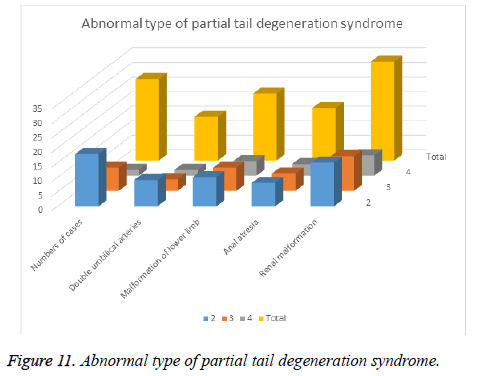
There were 28 cases (46.67%) of patients with partial caudal regression syndrome, including 18 cases of 2 comprehensive deformities (30.00%), 8 cases of 3 comprehensive deformities (13.33%), 2 cases of 4 comprehensive deformities (3.33%). All the ultrasonic examination results were in accordance with postpartum results, with no significant difference (p>0.05). Figure 11 illustrated the results.
Discussions
The aetiological agent of VACTERL syndrome and partial caudal regression syndrome is indefinite [10,11]. Stevenson et al. think the following factors may cause malformation. 1. Malformation generates in embryo after affected by chronic teratogenic conditions such as drugs, maternal hormone levels, the environment, etc. 2. The one category of first appeared malformation interferes the normal development of other anatomical structures, namely malformed sequence or malformation caused by cascade malformation. 3. Single gene mutation that occurs in fetus causes multiple anatomical systems involved in and thus abnormality occurs. 4. Gestational diabetes, high concentrations of lead intake in gravidae etc. These factors have close relations with the generation of VACTERL syndrome and partial caudal regression syndrome in fetuses. The number of categories of VACTERL syndrome and partial caudal regression syndrome in fetuses varies randomly. So it is of important significance of comprehending malformation characteristics of VACTERL syndrome and partial caudal regression syndrome in fetuses and seeking them one by one in prenatal examination to correctly diagnose VACTERL syndrome and partial caudal regression syndrome [12,13].
Multiple foreign scholars have studied and reported more than 400 cases of fetuses with VACTERL syndrome and partial caudal regression syndrome. Botto et al. have studied 287 cases. Rittler et al. have studied 21 cases. Weaver et al. have studied 46 cases. Solomon et al. have studied 60 cases. The results of the studies were statistically analysed. The incidence of spine abnormality was between 60%~80%, anal atresia between 55%~90%, tracheoesophageal abnormality between 50%~80%, cardiac malformation between 40%~80%, malformation of limbs between 40%~50%, and renal malformation between 50%~80% [14,15]. In the study results of our hospital, there were 18 cases of anal atresia (30.00%), 26 cases of spine abnormalities (43.33%), 24 cases of malformation of upper limb (40.00%), 23 cases of malformation of lower limb (38.33%), 34 cases of renal malformation (56.67%), 46 cases of cardiac malformation (76.67%), 18 cases of tracheoesophageal fistula or esophageal atresia (30.00%), 9 cases of double umbilical arteries (15.00%). In the patients with VACTERL syndrome, there were 41 cases of 3 comprehensive deformities (68.33%), 15 cases of 4 comprehensive deformities (25.00%), 4 cases of 5 comprehensive deformities (6.67%), without 6 comprehensive deformities. There were 28 cases (46.67%) of patients with partial caudal regression syndrome, including 18 cases of 2 comprehensive deformities (30.00%), 8 cases of 3 comprehensive deformities (13.33%), 2 cases of 4 comprehensive deformities (3.33%).
Compared with the foreign reports, the incidences of anal malformation, spine malformation and tracheoesophageal abnormality in our study were lower. The probable reasons might be as follows. 1. Anal atresia belongs to unconventional examination contents. Missed diagnosis will occur when intestinal dilatation is not manifested during examination to fetuses [16]. 2. There are no direct signs of tracheoesophageal abnormality in prenatal ultrasonic examination. It can only be indicated by other signs such as small or no magenblase, hydramnios etc. [17]. 3. Only median sagittal section of spine is examined in prenatal routine ultrasound examination. Single hemi-vertebral spinal deformity without significant scoliosis is not easy to diagnose. But this is facile to be diagnosed by X-ray or MRI after these fetuses with spinal malformation are born. Therefore, prenatal ultrasound diagnosis has certain limitations, which has not diagnosed all the malformations of the fetuses [18].
When the fetuses with VACTERL syndrome and partial caudal regression syndrome have malformation of limbs, other malformation complications will usually occur. This causes death of the fetuses in duration of pregnancy. The observation of malformation of limbs on fetus will be interfered by fetal position and amniotic fluid. Continuous sequence ultrasound tracking method or targeted ultrasound screening need to be conducted to enhance the accuracy of diagnosis [19,20]. Continuous sequence tracking method was employed in the limb examination on the fetuses in our study. So the detection rate of limb malformation in fetus was higher than related articles, which had correlations with normalized prenatal examination.
The fetuses born with VACTERL syndrome and partial caudal regression syndrome were treated by different treatment programs with different malformations. At present, surgical therapy on-going was mainly for anal atresia, oesophageal atresia, cardiac malformation etc. If these malformations could be discovered and treated early, the late survival rate of most fetuses would be enhanced [21].
In summary, the prenatal ultrasonic diagnosis of VACTERL syndrome and partial caudal regression syndrome is of great value, which could guide obstetric treatment and was worthy of promotion clinically.
References
- Kim PCW, Mo R, Hui C. Murine models of VACTERL syndrome: Role of sonic hedgehog signaling pathway. J Pediatr Surg 2001; 36: 381-384.
- Berrebi D, Lebras MN, Belarbi N. Bilateral adrenal neuroblastoma and nephroblastoma occurring synchronously in a child with Fanconis anemia and VACTERL syndrome. J Pediatr Surg 2006; 41: 11-14.
- Shaw-Smith C. Oesophageal atresia, tracheo-oesophageal fistula, and the VACTERL association: review of genetics and epidemiology. J Med Gene 2006; 43: 545-554.
- Solomon BD. VACTERL/VATER Association. Orphanet J Rare Dis 2011; 6: 56.
- Padmanabhan R. Retinoic acid-induced caudal regression syndrome in the mouse fetus. Reprod Toxicol 1998; 12: 139-151.
- Adra A, Cordero D, Mejides A. Caudal regression syndrome: etiopathogenesis, prenatal diagnosis, and perinatal management. Obstetr Gynecol Survey 1994; 49: 508.
- Chen PW, Chen XL, Yang XH. Prenatal diagnosis of caudal regression syndrome by ultrasound and review of the literature. Zhonghua Yixue Chaosheng Zazhi/ Chinese J Med Ultrasound (Electronic Version) 2011; 8: 70-73.
- Fahmy MAB. Caudal regression syndrome and sacral agenesis//rare congenital genitourinary anomalies. Springer Berlin Heidelberg 2015; 231-235.
- Zeidler C. Heterozygous FGF8 mutations in patients presenting cryptorchidism and multiple VATER/VACTERL features without limb anomalies. Birth Defects Res A Clin Mol Teratol 2014; 100: 750-759.
- Friedland-Little JM, Hoffmann AD, Ocbina PJR. A novel murine allele of Intraflagellar Transport Protein 172 causes a syndrome including VACTERL-like features with hydrocephalus. Human Mol Gene 2011; 241.
- Semba K, Ki Y. Etiology of caudal regression syndrome. Human Genet Embryol 2013; 3: 2161-2436.
- Kanasugi T, Kikuchi A, Matsumoto A. Monochorionic twin fetus with VACTERL association after intracytoplasmic sperm injection. Congenital Anomalies 2013; 53: 95-97.
- Brosens E. Clinical and etiological heterogeneity in patients with tracheo-esophageal malformations and associated anomalies.Eur J Med Genet 2014; 57: 440-452
- Solomon BD, Raam MS, Pineda-Alvarez DE. Analysis of genitourinary anomalies in patients with VACTERL (Vertebral anomalies, Anal atresia, Cardiac malformations, Tracheo-Esophageal fistula, Renal anomalies, Limb abnormalities) association. Congenital Anomalies 2011; 51: 87-91.
- Moore SW. Associations of anorectal malformations and related syndromes. Pediatr Surg Int 2013; 29: 665-676.
- Perlman S, Bilik R, Leibovitch L, Katorza E, Achiron R. More than a gut feeling-sonographic prenatal diagnosis of imperforate anus in a high-risk population. Prenat Diagn 2014; 34: 1307-1311.
- Haak MC, Lakeman P, Cohen-Overbeek TE. Tracheal agenesis: approach towards this severe diagnosis. Case report and review of the literature. Eur J Pediatr 2012; 171: 425-431.
- Reutter H. Evidence for annular pancreas as an associated anomaly in the VATER/VACTERL association and investigation of the gene encoding pancreas specific transcription factor 1A as a candidate gene. Am J Med Genet A 2014; 164: 1611-1613.
- Xu H, Wang Y, Chen Y. Clinical application of ultrasound diagnosis on fetal hand-foot deformity with a systematic continuous sequence approach. Zhonghua Yixue Chaosheng Zazhi/Chinese J Med Ultrasound (Electronic Version) 2011; 8: 1702-1712.
- Xu R, Xie T, Ohya J. Automatic real-time tracking of fetal mouth in fetoscopic video sequence for supporting fetal surgeries //SPIE Medical Imaging. Int Soc Optics Photonics 2013; 86710-86710.
- Orioli IM, Amar E, Arteaga-Vazquez J. Sirenomelia: an epidemiologic study in a large dataset from the International Clearinghouse of Birth Defects Surveillance and Research, and literature review. Am J Med Gene Part C: Seminars Medical Genetics Wiley Subscription Services Inc. Wiley Company 2011; 157: 358-373.
