Case Report - Current Pediatric Research (2017) Volume 21, Issue 1
Prenatal diagnosis of pentalogy of cantrell in a case with craniorachischisis
Shagufta Wahab1, Biswajit Sahoo2, Somit Mittal2, Rizwan Ahmad Khan31Associate Professor, Department of Radiodiagnosis, JN Medical College Hospital, AMU, Aligarh, India.
2Department of Radiodiagnosis, JN Medical College Hospital, AMU, Aligarh, India.
3Associate Professor, Department of Pediatric Surgery, JN Medical College Hospital, AMU, Aligarh, India.
- *Corresponding Author:
- Rizwan Ahmad Khan
Kashana-E-Wahab, Street No. 4
Iqra Colony, Near Iqra Public School
Aligarh 202002, India.
Tel: +919410210281
E-mail: drrizwanahmadkhan@yahoo.co.in
Accepted date: November 24, 2016
Abstract
Pentalogy of Cantrell as the name suggests involves combination of five complex congenital anomalies affecting midline abdomen. It is a complex and rare congenital anomaly which in extreme condition is not compatible with life specially if associated with other complex anomalies. Herein we report an antenatal detection of such a case with associated cranioschisis. Through this case we would like to emphasize the importance of antenatal care and antenatal sonographic screening.
Keywords
Pentalogy of Cantrell, Cranioschisis, Prenatal.
Introduction
The pentalogy of Cantrell was first described by Cantrell et al in 1958, who reported 5 cases with this anomaly [1]. The findings included a lower sternum defect, a midline supraumblical thoracoabdominal wall defect, a defect of the anterior diaphragm, a defect of the diaphragmatic pericardium and congenital cardiac anomalies [1,2]. The pentalogy is associated with very poor prognosis and therefore the need for prenatal diagnosis arises [3,4]. The overall prognosis and survival depends on extent of the ventral wall, sternal and cardiac defects. But when there is association of anomalies of central nervous system, the prognosis may become even poorer. Association of craniorachischisis to the pentalogy of Cantrell is extremely rare and has been reported in very few cases. In this report we describe a case of Pentalogy of Cantrell with craniorachischisis.
Case Report
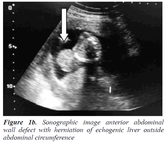
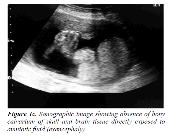
A 30 year old pregnant woman came for routine antenatal ultrasonography at around 19 weeks’ menstrual age. Transabdominal sonography was performed which revealed a single live fetus with femur length of 19 wks corresponding to the menstrual age. Gastroschisis with herniation of liver and ectopia cordis were seen (Figures 1a and 1b). The other sonographic findings were spinal dysraphism and exencephaly (Figure 1c); however there was no polyhydramnios and the facial structures were normal with no evidence of cleft lip or palate. On the basis of sonographic findings; we diagnosed the case with the rare disorder of fetus with pentalogy of Cantrell with craniorachischisis. The diagnosis was discussed with the parents and the prognosis was explained to them. The pregnancy was terminated and a male fetus was delivered. Ectopiacordis, gastroschisis with herniation of liver, lower sternal defect and exencephaly were observed in the expelled fetus.
Discussion
The Pentalogy of Cantrell is an extremely rare phenomenon with a prevalence of about 1 per 65000 live births and classified as a developmental defect of midline anterior thoracoabdominal wall [2,4-6]. It is thought to result from an abnormal migration of the sternal anlage and myotomes in the early 6th to 7th week of gestation however the exact pathology behind is still not very clear but mutation of TAS gene, is mentioned in some of the previous studies [3]. The classical pentalogy of Cantrell that was first described by Cantrell et al1 in 1958 had five features: Midline supraumbilical thoraco-abdominal wall defect; lower sternal defect; anterior diaphragmatic defect; diaphragmatic pericardial defect, ectopia cordis and intracardiac anomalies [2,7].Till now there have been few case reports describing the full spectrum having all of the five defects. Incomplete forms or variants with lesser number of defects of the pentalogy are more common. However a confident diagnosis can be made on the basis of the sonographic diagnosis of anterior abdominal wall defect and ectopia cordis which are the hallmark4of this rare syndrome [8-10].
Previous case reports show association of this syndrome with limb anomalies, craniofacial anomalies (cleft lip, cleft palate, hydrocephalus), vertebral anomalies; cystic hygroma. Cardiac anomalies consist of ventricular septal defect (in 100% of cases), atrial septal defect (52%), pulmonary stenosis (33%) and Tetralogy of Fallot (20%) [2,5]. Our case is a variant of pentalogy of Cantrell as it lacked any gross intracardiac defects however presence of exencephaly with spinal dysraphism makes our case one of the rarest. In addition our case is associated with gastroschisis not omphalocele. Exencephaly is characterized by absence of bony calvarium. Craniorachischisis is due to complete failure of the neurulation process and characterized by open cranial defect (anencephaly or exencephaly) continuing with the open spine (spinal dysraphism) [2,6,11]. However our case was not associated with polyhydramnios.
Antenatal diagnosis of this pentalogy of Cantrell is very difficult before 12 weeks of gestation, because there is physiological herniation of bowel out of abdomen in fetal development at this time. Differential diagnosis of fetal abdominal wall defect after 12 weeks is omphalocele, pentalogy of Cantrell and gastroschisis [12,13]. If midline abdominal wall defect is present together with other abnormalities specially ectopia cordis one should consider pentalogy of Cantrell.
The prognosis depends on the severity of the anomalies. The prognosis is poorer in patients with the complete form of pentalogy of Cantrell and in cases with other associated anomalies. Remarkably intracardiac defects do not seem to affect prognosis much.
Conclusion
Pentalogy of Cantrell (PC) is a congenital malformation syndrome resulting from defective development of the septum transversum and presents as midline thoracoabdominal wall defect. Our case of the pentalogy of Cantrell is interesting and rare because the fetus having herniation of liver and ectopia cordis and associated spinal dysraphism and exencephaly had rarely been reported in association with this syndrome. Through this case we underline the importance of antenatal sonography for general population and also for the ultrasonologist for keeping in mind the possibility of associated dorsal abnormalities in a patient with large ventral anomaly.
References
- Cantrell JR, Haller JA, Ravitch MM. A syndrome of congenital defects involving the abdominal wall, sternum, diaphragm, pericardium and heart. Surg Gynecol Obstet 1958; 107: 602-614.
- Van Hoorn JH, Moonen RM, Huysentruyt CJ, et al. Pentalogy of cantrell: Two patients and a review to determine prognostic factors for optimal approach. Eur J Pediatr 2008; 167: 29-35.
- Parvari R, Weinstein Y, Ehrlich S, et al. Linkage localization of the thoraco-abdominal syndrome (TAS) gene to Xq25-26. Am J Med Genet 1994; 49: 431-434.
- Angtuaco TL. Fetal anterior abdominal wall defect. In: CalenPW, editor. Ultrasonography in obstetrics and gynecology. Philadelphia: WB Saunders 2000. P 500.
- Cantrell JR, Haller JA, Ravitch MM. A syndrome of congenital defects involving the abdominal diaphragm, pericardium and heart. Surg Gynecol Obstet 1958; 107: 602-614.
- Monteagudo A, Timor-Tritsch IE: Fetal neurosonography of congenital brain anomalies. In: Timor-Tritsch IE, Monteagudo A, Cohen HI, editors. Ultrasonography of the prenatal and neonatal brain. New York: McGraw-Hill 2001. 164.
- McMahon CJ, Taylor MD, Cassady CI, et al. Diagnosis of pentalogy of cantrell in the foetus using magnetic resonance imaging and ultrasound. Pediatr Cardiol 2007; 28: 172?175
- Liang RI, Huang SE, Chang FM. Prenatal diagnosis of ectopia cordis at 10 weeks of gestation using two-dimensional and three-dimensional ultrasonography. Ultrasound Obstet Gynecol 1997; 10: 137-139.
- Oka T, Shiraishi I, Iwasaki N, et al. Usefulness of helical CT angiography and MRI in the diagnosis and treatment of pentalogy of Cantrell. J Pediatr 2003; 142: 84.
- Aslan A, Karagüzel G, Unal I, et al. Two rare cases of the pentalogy of cantrell or its variants. Acta Med Austriaca 2004; 31: 85-87.
- Chen CP, Hsu CY, Tzen CY, et al. Prenatal diagnosis of pentalogy of cantrell associated with hypoplasia of the right upper limb and ectrodactyly. Prenat Diagn 2007; 27: 86-87.
- Jnah AJ, Newberry DM, England A. Pentalogy of cantrell: Case report with review of the literature. Adv Neonatal Care 2015; 15: 261-268.
- Restrepo MS, Cerqua A, Turek JW. Pentalogy of cantrell with ectopia cordis totalis, total anomalous pulmonary venous connection and tetralogy of Fallot: A case report and review of the literature. Congenit Heart Dis 2014; 9: 129-134.


