Research Article - Journal of Clinical Dentistry and Oral Health (2023) Volume 7, Issue 1
Obturation Technique Used for Maxillary Canine- Single Cone Vs Lateral Compaction
Bala SS, Selvam D*Department of Conservative dentistry and endodontics, Saveetha Dental College and Hospitals, Saveetha Institute of Medical and Technical Sciences, Saveetha University, Chennai, India
- *Corresponding Author:
- Selvam D
Department of Conservative dentistry and endodontics
Saveetha Dental College and Hospitals
Saveetha Institute of Medical and Technical Sciences
Saveetha University, Chennai, India
E-mail: deepaks.sdc@saveetha.com
Received: 19-Dec-2022, Manuscript No. AACDOH-22-84041; Editor assigned: 20-Dec-2022, PreQC No. AACDOH-22-84041 (PQ); Reviewed: 04-Jan-2023, QC No. AACDOH-22-84041 (QC); Revised: 09-Jan-2023, Manuscript No. AACDOH-22-84041(R); Published: 16-Jan-2023, DOI: 10.35841/aacdoh-7.1.131
Citation: Bala SS, Selvam D. Obturation technique used for maxillary canine- single cone vs. lateral compaction. J Clinic Dent Oral Health. 2023;7(1):131
Abstract
Introduction: Obturation is used to seal and prevent reinfection of the cleansed, shaped, and disinfected root canal system. It has been stressed that excellent obturation should result in a good seal, which encompasses the apical, coronal, and lateral seals. There are several obturation procedures available, with no variation in long-term results, and no approach that eliminates leaking. The aim of the study is to determine the most commonly used Obturation technique for endodontic treatment of Maxillary permanent canines. Materials and method: In this retrospective study, the data was collected from the hospital database and further analysis was done and the results were tabulated. A statistical analysis of the collected data regarding the variance among the different obturation techniques used for the treatment of maxillary permanent canines. Results: The study sample consisted of 957 patients. Out of 957 patients, 52.42% underwent endodontic treatment for the maxillary left permanent canine, and 47.58% underwent endodontic treatment for the maxillary right permanent canine. When it comes to the Obturation technique used, 58.65% of the population used Lateral compaction technique and 41.35% of the population underwent Matched taper single cone technique. Discussion: While other techniques call for a wider flare and the unnecessary removal of sound enamel for easier instrumentation, cold lateral compaction offers caries- guided removal of tooth structure. Hence, it is the preferred obturation technique for maxillary canines. While each technique has its advantages and disadvantages, it is safe to say that most dentists employ the technique which they are more knowledgeable on. Other factors that contribute to the technique chosen include the anatomical intricacies of the affected tooth, armamentarium and material availability, cost, patient’s willingness to comply, etc. Conclusion: It can be concluded that Cold Lateral compaction is the most commonly used obturation technique to treat maxillary permanent canines.
Keywords
Obturation techniques, Lateral compaction, Innovative treatment, RCT, Canine RCT.
Introduction
Obturation is used to seal and prevent reinfection of the cleansed, shaped, and disinfected root canal system. It is now widely recognized that improperly obturatred teeth are frequently inadequately prepared [1]. Obturation without voids and to within 2.0 mm of the apex has been proven to have a significant positive impact on treatment outcome when evaluating obturatred roots [2]. It has been stressed that excellent obturation should result in a good seal, which encompasses the apical, coronal, and lateral seals. To accomplish this, a variety of materials and techniques have been devised; nevertheless, all of the materials and techniques are thought to show evidence of leakage to some degree. There are several obturation procedures available, with no variation in long-term results, and no approach that eliminates leaking [3]. The most commonly employed obturation techniques are described below:
Single Cone Technique: This is when a size-matched larger taper cone is used to precisely fit the canal preparation. This method is frequently used in conjunction with specialized filing systems. This method relies heavily on sealer and may not completely obturate the canal in three dimensions. The apical section, however, should be well-fitting. This approach is preferred by up to 49% of dentists, and experimental evidence suggests that it is comparable to lateral compaction [4].
Cold lateral compaction technique
The Final Working Length (FWL) and canal shape are calculated, and a master cone is selected, coated in sealer, and compressed laterally with finger spreaders. Until the obturation is complete, accessory cones will be applied. The core material and accessory cones remain separated as a result of this process, which does not generate a homogeneous mass. Sealers should be used to fill in the gaps as a result. Excessive force used when compacting GP might cause root fractures [5].
Warm Vertical Compaction: A master cone with the proper working length and canal size is selected. At this length, the cone should be able to withstand displacement. Once confirmed, the cone is sealed and inserted into the canal, where it is compacted vertically using a heated plugger until the apical 3-4 mm length of the canal is filled. Warm bits of core material are then used to backfill the canal system [6]. It is critical to evaluate the apical fill radiographically before backfilling; otherwise, correcting apically deficient fills is time consuming. The continuous wave compaction technique is a version of this. When commercial heating equipment is used, it specifies a process of warm vertical compaction [7]. The warm plugger is introduced to within 5-7 mm of the working length after fitting an adequate cone with sealer, either in a continuous ‘down-pack' motion or occasionally. A separation burst of heat is administered after a 10-second interval, while the softened GP cools, separating the apical GP. A flat-ended plugger is used to condense the warm GP apical to the point of severance, and the canal is subsequently ‘backfilled' using an injection technique [8].
Warm lateral compaction
A master cone is coated with sealer and introduced, matching to the working length and canal shaper. The core material is subsequently compacted laterally with a warm spreader. The process is then continued with the use of supplementary canals, and the compaction is done again. The heat causes the accessories to adhere to the core material, resulting in a more uniform mass of GP within the canal. The flexibility to control the working length is a benefit of this approach versus vertical techniques [9].
Carrier-based Thermoplasticized Technique: Warm GP or ‘Resilon'-coated plastic core carriers are placed to the working length into the canal. Before obturation, a blank core must be put into the canal to ensure length, taper, and fit. The canal should next be lightly coated with sealer, and the point should be heated in an oven before being cautiously but swiftly inserted into the canal. This procedure is quick and effectively obturates the canal, but true length control is poor, and there is a danger of both sealer and GP extrusion [10]. It is also common for the GP to ‘strip' off from the carrier before being fully implanted, resulting in an inadequate fill with voids and carrier contact with the root wall rather than the sealer and GP. In instances of retreatment or when a post is planned, removing cores can be problematic. Although specialized burs for removing the cores are available, they can be difficult to use [11].
Thermo mechanical Compaction: A sealer-coated master cone is attached to the working length and compacted with a manual spreader and then a rotary instrument running at 5,000 to 10,000 rpm. The rotary instrument is covered with pre-warmed gutta-percha, which directs the GP and sealer into the canal and all its intricacies due to its more fluid state and friction between the compactor and the canal wall [12].
Plasticised GP techniques
Because the material can flow within the canal area, plastic approaches have the potential to improve obturation. Due to challenges in length control, injection procedures without a cold cone or GP plug at the apical constriction are not recommended. Despite the fact that GP is highly biocompatible, the clinician should avoid extrusions, particularly into the sinus or ID canal [13].
Our team has extensive knowledge and research experience that has translate into high quality publications [14-28].
The aim of the study is to determine the most commonly used Obturation technique for endodontic treatment of Maxillary permanent canines.
Materials and Methods
This retrospective study examined the records of patients from 01 June 2019 to 31st March 2021 who visited Saveetha Dental College and Hospitals. Ethical approval was taken from the institutional review board. The study population included patients who underwent endodontic treatment. The study sample included both male and female gender, predominantly south indians. Sample size was 10672, out of which 957 underwent endodontic treatment in their maxillary permanent canines. The necessary data such as age, gender, obturation technique used etcwas recorded. Incomplete patient data was excluded. Data was recorded in Microsoft Excel and exported to the statistical package of social science for Windows (SPSS) and subjected to statistical analysis. Chi Square test was used for comparison of groups.
Results
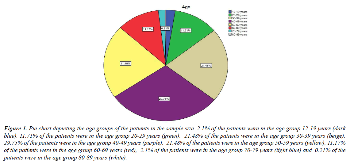
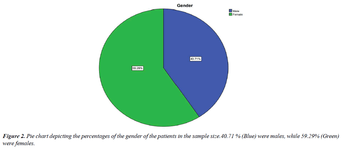
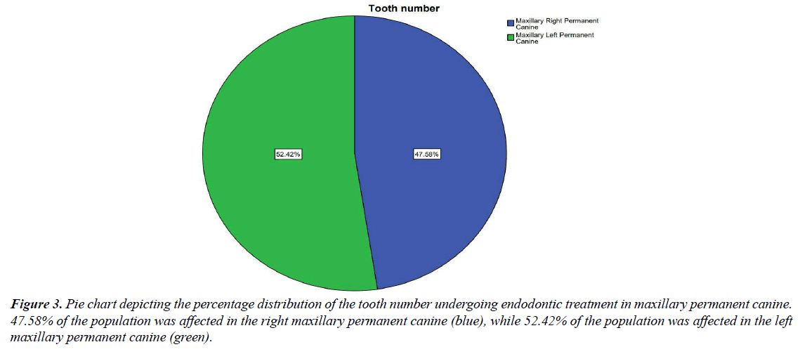
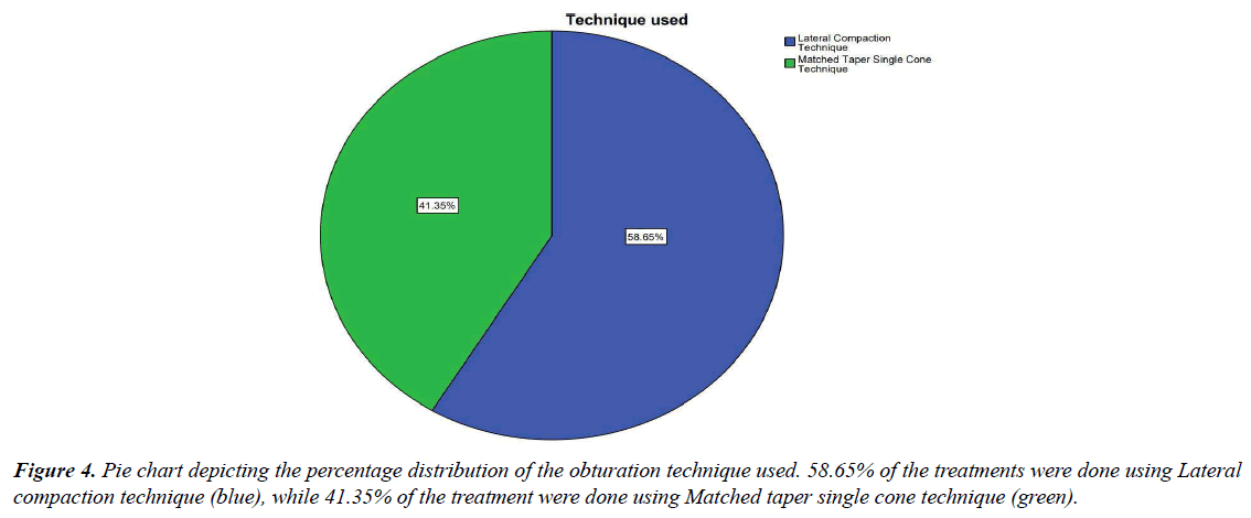
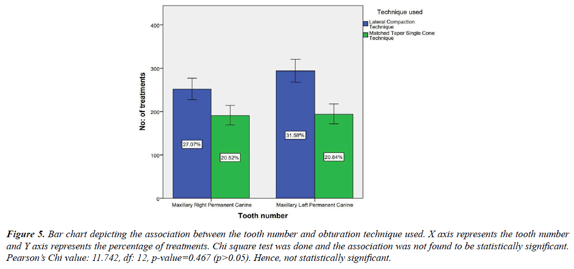
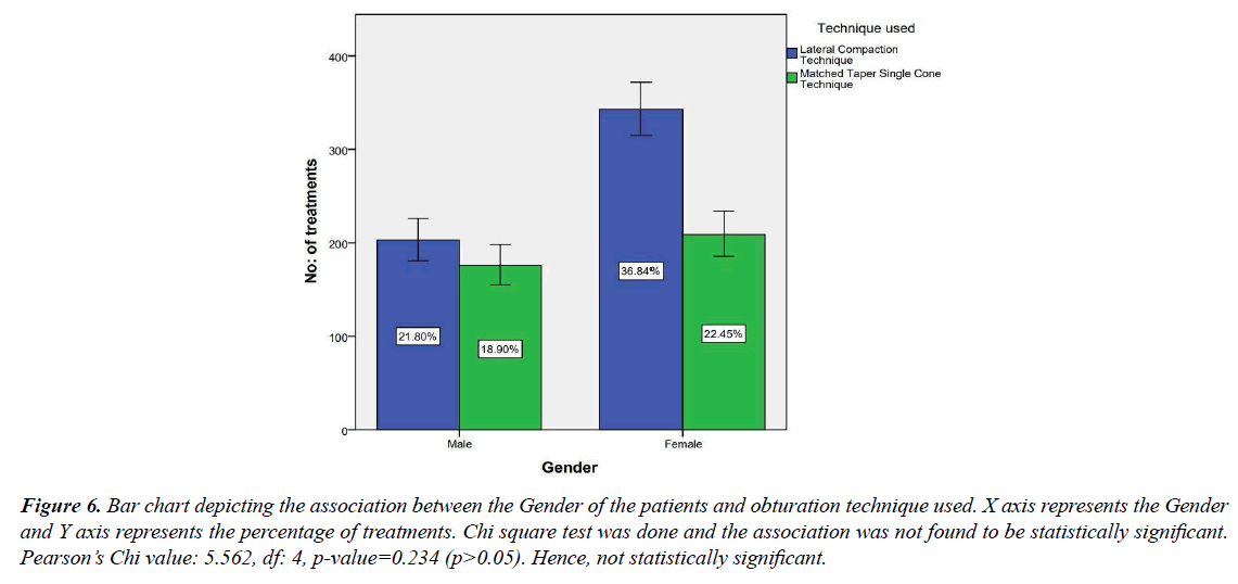
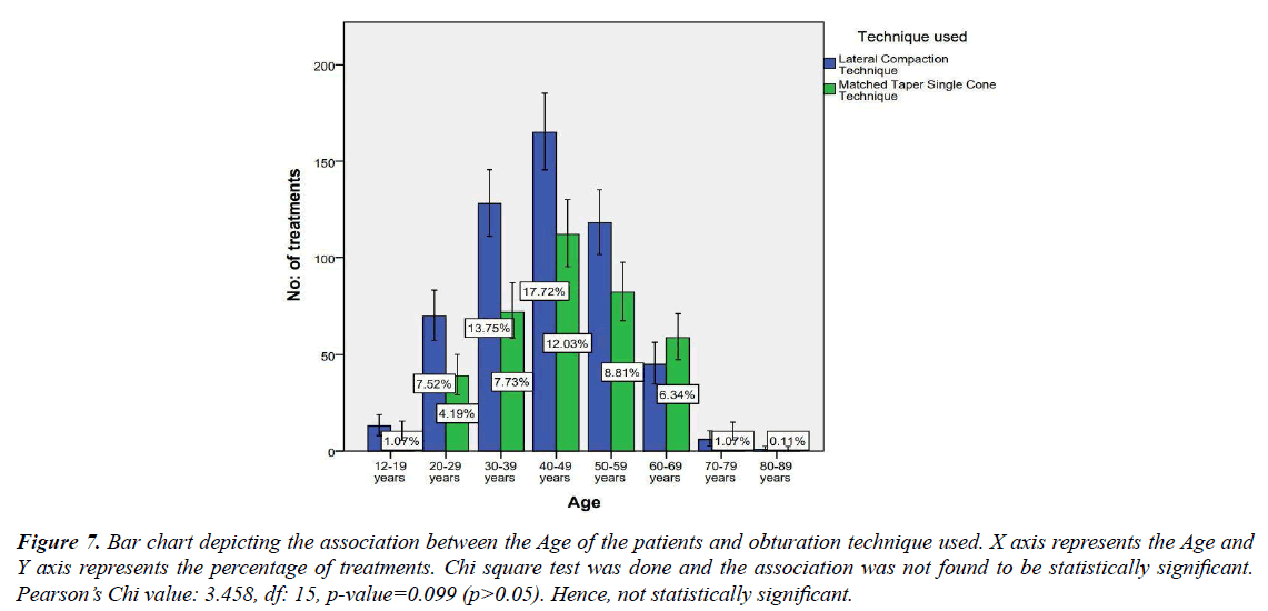
The study sample consisted of 957 patients. The patients were split into 8 age groups, the maximum patients belonging to 40-49 years of age (Figure 1). The population consisted of 59.29% females and 40.71% males (Figure 2). Out of 957 patients, 52.42% underwent endodontic treatment for the maxillary left permanent canine, and 47.58% underwent endodontic treatment for the maxillary right permanent canine (Figure 3). When it comes to the Obturation technique used, 58.65% of the population used Lateral compaction technique and 41.35% of the population underwent Matched taper single cone technique (Figure 4). The correlation between the age of the patient and the obturation technique used was not found to be statistically significant, meaning the technique is not dependent on the age of the patient (Figure 5). The correlation between the tooth number and the technique used was also not found to be statistically significant, meaning there is no association between the technique employed and the side of the affected tooth (Figure 6). The association between the Gender of the patient and the technique used was not found to be statistically significant (Figure 7).
Figure 1: Pie chart depicting the age groups of the patients in the sample size. 2.1% of the patients were in the age group 12-19 years (dark blue), 11.71% of the patients were in the age group 20-29 years (green), 21.48% of the patients were in the age group 30-39 years (beige), 29.75% of the patients were in the age group 40-49 years (purple), 21.48% of the patients were in the age group 50-59 years (yellow), 11.17% of the patients were in the age group 60-69 years (red), 2.1% of the patients were in the age group 70-79 years (light blue) and 0.21% of the patients were in the age group 80-89 years (white).
Figure 3: Pie chart depicting the percentage distribution of the tooth number undergoing endodontic treatment in maxillary permanent canine. 47.58% of the population was affected in the right maxillary permanent canine (blue), while 52.42% of the population was affected in the left maxillary permanent canine (green).
Figure 5: Bar chart depicting the association between the tooth number and obturation technique used. X axis represents the tooth number and Y axis represents the percentage of treatments. Chi square test was done and the association was not found to be statistically significant. Pearson’s Chi value: 11.742, df: 12, p-value=0.467 (p>0.05). Hence, not statistically significant.
Figure 6: Bar chart depicting the association between the Gender of the patients and obturation technique used. X axis represents the Gender and Y axis represents the percentage of treatments. Chi square test was done and the association was not found to be statistically significant. Pearson’s Chi value: 5.562, df: 4, p-value=0.234 (p>0.05). Hence, not statistically significant.
Figure 7: Bar chart depicting the association between the Age of the patients and obturation technique used. X axis represents the Age and Y axis represents the percentage of treatments. Chi square test was done and the association was not found to be statistically significant. Pearson’s Chi value: 3.458, df: 15, p-value=0.099 (p>0.05). Hence, not statistically significant.
Discussion
Root canal therapy's ultimate biologic goal is to prevent or cure apical periodontitis. Root canal treatment seeks to achieve this goal through debridement, disinfection, and obturation of the root canal system when the pulp is made non–viable [29]. Obturation is used to create a fluid-tight barrier around the periradicular tissues to protect them from bacteria in the oral cavity. While achieving a perfect airtight or hermatic seal is unrealistic, every effort should be taken to achieve this goal [30]. The goals of an obturation are as below:
Prevent microorganisms or possible nutrients from accessing the dead space of the root canal system through coronal leakage [31].
Prevent periapical or periodontal fluids from penetrating into the root canals and nourishing bacteria [32].
To prevent their multiplication and pathogenicity, enshrine any remaining microorganisms that have survived the debridement and disinfection stages of treatment [33].
The appropriate location, cleaning, shape, and obturation of the root canal system is an adequate measure for root canal therapy's long-term success. As a result, if any of these criteria are disregarded, the treatment outcome may be jeopardised, leading to post-treatment infection, discomfort, and/or complications in the treated tooth. The maxillary canines are the longest teeth in the human dental arch, which is very well known. They're flattened in the cervical area and should be flared to allow for proper instrumentation [34]. Previous studies conducted by Er, et al. have concluded that Thermomechanical compaction using System B can be very well used for endodontic treatment of maxillary canines and that the heat in the compaction did no potential harm to the maxillary canine models [35]. Hussain et al had concluded that the endodontic treatment of long canines can be proceeded using Instrument Cable Removal technique [36].
While each technique has its advantages and disadvantages, it is safe to say that most dentists employ the technique which they are more knowledgeable on. Other factors that contribute to the technique chosen include the anatomical intricacies of the affected tooth, armamentarium and material availability, cost, patient’s willingness to comply, etc. [37].
A study conducted by Oh et al has concluded that when compared to Orthograde MTA obturation, Cold Lateral Compaction technique was found to have lower filling densities [38]. This is in contradiction to the current study, where lateral compaction is the most commonly used obturation technique. This can be attributed to the easiness of handling the instruments in Cold lateral compaction. Lateral compaction is the technique employed by Yousef et al for the endodontic treatment of a 32 mm long maxillary canine, due to this technique offering sufficient root canal cleaning, shape, and obturation, but also allowed for the preservation of the tooth's residual structure [39].
Thermoplasticized GP techniques run the risk of overheating the tooth structure and causing damage to the periodontium. All warm techniques have to be controlled with precision and accuracy as they pose a risk of heating the tooth beyond repair, causing fractures, cracks and perforations [40]. Prior to obturation, the root canal system must be chemo-mechanically prepared. The root canal system must be obturated to the correct length and without voids in order to be successful. The coronal seal also contributes to this achievement. There is no particular approach that is superior to another, and case selection is crucial. Recent advancements have sought to make obturating canals easier; nevertheless, due to the constraints of in vitro research in this area, patience is required in order for meaningful clinical trials to arise [41]. While other techniques call for a wider flare and the unnecessary removal of sound enamel for easier instrumentation, cold lateral compaction offers caries- guided removal of tooth structure. Hence, it is the preferred obturation technique for maxillary canines [42].
Conclusion
It can be concluded that Cold Lateral compaction is the most commonly used obturation technique to treat maxillary permanent canines. This is due to the easiness of handling the instruments, sufficient root canal cleaning, shape, and obturation, but also allowed for the preservation of the tooth's residual structure.
Acknowledgements
The authors are thankful to Saveetha Dental College for providing a platform to express our knowledge.
Author contributions
Santhosh Bala contributed to data collection, analysis and interpretation and drafting of the article. Deepak S contributed to the critical revision of the article.
Conflict of interest
Authors declare no potential conflicts of interest.
Source of funding
The present study was supported by Saveetha Dental College, Saveetha Institute of Medical and Technical Sciences, Saveetha University.
References
- Darcey J, Roudsari RV, Jawad S, et al. Modern endodontic principles. Part 5: Obturation. Dent Update. 2016;43(2):114–116.
- Wu MK, Fan B, Wesselink PR. Leakage along apical root fillings in curved root canals. Part I: Effects of apical transportation on seal of root fillings. J Endod. 2000;26(4):210–216.
- Ng YL, Mann V, Rahbaran S, et al. Outcome of primary root canal treatment: Systematic review of the literature: Part 2. Influence of clinical factors. Int Endod J. 2008;41(1):6-31.
- Grossman LI, Oliet S. Correlation of clinical diagnosis and bacteriologic status of symptomatically involved pulps. Oral Surg Oral Med Oral Pathol. 1968;25(2):235-238.
- Biggs SG, Knowles KI, Ibarrola JL, et al. An in vitro assessment of the sealing ability of resilon/epiphany using fluid filtration. J Endod. 2006;32:759–761.
- Aqrabawi JA. Outcome of endodontic treatment of teeth filled using lateral condensation versus vertical compaction (Schilder’s technique). J Contemp Dent Pract. 2006;7(1):17–24.
- Ng YL, Mann V, Rahbaran S, et al. Outcome of primary root canal treatment: systematic review of the literature: Part 1. Effects of study characteristics on probability of success. Int Endod J. 2007;40(12):921–939.
- Tay FR, Pashley DH. Monoblocks in root canals: A hypothetical or a tangible goal. J Endod. 2007;33(4):391–398.
- de Chevigny C, Dao TT, Basrani BR, et al. Treatment outcome in endodontics: The Toronto study--phase 4: Initial treatment. J Endod. 2008;34(3):258–263.
- Peng L, Ye L, Tan H, et al. Outcome of root canal obturation by warm gutta-percha versus cold lateral condensation: A meta-analysis. J Endod. 2007;33(2):106–109.
- Whitworth JM, Baco L. Coronal leakage of sealer-only backfill: An in vitro evaluation. J Endod. 2005;31(4):280–282.
- Gordon MPJ, Love RM, Chandler NP. An evaluation of .06 tapered gutta-percha cones for filling of .06 taper prepared curved root canals. Int Endod J. 2005;38(2):87–96.
- Tamse A, Tsesis I, Rosen E. Vertical root fractures in dentistry. Springer 2015.
- Muthukrishnan L. Imminent antimicrobial bioink deploying cellulose, alginate, EPS and synthetic polymers for 3D bioprinting of tissue constructs. Carbohydr Polym. 2021;260:117774.
- PradeepKumar AR, Shemesh H, Nivedhitha MS, et al. Diagnosis of vertical root fractures by cone-beam computed tomography in root-filled teeth with confirmation by direct visualization: A systematic review and meta-analysis. J Endod. 2021;47(8):1198–1214.
- Chakraborty T, Jamal RF, Battineni G, et al. A review of prolonged post-COVID-19 symptoms and their implications on dental management. Int J Environ Res Public Health. 2021;18(10).
- Muthukrishnan L. Nanotechnology for cleaner leather production: A review. Environ Chem Lett. 2021;19(3):2527–2549.
- Teja KV, Ramesh S. Is a filled lateral canal: A sign of superiority? J Dent Sci. 2020;15(4):562.
- Narendran K, MS N, Sarvanan A. Synthesis, characterization, free radical scavenging and cytotoxic activities of phenylvilangin, a substituted dimer of embelin. Indian J Pharm Sci. 2020;82(5):909-912.
- Reddy P, Krithikadatta J, Srinivasan V, et al. Dental caries profile and associated risk factors among adolescent school children in an urban South-Indian City. Oral Health Prev Dent. 2020;18(1):379–386.
- Sawant K, Pawar AM, Banga KS, et al. Dentinal microcracks after root canal instrumentation using instruments manufactured with different NiTi alloys and the SAF system: A systematic review. Adv Sci Inst Ser. 2021;11(11):4984.
- Bhavikatti SK, Karobari MI, Zainuddin SL, et al. Investigating the antioxidant and cytocompatibility of Mimusops elengi Linn extract over human gingival fibroblast cells. Int J Environ Res Public Health. 2021;18(13):7162.
- Karobari MI, Basheer SN, Sayed FR, et al. An in vitro stereomicroscopic evaluation of bioactivity between Neo MTA Plus, Pro Root MTA, BIODENTINE & glass ionomer cement using dye penetration method. Materials. 2021;14(12):3159.
- Rohit Singh T, Ezhilarasan D. Ethanolic extract of Lagerstroemia speciosa (L.) Pers., induces apoptosis and cell cycle arrest in HepG2 cells. Nutr Cancer. 2020;72(1):146-156.
- Ezhilarasan D. MicroRNA interplay between hepatic stellate cell quiescence and activation. Eur J Pharmacol. 2020;885:173507.
- Romera A, Peredpaya S, Shparyk Y, et al. Bevacizumab biosimilar BEVZ92 versus reference bevacizumab in combination with FOLFOX or FOLFIRI as first-line treatment for metastatic colorectal cancer: A multicentre, open-label, randomised controlled trial. Lancet Gastroenterol Hepatol. 2018;3(12):845–855.
- Raj R. β-Sitosterol-assisted silver nanoparticles activates Nrf2 and triggers mitochondrial apoptosis via oxidative stress in human hepatocellular cancer cell line. J Biomed Mater Res A. 2020;108(9):1899–1908.
- Kanniah P, Radhamani J, Chelliah P, et al. Green synthesis of multifaceted silver nanoparticles using the flower extract of Aervalanata and evaluation of its biological and environmental applications. Chem Select. 2020;5(7):2322–2331.
- Tomson RME, Polycarpou N, Tomson PL. Contemporary obturation of the root canal system. Br Dent J. 2014;216(6):315–322.
- Klevant FJ, Eggink CO. The effect of canal preparation on periapical disease. Int Endod J. 1983;16(2):68–75.
- Haapasalo M, Endal U, Zandi H, et al. Eradication of endodontic infection by instrumentation and irrigation solutions. Endodontic Topics. 2005;10:77–102.
- Ray HA, Trope M. Periapical status of endodontically treated teeth in relation to the technical quality of the root filling and the coronal restoration. Int Endod J. 1995;28(1):12–18.
- Byström A, Sundqvist G. Bacteriologic evaluation of the efficacy of mechanical root canal instrumentation in endodontic therapy. Scand J Dent Res. 1981;89(4):321–328.
- Ferreira JJ, Rhodes JS, Pitt Ford TR. The efficacy of gutta-percha removal using profiles. Int Endod J. 2001;35:267–74.
- Er O, Yaman SD, Hasan M. Finite element analysis of the effects of thermal obturation in maxillary canine teeth. Oral Surg Oral Med Oral Pathol Oral Radiol Endod. 2007;104(2):277–286.
- Awooda E, Faki A, Hassan N, et al. Root canal treatment of three - rooted maxillary second premolar done by undergraduate dental student: A case report. Br J Med Med Res. 2015;8:558–563.
- Park JW, Hong SH, Kim JH, et al. X-ray diffraction analysis of white ProRoot MTA and diadent bioaggregate. Oral Surg Oral Med Oral Pathol Oral Radiol Endodont. 2010;109(1):155–158.
- Oh S, Perinpanayagam H, Kum DJW, et al. Evaluation of three obturation techniques in the apical third of mandibular first molar mesial root canals using micro-computed tomography. J Dent Sci. 2016;11(1):95–102.
- Al-Dahman YH, Al-Qahtani SA, Al-Mahdi AA, et al. Endodontic management of mandibular premolars with three root canals: Case series. Saudi Endod J. 2018;8(2):133.
- Min KS. Appreciation to the scientific advisory board members in 2020 for their contributions to restorative dentistry and endodontics. Restor Dent Endod. 2021;46(1).
- Rahbaran S, Gilthorpe MS, Harrison SD, et al. Comparison of clinical outcome of periapical surgery in endodontic and oral surgery units of a teaching dental hospital: A retrospective study. Oral Sur Oral Med Oral Pathol Oral Radiol Endodont. 2001;91(6):700-709.
- De-Deus G, de Souza MCB, Sergio Fidel RA, et al. Negligible expression of arsenic in some commercially available brands of Portland cement and mineral trioxide aggregate. J Endod. 2009;35(6):887–890.
Indexed at, Google Scholar, Cross Ref
Indexed at, Google Scholar, Cross Ref
Indexed at, Google Scholar, Cross Ref
Indexed at, Google Scholar, Cross Ref
Indexed at, Google Scholar, Cross Ref
Indexed at, Google Scholar, Cross Ref
Indexed at, Google Scholar, Cross Ref
Indexed at, Google Scholar, Cross Ref
Indexed at, Google Scholar, Cross Ref
Indexed at, Google Scholar, Cross Ref
Indexed at, Google Scholar, Cross Ref
Indexed at, Google Scholar, Cross Ref
Indexed at, Google Scholar, Cross Ref
Indexed at, Google Scholar, Cross Ref
Indexed at, Google Scholar, Cross Ref
Indexed at, Google Scholar, Cross Ref
Indexed at, Google Scholar, Cross Ref
Indexed at, Google Scholar, Cross Ref
Indexed at, Google Scholar, Cross Ref
Indexed at, Google Scholar, Cross Ref
Indexed at, Google Scholar, Cross Ref
Indexed at, Google Scholar, Cross Ref
Indexed at, Google Scholar, Cross Ref
Indexed at, Google Scholar, Cross Ref
Indexed at, Google Scholar, Cross Ref
Indexed at, Google Scholar, Cross Ref
Indexed at, Google Scholar, Cross Ref
Indexed at, Google Scholar, Cross Ref
Indexed at, Google Scholar, Cross Ref
Indexed at, Google Scholar, Cross Ref
Indexed at, Google Scholar, Cross Ref
Indexed at, Google Scholar, Cross Ref
Indexed at, Google Scholar, Cross Ref
Indexed at, Google Scholar, Cross Ref
Indexed at, Google Scholar, Cross Ref
Indexed at, Google Scholar, Cross Ref
