- Biomedical Research (2014) Volume 25, Issue 2
Morphometric analysis of penis development in human fetuses.
Mehmet Tugrul Yilmaz, Duygu Akin, Anil Didem Aydin Kabakci, Gokalp Sahin, Aynur Emine Cicekcibasi*Department of Anatomy, Meram Faculty of Medicine, Necmettin Erbakan University, Konya, Turkey
- *Corresponding Author:
- Aynur Emine Cicekcibasi
Department of Anatomy
Meram Faculty of Medicine
Necmettin Erbakan University
42080 Meram, Konya, Turkey
Accepted January 27 2014
Abstract
In this study, obtaining reference ranges for fetal penis length, penis diameter and femur lengt were aimed. The study was conducted on fifty male fetuses ranging between 14 and 37 weeks of gestational age which was determined using CRL measurement, belonging to Meram Faculty of Medicine, University of Necmettin Erbakan. Data related to penis length, penis diameter and femur length among male fetuses were determined. In 2nd and 3rd trimesters, mean values were calculated 6.62 ±3.06 mm and 16.58±4.82 mm for penis length, 3.39±1.38 mm and 7.99±2.07 mm for penis diameter, and 40.72± 9.48 mm and 73.52±11.58 mm for femur length, respectively. The data were increased by gestational age at rates of %70 (r2=0.69), %81 (r2=0.81), and %90 (r2=0.89), respectively. The relationship between gestational age and all those parameters were statistically meaningful (p<0.01). The reference ranges were determined using the Least Squares regression analysis as follows:
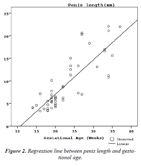
Penis length (mm)= 0.763 x Gestational Age (week) - 8.230
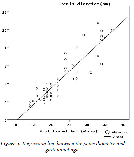
Penis diameter (mm)= 0.373 x Gestational Age (week) - 3.947
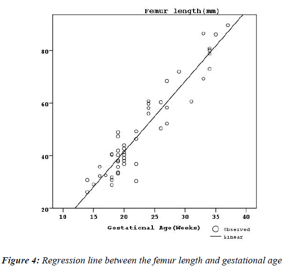
Femur length (mm)= 2.681 x Gestational Age (week) - 12.079
A normogram belonging to fetal penis was formed which may help to clinicians during ultrasonographic diagnose of genetical anomalies, endocrinological problems and fetal gender determination.
Keywords
Penis length, Penis diameter, Human fetus
Introduction
Discerning the sex of a fetus before birth is important to not only satisfy the curiosity of parents but also to followup the development of normal fetal gender during the prenatal ultrasonographic [1-2]. In addition, the early diagnosis of developmental defects related to fetal genitalia may guide to physician to identify some anomalies such as genetic disorders or endocrinological problems. More than 70 possibilities of genetic / chromosomal abnormalities can be considered in a common condition like ambiguous genitalia, micropenis/hypogenitalism [3-6]. Various abnormalities such as hypospadias, ambiguous genitalia and cloacal abnormalities related to fetal perineum and phallus have been defined via sonographic imaging in prenatal period [7-9]. Determining the gender is highly important in lower urinary tract anomalies, particularly for intrauterine diagnosis and course of bladder outflow obstruction [10]. The aim of this study is to evaluate morphometric aspects of fetal penile development and form a reference range for dimensions of fetal penis and femur.
Materials and Methods
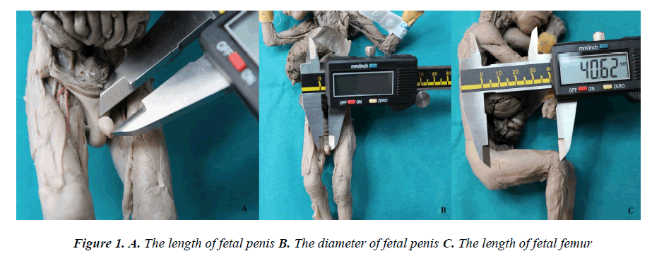
This study has been conducted on fetus collection of Department of Anatomy, Meram Faculty of Medicine, University of Necmettin Erbakan. Fetuses with abnormal genitourinary system and morphological anomalies that can be diagnosed by inspections were excluded. After receiving approval from the Institutional Review Board (Ref: 2013/349), 50 male fetuses ranging from 14 to 37 gestational weeks (37 second trimester – 13 third trimester) were classified according to the age determination method of Polin and Fox [11]. Measurements were taken three consecutive times by one person. In order to reduce the errors belonging to measurement technique, mean values were calculated by using readings that were taken from each fetus individually. Digital caliper was used to measure dimensions. Penis length; while penis being positioned with an angle of 90 degrees, was measured from tip (glans penis) to the point where it joins scrotum (radix penis) [12]. Penis diameter; was measured from the thickest point in the same position mentioned before. Femur length; the distance between the tip of the major trochanter and the lowest point of the lateral condylus was measured (Figure 1).
The obtained data were evaluated by using SPSS 15.0 (Statistical Package for Social Sciences). Data were analyzed by both descriptive (mean value, standard deviation, maximum and minimum values, percentages) and quantitative statistical methods. Results were evaluated statistically in %95 confidence interval and differences were accepted significant if p<0.01.
Results
Statistically, penis length, penis diameter and femur length was found to be significantly different between 2nd and 3rd trimesters was determined to have increased (p<0.01) (Table 1). In 2nd and 3rd trimesters, mean values were calculated 6.62 ±3.06 mm and 16.58±4.82 mm for penis length, 3.39±1.38 mm and 7.99±2.07 mm for penis diameter, and 40.72± 9.48 mm and 73.52±11.58 mm for femur length, respectively (Table 1).
Measurements that belong to the length and diameter of penis and the length of femur were increasing by advancing gestational age, at rates of %70 (r2=0.69), %81 (r2=0.81), and %90 (r2=0.89), respectively. The relationship between gestational age and all those parameters was statistically meaningful (p<0.01) (Tables 2-4).
The reference ranges on penis length, penis diameter and femur length were determined using the Least Squares regression analysis as follows (Figures 2-3-4):
Penis length (mm)= 0.763 x Gestational Age (week) - 8.230 Penis diameter (mm)=0.373 x Gestational Age (week)- 3.947 Femur length (mm)=2.681xGestational Age (week) - 12.079
Discussion
In fetal gender assignation, ultrasonography is an important tool. In our study, series of normal penis length were obtained from fetuses ranging between 14 and 37 gestational weeks since masculinization of external genital organs starts at 14th week of gestational age according to previous sonographic studies [2,12-15]. The scale of the normal values related to penis for various gestational weeks is important to diagnose male genital developmental disorders such as genitourinary system anomalies, hormonal defects and genetic syndromes [12].
The penis size has been a subject of interest and research of many people through penile sizes becomes the center of focus during the history of humanity. In addition, since it is considered as a taboo and privacy in society, it attracts many researchers and becomes a research topic, it still remains as an unclear mystical topic [10,12,13,15-20]. Micropenis, is a term which is used to define unusually small but structurally normal penis [21]. Penis length less than 2.5 standard deviation is widely accepted as the criteria to define micropenis [21-22]. There are studies that investigated the effects of ethnicity to the definition of the micropenis. Phillip et al. [16] reported that penis length did not show a significant difference related to ethnic origins among 268 Jewish and Bedouin male newborns (2 and 3 days old). According to their study, the mean penis length in Jewish and Bedouin newborns were 36.1±4.4 mm and 37.1±4.7 mm, respectively. Nevertheless, Lian et al. [17] demonstrated that there was a significant difference for the length of penis between two ethnic origins in the study which was conducted on Indian and Chinese infants. The penis length and diameter were measured on 105 newborns (from 38 to 42 weeks of gestation) with various ethnic origins by Cheng and Chanoine [18]. The mean data of penis length and the penis diameter for Caucasian, Chinese and East India newborns were 3.4 cm, 3.1 cm, 3.6 cm and 1.13cm, 1.07cm, 1.14cm, respectively. Ting and Wu [19] studied the stretched penis length and testicle volume on 195 Malaysian, 129 Chinese and 16 Indian full-term newborns and the mean length of penis was found 35.2±3.9 mm, 35.2±4.0 mm, and 37.5±4.5 mm, respectively.
Zalel et al. [12] observed using ultrasonography that the penis length was 3.88 mm and 23.77 mm at gestational ages of 14th and 38th weeks, respectively and found a positive correlation between gestational age and length of penis. In the same way, the mean fetal penis length was measured as 6 mm and 26.4 mm at gestational ages of 16th and 38th weeks by Johnson and Maxwell [10] and it showed a significant increase with gestational age. Feldman et al. [15] measured the stretched length and diameter of penis on 76 normal premature and 37 full-term newborns and found mean values of 3.5±0.7 cm and 1.1 ±0.2 cm among subjects, respectively. Although many methods have been tried to measure the penis length, it was observed that the most useful and repeatable measurement was in the stretched position. In our study, the mean penis length was measured between 3.31mm and 22.07mm among 50 male fetuses ranging from 13th to 37th gestational weeks. Johnson and Maxwell [10] stated a significant increase in penis length with increasing gestational age (p<0.005) in a study which was conducted on 95 normal fetuses belonging to 16th-38th gestational weeks. The formula of regression for the mean penis length was 4.1754 – 0.2198 x age + 0.0212 x age2 and formula of standard deviation was SD = 0.0097 + 0.0818 x age.
The regression equation was identified as 0.121 x Gestational Age + 0.277 on 419 male fetuses ranging from 14th to 38th weeks of gestation by Zalel et al. [12]. Danon et al. [23] found that the regression formula of fetal penis length was 0.076 x Gestational Age - 0.819 on 100 male fetuses ranging between 22th to 36th gestational weeks. Tuladhar et al. [20] determined the regression equation as 0.16 x Gestational Age - 2.27 on 192 male newborns with gestational ages lower than 37 weeks.
As stated earlier we found that penis length, diameter and femur length increased significantly with gestational age (p<0.05). Besides, the regression formulas for penis sizes and femur length were detected as seen in the following table (Table 5). The relationship between penis length and various body parts was investigated in many studies. No connection was found between two parameters in a study which examined the relationship of penis length with foot length [24]. In another study conducted on Greek males, the penis length was found longer in cases with the longer index finger. Height, weight, and body mass index found to be related with penis length in Italian population [25,26].
In our study, we found that there was a strong correlation among the parameters of fetal penis length, diameter and femur length (P<0.05) and the relationship between penis diameter and femur length (r2=0.957) was stronger than between penis length and femur length (r2=0.892). A nomogram for fetal penis sizes, being useful for early diagnosis of genital developmental disorders, anomalies related to genitalia, genetic and endocrinological problems, was created as a result of this study. We believed that this nomogram will be helpful for situations related to genital developmental disorders and termination of pregnancy.
Acknowledgments
The authors wish to thank to the donors for the fetuses used in the study.
Conflict of interest
The authors have no conflicts of interest.
Ethical Review Committee Statement
This study conformed to the Helsinki Declaration.
References
- Achiron R, Pinhas-Hamiel O, Zalel Y, Rotstein Z, Lipitz S. Development of fetal male gender: prenatal sonographic measurement of the scrotum and evalua- tion of testicular descent. Ultrasound Obstet Gynecol 1998; 11(4): 242–245.
- Soriano D, Lipitz S, Seidman DS, Maymon R, Ma- shiach S, Achiron R. Development of the fetal uterus between 19 and 38 weeks of gestation: in-utero ultra- sonographic measurements. Hum Reprod 1999; 14(1):215–218.
- Jones K, Ambiguous genitalia and micropenis. In: Smith DW, eds. Recognizable Patterns of Human Mal- formations. W.B. Saunders, Philadelphia 1997; 835–836.
- Hartke DM, Palmer JS. Commonly encountered con- genital and acquired anomalies of the penis. Drugs To- day 2006; 42(2): 121-126.
- Lee JH, Ji YH, Lee SK, Hwang HH, Ryu DS, Kim KS, Choo HS, Park S, Moon KH, Cheon SH, Park S. Change in penile length in children: preliminary study. Korean J Urol 2012; 53(12): 870-874.
- Bouvattier C. How and when to evaluate hypospadias?Arch Pediatr 2013; 20 Suppl 1: 5-10.
- Sides D, Goldstein RB, Baskin L, Kleiner BC. Prenatal diagnosis of hypospadias. J Ultrasound Med 1996; 15(11): 741-746.
- Cooper C, Mahony BS, Bowie JD, Pope II. Prenatal ultrasound diagnosis of ambiguous genitalia. J Ultra- sound Med 1985; 4(8): 433–436.
- Sivan E, Koch S, Reece EA. Sonographic prenatal diagnosis of ambiguous genitalia. Fetal Diagn Ther 1995; 10(5): 311-314.
- Johnson P, Maxwell D. Fetal penile length. Ultrasound Obstet Gynecol 2000; 15(4): 308-310.
- Hensinger RN. Standarts and measurements: Fetus and neonate: In: Polin RA, Fox WW(eds.): Fetal And Neo- natal Physiology. W.B. Saunders Co., Philadelphia 1992; 1687-1696.
- Zalel Y, Pinhas-Hamiel O, Lipitz S, Mashiach S, Achi-ron R. The development of the fetal penis--an in utero sonographic evaluation. Ultrasound Obstet Gynecol 2001; 17(2): 129–131
- Efrat Z, Akinfenwa OO, Nicolaides KH. First-trimester determination of fetal gender by ultrasound. Ultra- sound Obstet Gynecol 1999; 13(5): 305-307.
- Emerson DS, Felker RE, Brown DL. The sagittal sign. An early second trimester sonographic indicator of fe- tal gender. J Ultrasound Med 1989; 8(6): 293–297.
- Feldman KW, Smith DW. Fetal phallic growth and penile standards for newborn male infants. J Pediatr 1975; 86(3): 395-398.
- Phillip M, De Boer C, Pilpel D, Karplus M, Sofer S. Clitoral and penile sizes of full term newborns in two different ethnic groups. J Pediatr Endocrinol Metab 1996; 9(2): 175-179.
- Lian WB, Lee WR, Ho LY. Penile length of newborns in Singapore. J Pediatr Endocrinol Metab 2000; 13(1): 55-62.
- Cheng PK, Chanoine JP. Should the definition of mi- cropenis vary according to ethnicity? Horm Res 2001; 55(6): 278-281.
- Ting TH, Wu LL. Penile length of term newborn in-fants in multiracial Malaysia. Singapore Med J 2009; 50(8): 817-21.
- Tuladhar R, Davis PG, Batch J, Doyle LW. Establish- ment of a normal range of penile length in preterm in- fants. J Paediatr Child Health 1998; 34(5): 471-473.
- Lee PA, Mazur T, Danish R, Amrhein J, Blizzard RM, Money J, Migeon CJ. Micropenis. I. Criteria, etiologies and classification. Johns Hopkins Med J 1980; 146(4): 156-163.
- Tsang S. When size matters: a clinical review of patho- logical micropenis. J Pediatr Health Care 2010; 24(4): 231-240.
- Danon D, Ben-Shitrit G, Bardin R, Machiach R, Var- dimon D, Meizner I. Reference values for fetal penile length and width from 22 to 36 gestational weeks. Pre- nat Diagn 2012; 32(9): 829-832.
- Shah J, Christopher N. Can shoe size predict penile length? BJU Int. 2002; 90(6): 586-587.
- Spyropoulos E, Borousas D, Mavrikos S, Dellis A, Bourounis M, Athanasiadis S. Size of external genital organs and somatometric parameters among physi- cally normal men younger than 40 years old. Urology 2002; 60(3): 485-489.
- Siminoski K, Bain J. The relationships among height, penile length, and foot size. Annals of Sex Research 1993; 6(3): 231-235.


