- Biomedical Research (2012) Volume 23, Issue 2
Morphometric analysis of hard palate in south Indian skulls
Antony Sylvan D’Souza, H. Mamatha* and Nayak JyothiDepartment of Anatomy, Kasturba Medical College, Manipal, Karnataka, India.
- *Corresponding Author:
- H. Mamatha
Department of Anatomy Kasturba Medical College
Manipal-576104 Karnataka, India
Accepted date: September 12 2011
Abstract
In the present study 40 dry skulls from the Kasturba Medical College Manipal were examined to obtain the morphometric data of the hard palate. Mean palatal length, breadth and height for the total sample was 49.13mm, 4.04mm, and 0.8mm respectively. The contributions from the premaxilla for the hard palate being 9.4mm and palatine process 39.76mm.The palatine index showed that 40% of the skulls had wide (brachystaphyline),37.5% narrow (leptostaphyline), and 22.5% intermediate (mesostaphyline) palates. The greater palatine foramen was found to be at the level of third molar in 75%, in between second and third molars in 22.5%,and at the level of second molar in 2.5%.The distance from greater palatine foramen to middle maxillary suture was about 14mm.In majority of the skulls (62.5%) only one lesser palatine foramen was found and about 30% of the skulls had two lesser palatine foramina and 7.5% of skulls had 3 lesser palatine foramina. These data will be helpful in comparing these skulls to those from various regions as well as comparing the skulls of different races.
Keywords
Hard palate, greater palatine foramen, lesser palatine foramen.
Introduction
Measurement studies of the cranial bones play an important role in the analysis of skeletal variations, in determining the population history and classification, in studying the relationships between population and in investigations of adaptive and behavioral significance of bone morphology [1] .
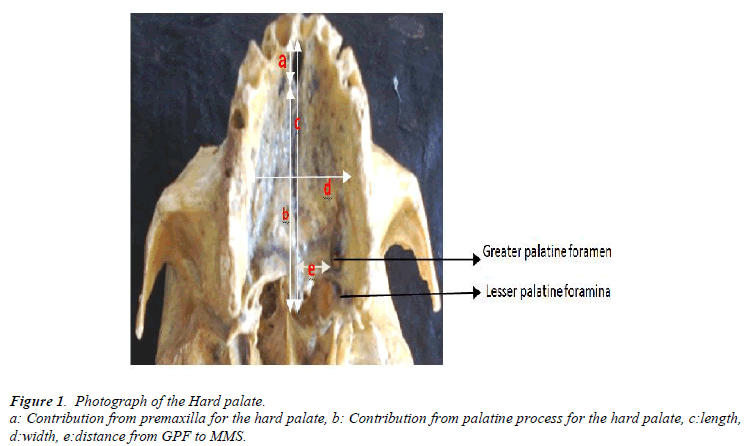
The hard palate is an essential region of the skull formed by the palatine processes of the maxillae and two horizontal plates of the palatine bones which are linked by a cruciform suture formed at the junction of the four described bones [2,3]. The hard palate plays an important role in the passive articulation of speech, therefore osteological and morphological variations in the bony palate is of great clinical significance. In adults the junction between the primitive and the permanent palate is represented by the incisive fossa. So for the formation of the palate both premaxilla and permanent palate contribution is paramount [2]; which may be altered in conditions like cleft palate. In the present study we have measured and analyzed the contribution of the premaxilla and maxillary processes to the bony palate.
The aim of the present study was to determine the palatine length, breadth, height, contribution from premaxilla and palatine processes for the hard palate, position of greater palatine foramen [GPF], and variations in the number of lesser palatine foramina. The observations of the present study can be utilized for the ethnic and racial classification of crania and anthropological studies.
Materials and Methods
The study was conducted in 40 dried, unsexed Indian skulls from Kasturba Medical College Manipal. All the skulls were normal and edentulous
Each skull was measured for the following by using the Vernier Caliper
• Palatal length, breadth and height
• Distance from the Middle maxillary suture to GPF.
• Contribution from the pre-maxilla and the palatine process for the hard palate
• Position of greater palatine foramen and number of lesser palatine foramina.
Measurements were made to the nearest millimeter in the following manner. Length was the distance between orale anteriorly (the orale is the point at the anterior end of the incisive suture located between the sockets of the two medial maxillary incisors) and the posterior nasal spine posteriorly. Width was the distance of the inner borders of the sockets of the upper second molars, endomolaria. Height was the maximum arching of palate from the line connecting the two endomolaria.
• Palatine index was determined by using the for mula: Breadth/Length X 100
• Palatine height index was determined by using
• theformula: Height/Breadth X 100.
All the findings were tabulated and statistically analyzed.
Results
In majority of the skulls (75%), the GPF’s were opposite to the third molar, 22.5% of foramina were in between the second and third molars, and 2.5% were at the level of second molar.
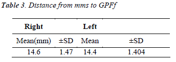
The distance from GPF to midline on the right side was14.6mm±1.47mm(mean±SD)and 14.4mm±1.404mm (mean±SD) on the left side.(Table 3)
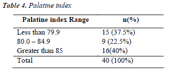
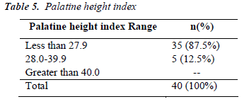
The palatine index showed that 40% of total sample of skulls has wide (Brachystaphyline), 37.5% narrow (leptostaphyline) and 22.5% intermediate (mesostaphyline) palates. The palatine height index showed that 87.5% skulls had low (chamestaphyline), 12.5% intermediate (orthostaphyline).
In majority of the skulls(62.5%) only one lesser palatine foramen(LPF) was found, and about 30% skulls had 2 LPF, and 7.5% skulls had 3 LPF.
Discussion
In the previous studies, Ajmani [4] observed that the location of the GPF opposite to the third molar in 64% of adult Indian skulls, Saralaya & Nayak [5] observed in 74.6% of the skulls, Bruno R et al., [6] observed in 54% of the skulls. In a study on Kenyan skulls 76% of cases showed that location of GPF opposite to the maxillary third molar, in comparison to our study which was seen in 75% of the skulls. Our study also showed that the location of GPF was intermediate between second and third molar in 22.5% and opposite to second in 2.5% of the skulls.
The distance from the MMS to GPF also showed the variations in the present study. According to Westmoreland and Blanton [7], the distance from GPF to MMS on the right had a mean of 14.8mm and 15.0mm on the left. Ajmani [4] reported a distance of 15.4mm from the saggital plane in Nigerian skulls and 14.7mm on the right and 14.6mm on the left in Indian skulls. Saralaya and Nayak [5] found 14.7mm on both sides. Bruno R Chrcanovic [6] reported 14.68mm on the right side and 14.44mm on the left side which was very similar to those of the present study. Variability in location of the foramen may be due to sutural growth occurring between the maxilla and palatine bone. The anteroposterior dimension of the palate increases with the eruption of the posterior teeth [8].
On a study on Kenyan African skulls by Hassanali [9], the palatine index showed that 43.2% of the total (brachystaphyline). Palatine height index showed that 40% skulls had low (chamestaphyline), 57% intermediate (orthostaphyline), and 3% deep (hypsistaphyline) palates, where as in our study we found that 40% of skulls had wide (brachystaphyline),37.5% narrow (leptostaphyline), 22.5% intermediate(mesostaphyline) palates. Palatine height index showed that 87.5% of the skulls had low (chamestaphyline) and 12.5% intermediate (orthostaphyline) palates.
Saralaya and Nayak [5] observed bilateral symmetry in the number of LPF in 40% of skulls. Hassanali [9] observed two to five LPF in 40% of skulls. In our study 62.5% of the skulls had 1 lesser palatine foramen.
Conclusion
Exact location of the greater palatine foramen is important for the clinicians when administrating certain types of local anesthesia as well as performing certain surgical procedures in the hard and soft palate. These findings may be helpful clinically, fabricating maxillary complete dentures for edentulous patients. This data will also be helpful in comparing the Indian skulls with those from various other regions as well as skulls of different races.
References
- Djuric-Srejic M. LeticV, Zivanovic S. New Facial measurement used in the determination of palatine in- dex and thickness of the mandibular body index. Hu- man evolution 2001; 16 (24): 143-149
- Williams PL ,Warwick R, Dyson M, Bannster H. Gray’s Anatomy.37th edition,London, 1989: 354.
- DuBrul EL. Sicher and DuBrul’s oral anatomy.8thedition,Ishiyaku Euro America,St Louis1988; 269- 284.
- Ajmani ML. Anatomical variation in position of the greater palatine foramen in the adult human skulls. J Anat 1994; 184: 635-637.
- Saralaya V, Nayak SR .The relative position of the greater palatine foramen in dry Indian skulls. Singapore Med J 2007; 48: 1143-1146.
- Bruno R. Chrcanovic, Antonio L. N. Custodio. Ana- tomical variation in the position of the greater palatine foramen. Journal of Oral Science 2010; 52 (1): 109-113.
- Westmoreland EE, Blanton PL. An analysis of the vari- ations in position of the greater palatine foramen in the adult human skull, Anat Rec 1982; 204: 383-388.
- Slavkin HC, Canter MR, Canter SR. An anatomical study of the pterygomaxillary region in the craniums of infants and children. Oral Surg 1966; 21: 225-235.
- Hassanali J, Mwaniki D. Palatal analysis and osteology of the hard palate of the Kenyan Africanskulls, Ant Rec 1984; 209: 273-280.


