Research Article - Biomedical Research (2017) Volume 28, Issue 19
Modified extended Karapandzic flap for large lower lip reconstruction
Sun Yuhang, Jingzhen Song, Jianchao He, Jing An, Haojun Yuan and Shujing Qi*
Affiliated Hospital of Hebei University of Engineering, PR China
Accepted date: September 23, 2017
Abstract
We recently repaired a modified extended Karapandzic flaps to reconstruct total or near total lower lip defects. The lower lip reconstructed by this method has good appearance and this method is easy to be master.
Keywords
Lip reconstruction, Modified extended Karapandzic flap, Depressor anguli oris
Introduction
Reconstruction of total full-thickness lip defect is a challenging task. There are many methods to finish the hard work. For example, cheek advanced flap, Gillies fan flap, McGregor fan flap, Karapandzic technique, nasolabial flap, free flap and so on. We recently repaired total or near total full-thickness lower lip defect by our modified extended Karapandzic flaps and got satisfactory results. Now we report this new method as follows.
Surgical Techniques
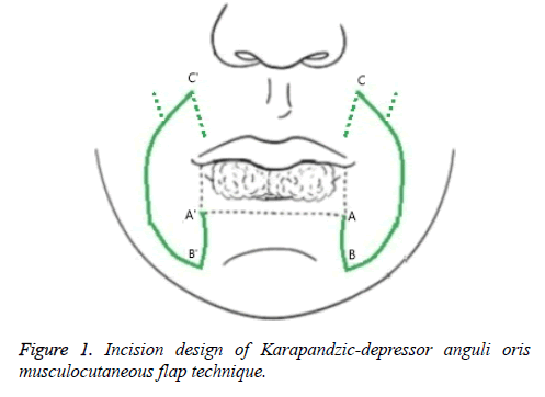
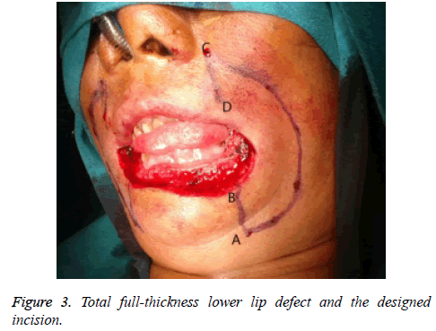
First, make a vertical full-thickness incision along the vertical edge of the lip defect. The length of this incision is the width of our modified extended Karapandzic flaps. Second, make a cambered incision from the end point of the first vertical incision to the lateral of the nasal alar. The lower part of this cambered which is inferior to the commissure is a fullthickness incision and the upper part superior to the commissure is cutaneous incision. Third, make blunt dissection in the subcutaneous and the muscle layer to release the extended Karapandzic flap. Do preserve the nerve and vessel branches in this step. Last, rotate and advance the flap into the defect, sutures the flap layer by layer, and closes the donor site layer by layer.
We should pay attention to the following matters when harvest the modified extended Karapandzic flap.
1) The width of the flap is equal to the width of the lip defect. In patients whose buccal skin is flabby, the width of the flap should be a little greater than the width of the defect in order to offset the volume lack of the new reconstructed lip which is always caused by flap retraction or incision scar contracture. The biggest width of the flap is 2.5 cm and it can be 3.0 cm in elder patients whose skin are relaxed.
2) Bluntly dissect the orbicular muscle above the commissure in order to avoid the damage of the nerve and blood vessel bundles.
3) Maintain the integrity of the modiolar, especially pay attention to the continuity of the modiolar and depressor anguli oris muscle.
4) Keep the integrity of the oral mucosa. Neither the mucosa above the commissure or lower the commissure will not be cut open.
Case Reports
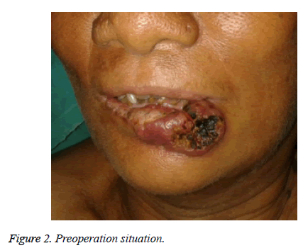
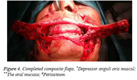
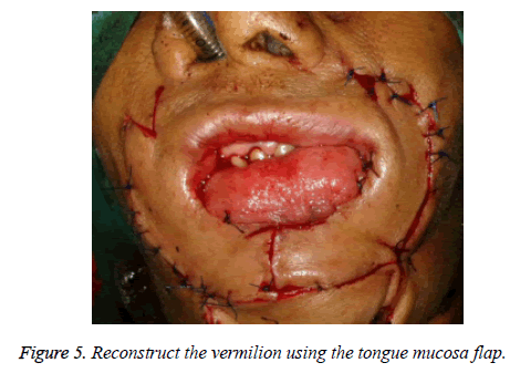
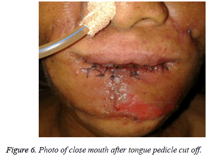
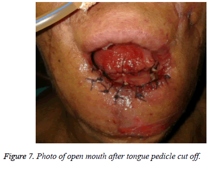
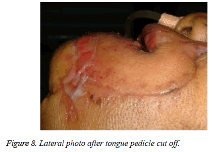
A 65 y old female came to B.P. Koirala Memorial Cancer Hospital of Nepal for the squamous cell carcinoma of her total lower lip. The lesion involved total lower lip and part skin of the chin (Figures 1 and 2). Under general anesthesia, we make a peripheral extended resection first. Then we use this modified extended Karapandzic flaps to rebuild the lower lip and use tongue mucosa flap to rebuild the vermilion (Figures 3 and 4). The new reconstructed lip shows a good appearance with good oral function and without microstomia and sialorrhea after the principal of tongue flap was cut off (Figures 5-7).
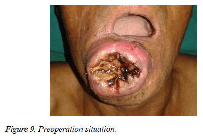
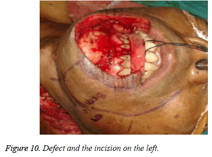
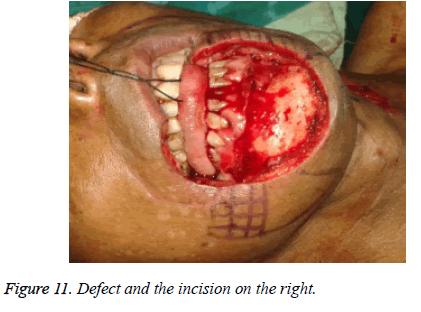
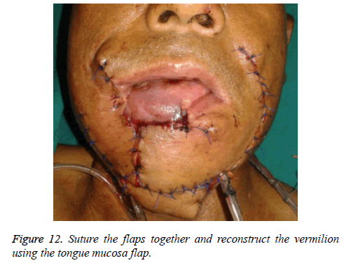
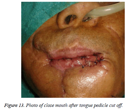
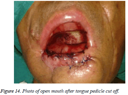
A 57 y old male came to B.P. Koirala Memorial Cancer Hospital of Nepal for a recurrent lower lip squamous cell carcinoma. Almost half of his lower lip was excised. And he then was given a lower lip reconstruction surgery immediately by means of a non-standard Karapandzic technique in which all the neurovascular bundles below the incision were cut off. We found that his recurrent tumor involved all the lower lip and part of the upper lip and an incision scar was located around the mouth. The incision scar reached the point below the left commissure in the left and the point outside the right alae nasi in the right. We gave the patient an extended resection and designed two flaps to rebuild the lower lip (Figures 8-10). One flap on the left was Karapandzic-depressor anguli oris musculocutaneous flap. The other flap on the right was depressor anguli oris musculocutaneous flap only because of the vascular damage in the first non-standard reconstruction surgery. The tongue mucosa flap was used to reconstruct the vermilion just the same as the first patient (Figure 11). The new lower lip looks well and has no functional problem even though the defect after extended resection involved part of upper lip and right cheek except the total lower lip (Figures 12-14).
Discussion
Matthew et al. [1] reported that they used extended Karapandzic flap and achieved good results in 2011. But the central section of the lower lip reconstructed through the extended Karapandzic flap was in a dynamic state [2]. We make the “dog ear” of the Matthew's extended Karapandzic flap which should be wasted into our modified extended Karapandzic flap. So more depressor anguli oris and skin is contained in the flap. This promotion increases the tissue used to reconstruct the lower lip. Depressor anguli oris musculocutaneous flap has been studied by many scholars [3-8]. This flap was perfected by Tobin [9], Takatoshi [10], and Miguel [11]. It was affirmed that even if the nerve branches were cut, the sensory and the sphincteric function of reconstructed lips would come back in the next six months. This modified extended Karapandzic flaps combine two techniques. The upper part of our modified extended Karapandzic flap is a traditional Karapandzic flap in deed which makes sharp skin incision only, and release orbicularis muscle bluntly in order to save blood vessels and nerve bundles, and maintain the integrity of the mucous membrane. The lower part of our method which is inferior to the commissure is depressor anguli oris musculocutaneous flap in fact. Below the level of commissure, more depressor anguli oris is used to reconstruct the orbicularisoris muscle ring compared with Matthew's design.
This technique is similar to traditional extended Karapandzic technique and is easy to master. The upper lip and new reconstructed lower lip are balanced by the ring incision in some extent.
Our integrated surgical design applies for total or near total full thickness lower lip defect cases. But if the defect involving the cheek or chin, the flap cannot be adopted. Free flaps are suit for this lip defect with cheek or chin defect cases [12-15]. In addition, the function of the depressor anguli oris muscle should be checked first to determine whether there is a serious muscle atrophy which will reduce the function of these muscles in the reconstructed lip.
References
- Hanasono MM, Langstein HN. Extended Karapandzic flaps for near-total and total lower lip defects. Plast Reconstr Surg 2011; 127: 1199-1205.
- Yotsuyanagi T, Nihei Y, Yoloi K, Sawada Y. Functional reconstruction using a depressor anguli oris myocutaneous flap for large lower lip defects, especially for elderly patients. Plast Reconstr Surg 1999; 103: 850-856.
- Macfee WF. The surgical treatment of cancer of the nose, with emphasis on methods of repair. Ann Surg 1954; 140: 475-496.
- Cameron RR, Latham WD, Dowling JA. Reconstructions of the nose and upper lip with nasolabial flaps. Plast Reconstr Surg 1973; 52: 145-150.
- Spear SL, Kroll SS, Romm S. A new twist to the nasolabial flap for reconstruction of lateral alar defects. Plast Reconstr Surg 1987; 79: 915-920.
- Guero S, Bastian D, Lassau JP, Csukonyi Z. Anatomical basis of a new naso-labial island flap. Surg Radiol Anat 1991; 13: 265-270.
- Wolfe SA. Eyelid reconstruction. Clin Plast Surg 1978; 5: 525-531.
- Thaller SR, Kim S, Wildman M, Patterson H. Microdissection of the nasolabial axial myocutaneous flap. Ear Nose Throat J 1991; 70: 93-96.
- Tobin GR, ODaniel TG. Lip reconstruction with motor and sensory innervated composite flaps. Clin Plast Surg 1990; 17: 623-632.
- Takatoshi Y, Yoshihiro N, Katsunori Y. Functional reconstruction using depressor anguli oris musculocutaneous flap for large lower lip defects, especially for elderly patients. Plast Reconstr Surg 1999; 103: 850-856.
- Miguel SN, Helton TC, Lydia MF, Julio H, Sabrina RT. Utilization of the depressor anguli oris musculocutaneous flap for lip reconstruction. Ann Plast Surg 2000; 44: 23-28.
- Bayramiasli M, Numanoaylu A, Tezel E. The mental V-Y island advancement flap in functional lower lip reconstruction. Plast Reconstr Surg 1997; 100: 1682-1690.
- Jeng SF, Kuo YR, Wei FC, Su CY, Chien CY. Reconstruction of concomitant lip and cheek through-and through defects with combined free flap and an advancement flap from the remaining lip. Plast Reconstr Surg 2004; 113: 491-498.
- Jeng SF, Kuo YR, Wei FC, Su CY, Chien CY. Total lower lip reconstruction with a composite radial forearm-palmaris longus tendon flap: a clinical series. Plast Reconstr Surg 2004; 113: 19-23.
- Yoshida T, Sugihara T, Ohura T, Minakawa H, Igawa H. Double cross lip flaps for reconstruction of the lower lip. J Dermatol 1993; 20: 351-357.
