Research Article - Journal of Medical Oncology and Therapeutics (2016) Volume 1, Issue 3
Mobile Left Atrial Mass-Clot or Left Atrial Myxoma
Suraj Wasudeo Nagre*
Associate Professor, Department of CVTS, Grant Medical College, Mumbai, India.
- *Corresponding Author:
- Dr. Suraj Wasudeo Nagre
Associate Professor, Department of CVTS, 31
Trimurti Building, J J Hospital Compound
Byculla, Mumbai, India.
Tel: 09967795303
E-mail: surajnagre@yahoo.com
Accepted date: November 10, 2016
DOI: 10.35841/medical-oncology.1.3.94-96
Visit for more related articles at Journal of Medical Oncology and TherapeuticsAbstract
Mobile masses within the left atrial cavity are commonly caused by organized thrombi or left atrial myxoma. The use of transesophageal echocardiography has provided means for differentiation between the two conditions. We report a case of a left atrial mass in a middle age male patient having past history of closed mitral commissurotomy presented with mitral stenosis and atrial fibrillation. On transthoracic echocardiography a round mass appeared freely mobile in left atrium but did not prolapse between the mitral leaflets due to the tightly stenosed valve orifice. In the presence of mitral stenosis, atrial fibrillation and dilated left atrium, the appearance of the mass was in keeping with mobile thrombus but possibility of left atrial myxoma cannot be ignored. Consequently, the patient was referred for surgical treatment and the mass removed through left atriotomy. Microscopic examination revealed a highly organized thrombus. We conclude that TTE is still a reliable tool in the diagnosis of large mobile atrial thrombi; TEE may help to differentiate clot from atrial myxoma but histopathological examination is a gold standard.
Keywords
Left atrium, Mitral stenosis, Mitral valve replacement, CMC-closed mitral commissurotomy.
Abbreviations
MS: Mitral Stenosis; LA: Left Atrium; CPB: Cardiopulmonary By-Pass
Introduction
Nearly every fifth mitral stenosis patient presents with clot in the left atrium. Most clots are located in the left atrial appendage, but atrial appendage clot can also extend to the left atrial cavity [1-3]. Left atrial clot in operated case of closed mitral commissurotomy is very rare to found as left atrial appendage was tied during CMC and not reported in literature. Left atrial clot without mitral disease is rare [2]. The diagnosis of a left atrial clot should be regarded as an urgent indication for preventive surgery [4,5]. The differential diagnosis for an intra-cavitary cardiac mass includes thrombus, myxoma, lipoma and non-myxomatous neoplasm. Cardiac myxomas are uncommon tumours found in 0.5 million population per year with left atrium being the predominant site. Over 72% of primary cardiac tumors are benign. In adults, the majority of benign lesions are myxomas.
Case Report
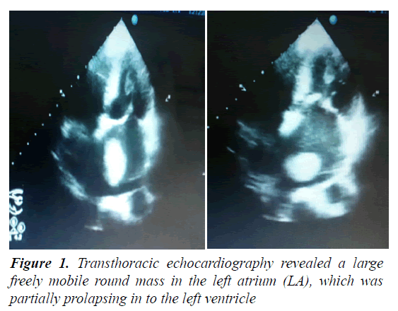
A 38 year old male with past history of closed mitral commissurotomy ten years back presented with symptoms of shortness of breath, palpitation and facial puffiness. He had longstanding mitral restenosis and atrial fibrillation and was classified as NYHA-III. There was no history of stroke or embolic event in the past. Physical examination revealed a mid-diastolic murmur at the apex. Her ECG showed an atrial fibrillation rhythm, chest x-ray revealed cardiomegaly with dilated LA, Transthoracic echocardiography revealed a large freely mobile round mass in the left atrium (LA), which was partially prolapsing in to the left ventricle (Figure 1). The mass was moving freely in the LA and was bouncing back after striking the mitral valve leaflets like a ball in a pinball machine. Patient was scheduled for surgical removal of the mass along with mitral valve replacement under cardiopulmonary bypass. Intra-operative transesophageal echocardiography (TEE) confirmed the pre-operative findings.
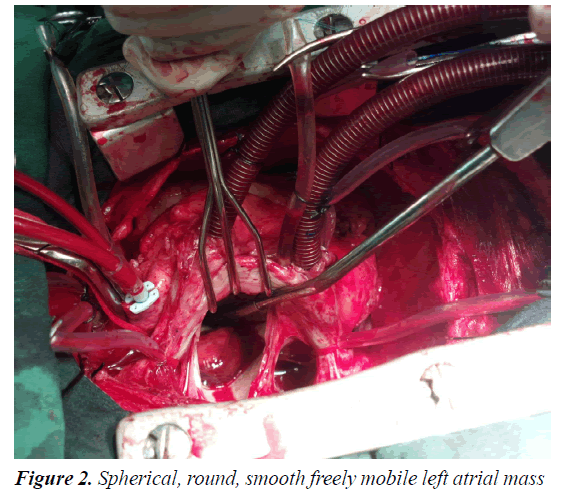
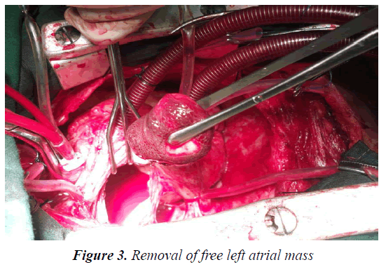
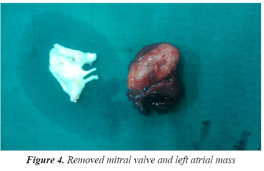
At surgery, a large spherical pink colored mass (Figure 2) with a smooth surface was removed (Figure 3). Evidence of peduncle or wall attachment was not demonstrated during surgery. Though this patient has past history of CMC, intraopt we found left atrial appendage opening into left atrium through small partially closed opening. We closed it internally with prolene 4-0 suture. Stenosed mitral valve removed with preservation of posterior mitral leaflet (Figure 4). Mitral valve replacement done with mechanical valve by intermittent 2-0 pledgetted ethibond sutures in horizantal mattress pattern with pledgett on left atrial side. Minimal inotropic support was used during weaning from bypass. Postoperatively, the patient remained hemodynamically stable. Patient ventilated for 5 h and had minimal blood loss. ICU stay was two days, during which intensive chest physiotherapy was performed. Histopathology examination of the mass revealed features consistent with thrombus. The patient was discharged after 8 days on tablet warfarin 5 mg with INR of 2.6.
Discussion
The probable predisposing factors for thrombus formation are mitral valve disease, non-valvular atrial fibrillation, severe LV dysfunction, and other causes of atrial contractile failure [6]. Among these factors, mitral valve disease or atrial fibrillation are the most common. Thrombus formation in the presence of sinus rhythm is rare. Any condition predisposing to low flow state can lead to thrombus formation. Thus, in the present patient the risk factors for thrombus formation included atrial fibrillation and LV dysfunction. Patients with LA thrombus are at a high risk for thromboembolic events and sudden death as a freely mobile LA thrombus can occlude the mitral valve by a ball-valve mechanism.
The differential diagnosis for an intra-cavitary cardiac mass includes thrombus, myxoma, lipoma and nonmyxomatous neoplasm [7,8]. Among them, cardiac myxoma is the most common benign primary tumor of the heart and generally found in the LA. On echocardiogram cardiac myxomas typically appear as a mobile mass attached to the endocardial surface by a stalk, usually arising from the fossa ovalis. Cardiac thrombi, which appear more frequently than cardiac myxomas are typically located more often in the LA or LAA and generally occur in patients with organic heart disease.
In the present patient, features were not consistent with thrombus as it was spherical and mobile, surface was smooth and echogenicity was more consistent with myxoma. The present thrombus may have been attached earlier, but got dislodged due to lysis at some stage. Other intra-cardiac masses such as lipoma or non-myxomatous neoplasm are rare. TEE is the extensive diagnostic tool to investigate suspected intra-cardiac mass involving the LA [8] and histopathological examination is gold standard for diagnosis of myxoma [9,10].
Conclusion
To the best of our knowledge this is the only case report of a mitral stenosis with left atrial mass in a patient already having history of closed mitral commissurotomy. In the present patient, features were not consistent with thrombus as it was spherical and mobile, surface was smooth and echogenicity was more consistent with myxoma. Transoesophageal echocardiography imaging provides detailed information of a mobile mass in the left atrium. Surgical removal of a mobile left atrial mass should be performed before the occurrence of a fatal embolism. We conclude that TTE is still a reliable tool in the diagnosis of large mobile atrial thrombi; TEE may help to differentiate clot from atrial myxoma but histopathological examination is gold standard.
Consent
Informed consent has been obtained.
Funding
No funding was required for this study.
Conflict of Interest
No potential conflict of interest exists.
References
- Colucci WS, Schoen FJ. In: Primary tumors of the heart, chapter 49. Heart Disease: A Textbook of Cardiovascular Medicine. 6th edition. Edited by Braunwald E, Zipes D, Libby P. Philadelphia, Pennsylvania, USA, W.B. Saunders Company 2001; 2: 1809-1819.
- Agmon Y, Khandheria BK, Gentile F, et al. Clinical and echocardiographic characteristics of patients with left atrial thrombus and sinus rhythm: Experience in 20643 consecutive transesophageal echocardiographic examinations. Circulation 2002; 105: 27-31
- Rost C, Daniel WG, Schmid M. Giant left atrial thrombus in moderate mitral stenosis. Eur J Echocardiogr 2009; 10: 358-359.
- Reynen K. Cardiac myxomas. N Engl J Med 1995; 333: 1610-1617.
- Amano J, Kono T, Wada Y, et al. Cardiac myxoma: Its origin and tumor characteristics. Ann Thorac Cardiovasc Surg 2003; 9: 215-221.
- Alam M, Sun I. Transesophageal echocardiographic evaluation of left atrial mass lesions. J Am Soc Echocardiogr 1991; 4: 323-330.
- Fujiwara M, Watanabe H, Oguma Y, et al. A free-floating left atrial thrombus develops intermittent entrapment in the mid-ventricle during diastole. Heart Vessels 2012; 27: 428-423.
- Ando T, Abe H, Ro D. Case of embolism due to a floating thrombus migrating from the left atrial appendage to the ostium of the celiac artery. Ann Vasc Dis 2012; 5: 229-232.
- Burke AP, Virmani R. Cardiac myxoma: A clinicopathologic study. Am J ClinPathol 1993, 100: 671-680.
- Yu K, Liu Y, Wang H, et al. Epidemiological and pathological characteristics of cardiac tumors: A clinical study of 242 cases. Interact CardiovascThorac Surg 2007; 6: 636-639.


