Research Article - Journal of Brain and Neurology (2017) Journal of Brain and Neurology (Special Issue 1-2017)
Methodical aspects of dopplerography of the main arteries in the circle of Willis.
- *Corresponding Author:
- Abdullaiev RY
Department of Ultrasound Diagnostics Kharkiv Medical Academy of Postgraduate Education Ukraine
E-mail: r.abdullaev@bk.ru
Accepted date: November 10, 2017
Citation: Abdullaiev RY, Sysun LA, Kalashnikov VI, et al. Methodical aspects of dopplerography of the main arteries in the circle of Willis. J Brain Neurol 2017;1(1):9-13.
Abstract
Introduction: Cerebral circulation receives 15-20% of the cardiac output Cerebral angiography is the main diagnostic method for cerebrovascular pathologies, however, is associated with invasiveness and the risk of dangerous complications after use of contrast media. Objective: To evaluate the techniques of transcranial dopplerography in triplex mode and to study the quantitative parameters of blood flow in the main arteries in the circle of Willis in healthy middle-aged people. Materials and methods: Transcranial Dopplerography (TCDG) was performed in color and energy Doppler mapping (CDM and EDM). The examination was conducted in 48 patients with unchanged carotid arteries, normal blood pressure and without any cardiovascular diseases. The age of the subjects was 41-60 years, among them 25 men and 23 women. Results: In 3 (6.25%) cases, the transtemporal window was not accessible from one side: in two 59 and 60 years old women and in one man aged 57. In 1 (2,08%) case the transtemporal window was absent from two sides in one man of 58 years. With the help of compression probes, the consistency of the connective arteries was evaluated. Venus Galen was able to visualize in 36 (75%) cases, Rosenthal vein - in 29 (60%), and direct sinus in 31 (64.5%) cases. All the main arteries had a monophasic spectrum with a narrow incision at the beginning of the diastole. Conclusion: Transcranial dopplerography in triplex mode justify the non-invasive approach to obtain quantitative parameters of blood flow, which can be used in various pathological conditions leading to disruption of cerebral hemodynamics.
Keywords
Cardiac output, Cerebral angiography, Ultrasonography, Transcranial Dopplerography (TCDG).
Introduction
The brain is one of the vital organs in the body and stable perfusion is essential to maintain its function. Cerebral circulation receives 15-20% of the cardiac output and is closely regulated to maintain perfusion in response to metabolic and physiological demands. The main cerebral distribution center for blood flow is the Circle of Willis [1]. Blood is delivered to the brain through the two internal carotid arteries that contribute 80% of the blood supply, and the two vertebral arteries that join intracranially to form the basilar artery. Each of the internal carotid arteries branches to form the middle and anterior cerebral arteries, which supply blood to the front and the sides of the brain (the frontal, temporal, and parietal regions of the brain). The basilar artery bifurcates into the right and left posterior cerebral arteries, which perfuse the back of the brain (the occipital lobe, cerebellum and the brain stem). The ring is completed by communicating arteries that connect the posterior and anterior cerebral arteries (via posterior communicating arteries) and the two anterior cerebral arteries (via the anterior communicating artery).
The outflow of venous blood from the vascular plexuses and deep sections occurs through a large cerebral vein flowing into a straight sine. Superficial veins of the brain that collect blood from the cerebral cortex flow into the upper sagittal sinus, cavernous, upper stony, etc. Through the sinuses of the dura mater, the blood flows into the internal jugular veins [2].
Cerebral angiography is the main method of diagnosing cerebrovascular pathologies. For a long time, radiocontrast angiography remained the "gold standard" status in the study of the vessels of the brain. However, the invasiveness of radiopaque angiography, the risk of dangerous complications in its implementation, and the development of new non-invasive diagnostic methods have led to the fact that this method is used less and less now [3].
At present, magnetic resonance angiography (MRA) has become more widely used, which actually claims its diagnostic capabilities for a safe alternative to radiopaque angiography [4]. Another method of investigation is spiral computer tomography angiography (CTTA). However, X-ray irradiation, the need to use contrast agents, makes this method less universal for wide application [5].
In recent years, transcranial "blind" - not visualizing dopplerography has become an integral part of monitoring cerebral hemodynamics during various surgical interventions [6]. However, the method does not allow you to directly visualize the blood flow and accurately determine the location of the recording of blood flow in the vessels in the circle of Willis. Ultrasonography in triplex mode makes it possible to visualize the vessels under study and to register the blood flow in the site of interest [7,8].
Objective
To systematize the technique of transcranial dopplerography in triplex mode and to study the quantitative parameters of blood flow in the main arteries in the circle of Willis in healthy middle-aged people.
Materials and Methods
Transcranial dopplerography (TCDG) was performed in triplex mode. Visualization of brain structures was carried out in the gray-scale, main vessels - in color Doppler and registration of quantitative parameters of blood flow - in spectral modes. The examination was conducted in 48 patients with unchanged carotid arteries, normal blood pressure and without any cardiovascular diseases. The age of the subjects was 41-60 years, among them 25 men and 23 women.
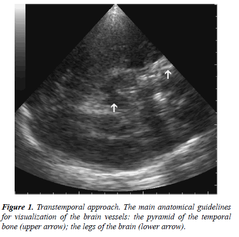
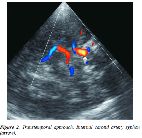
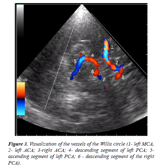
The study was conducted without preliminary preparation of the patient in a supine or sitting position. Scanning starts in Bmode with visualization of the brain structures from the transtemporal (through the scales of the temporal bone) and suboccipital (through the large occipital foramen) accesses. From the transtemporal access, the pyramid of the temporal bone and the structure of the brain of the central and hemispheric localization were visualized: with frontal lobes, anterior horns of the lateral ventricles, the third ventricle, visual bumps, temporal lobes, occipital lobes with high axial sections; at low axial sections - the basal parts of the frontal lobes, the interhemispheric fissure, the temporal lobes, the Sylvian cracks, the legs of the brain, the cerebellum, the occipital lobes. The pyramid of the temporal bone served as a reference for the identification of cerebral vessels (Figure 1). From the transtemporal access, the internal carotid siphon, middle, anterior and posterior cerebral arteries were visualized (Figures 2 and 3).
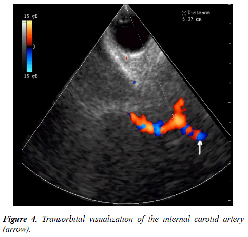
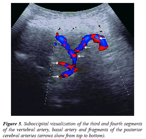
Transorbital access made it possible to visualize the internal carotid and orbital artery (Figure 4). Suboccipital access made it possible to visualize the third and fourth segments of the vertebral artery, basal artery and fragments of the posterior cerebral arteries (Figure 5).
Results
The advantages and disadvantages of the main approaches for the study of cerebral vessels: transtemporal, suboccipital and transorbital were determined.
Transocipital access in only 13 (27.1%) cases allowed visualization of the initial segments of the posterior cerebral arteries, in all 100% of cases - the basal artery and the fourth segment of the vertebral artery.
In 3 (6.25%) cases, the transtemporal window was absent from one side: in two women 59 and 60 years and in one man at the age of 57 years. In 1 (2,08%) case the transtemporal window was absent from two sides in one man of 58 years. The first segment of the middle cerebral artery was visualized in all cases of the ultrasound window, and the second segment in 84.1% of cases.
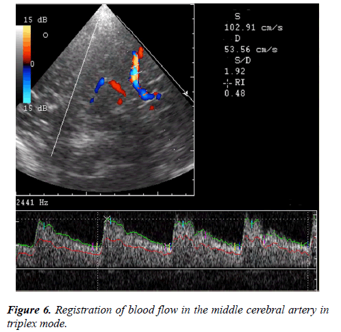
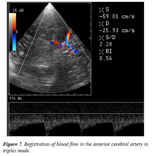
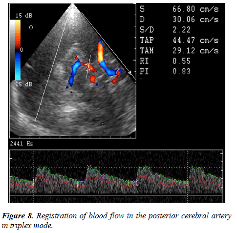
The Doppler spectrum of the blood flow was monophasic, in all cases of the onset of diastole a narrow incision was noted. The maximum systolic velocity (Vs), the end diastolic velocity (Vd), average systolic velocity (TAMX), the resistance index (RI) and the pulsativity index (PI) were determined. For the middle cerebral artery, Vs averaged 108.4 ± 6.1 cm/c, Vd – 44.5 ± 4.5 cm/s, TAMX – 77.1 ± 3.2 cm/s, PI – 0.83 ± 0,06, RI – 0.59+0.03 (Figure 6). These parameters for the anterior cerebral artery were: Vs – 68.3+5.2 cm/s, Vd – 30.1+3.2 cm/s, TAMX – 47.2+3.1 cm/s, PI – 0.81+0.04, RI – 0.56+0.03 (Figure 7). Dopplerometric parameters of blood flow in the posterior cerebral artery were as follows: Vs – 63.7+5.1 cm/s, Vd – 28.4+2.7 cm/s, TAMX – 43.1+2.9 cm/s, PI – 0.82+0.05, RI – 0.55+0.03 (Figure 8) (Table 1).
| Hemodynamic parameters | MCA | ACA | PCA |
|---|---|---|---|
| Vs (CM/C) | 108.4 ± 6.1 | 68.3 ± 5.2 | 63.7 ± 5.1 |
| Vd (CM/C) | 44.5 ± 4.5 | 30.1 ± 3.2 | 28.4 ± 2.7 |
| TAMX (CM/C) | 77.1 ± 3.2 | 47.2 ± 3.1 | 43.1 ± 2.9 |
| PI | 0.83 ± 0.06 | 0.81 ± 0.04 | 0.82 ± 0.05 |
| RI | 0.59 ± 0.03 | 0.56 ± 0.03 | 0.55 ± 0.03 |
Table 1: Hemodynamic parameters of the blood flow in the circle of Willis.
Discussion
The role of dopplerography in the study of the main cerebral vessels is shown in previously published scientific papers [9-11]. Tsivgoulis et al. showed that a systolic velocity greater than 180 cm/s or an increase of 20% during the day, a pulsatile index (PI) of more than 1.5 can be reliable signs of hemorrhagic stroke. Verlhac studied the possibilities of dopplerography in childhood, and in the work of Purkavastha et al. shows the averaged indices of systolic blood flow velocity in cerebral vessels in individuals in the large age range from 20 to 70 years.
Conclusion
Transcranial dopplerography in triplex mode allows to determine the anatomical landmarks of the arteries of the Willis circle. They are: the pyramid of the temporal bone, the central location of the interhemispheric fissure, the legs of the brain. With the help of a standardized method it is possible to register hemodynamic parameters of blood flow in the middle, anterior and posterior cerebral arteries. The greatest rate of blood flow is observed in the middle cerebral artery. The indices of resistance and pulsation of blood flow in each vessel differ slightly.
References
- Cebral JR, Castro MA, Soto O, et al. Blood-flow in the Circle of Willis from magnetic resonance data. J Eng Math. 2003;47(4):369-86.
- Valdez-Jasso D, Haider MA, Banks HT, et al. Viscoelastic mapping of the arterial ovine system using a Kelvin model. Submitted, 2007.
- Sawicki M, Bohatyrewicz R, walecka A, et al. CT Angiography in the Diagnosis of Brain Death. Pol J Radiol. 2014;79:417-21.
- Kim BJ, Kang GH, Ahn SH, et al. Magnetic Resonance Imaging in Acute Ischemic Stroke Treatment. J Stroke. 2014;16(3):131-45.
- Kumamaru KK, Hoppel BE, Mather RT, et al. CT Angiography: Current Technology and Clinical Use. Radiol Clin North Am. 2010;48(20):213-35.
- Lushyk UB. The ?blind? Doppler for clinical intellectuals. Istyna Research Center. 2017.
- Abdullaeiv RY. Dopplerography of the vessels of the neck and head. Kharkiv. 2010:128.
- Abdullaiev RY, Kalashnikov VI, Sysun LA, et al. Peculiarities of Arterial and Venous Hemodynamics with Transitorial Ischemic Attacks in the Vertebro-Basilary Basin. Am Res J Neurol. 2017;1(1):4-6.
- Tsivgoulis G, Alexandrov AV, Sloan MA. Advances in transcranial Doppler ultrasonography. Curr Neurol Neurosci Rep. 2009;9(1):46-54.
- Verlhac S. Transcranial Doppler in children. Pediatr Radiol. 2011;41(Suppl 1):S153-S65.
- Purkavastha S, Sorond F. Transcranial Doppler Ultrasound: Technique and Application. Semin Neurol. 2012;32(4):411-20.
