Image Article - Ophthalmology Case Reports (2020) Volume 4, Issue 2
Maternally inherited diabetes and deafness (MIDD)-associated macular dystrophy imaged with oct angiography.
Jaclyn L. Kovach*
Department of Clinical Ophthalmology, Bascom Palmer Eye Institute, University of Miami Miller School of Medicine, Naples, United States of America
- *Corresponding Author:
- Jaclyn L. Kovach
Bascom Palmer Eye Institute
Department of Clinical Ophthalmology
University of Miami Miller School of Medicine
United States of America
E-mail: jkovach@med.miami.edu
Citation: JL. Maternally inherited diabetes and deafness (MIDD)-associated macular dystrophy imaged with oct angriography Ophthalmol Case Rep. 2020;4(2):2-2.
Abstract
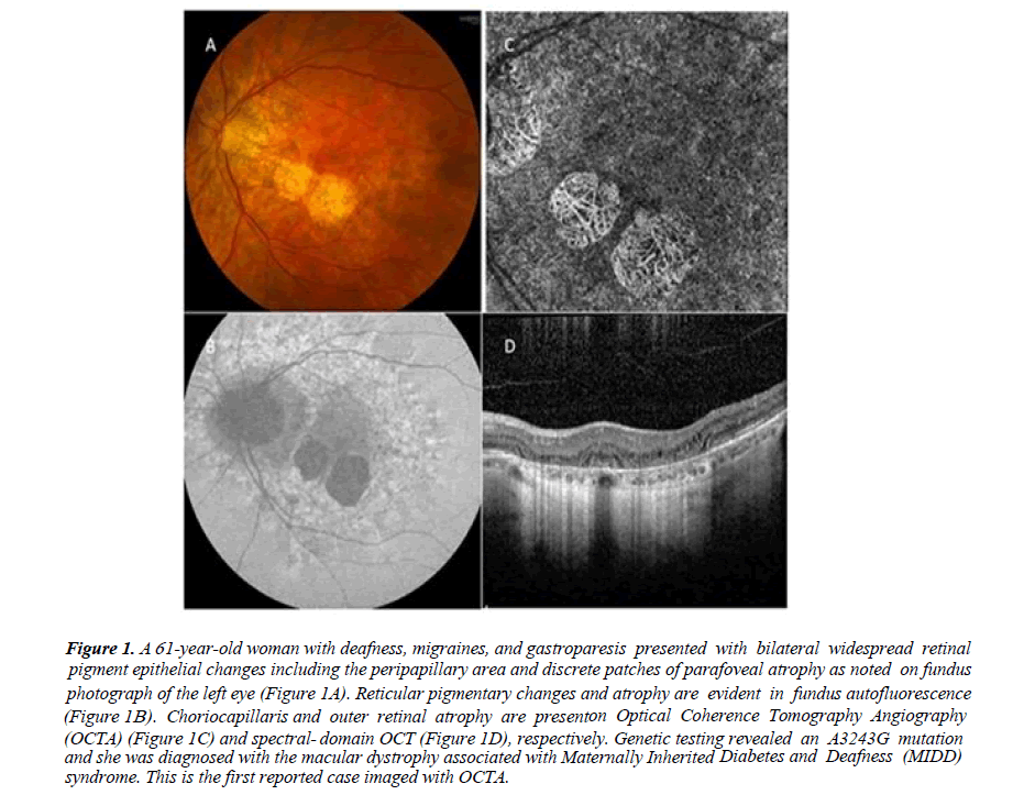
A 61-year-old woman with deafness, migraines, and gastroparesis presented with bilateral widespread retinal pigment epithelial changes including the peripapillary area and discrete patches of parafoveal atrophy as noted on fundus photograph of the left eye (Figure 1A). Reticular pigmentary changes and atrophy are evident on fundus autofluoresence (Figure 1B). Choriocapillaris and outer retinal atrophy are present on optical coherence tomography angiography (OCTA) (Figure 1C) and spectral domain OCT (Figure 1D), respectively. Genetic testing revealed an A3243G mutation and she was diagnosed with the macular dystrophy associated with maternally inherited diabetes and deafness (MIDD) syndrome. This is the first reported case imaged with OCTA.
Figure 1: A 1-year-old woman with deafness, migraines, and gastroparesis presented with bilateral widespread retinal pigment epithelial changes including the peripapillary area and discrete patches of parafoveal atrophy as noted on fundus photograph of the left eye (Figure 1A). Reticular pigmentary changes and atrophy are evident in fundus autofluorescence (Figure 1 ). Choriocapillaris and outer retinal atrophy are present on Optical Coherence Tomography Angiography (OCTA) (Figure 1C) and spectral-domain OCT (Figure 1D), respectively. Genetic testing revealed an A3243G mutation and she was diagnosed with the macular dystrophy associated with Maternally Inherited Diabetes and Deafness (MIDD) syndrome. This is the first reported case imaged with OCTA.
Declaration
The author has no conflict of interest with the material presented in this article.
