Short Communication - Journal of Parasitic Diseases: Diagnosis and Therapy (2016) Volume 1, Issue 1
Management of Ocular Setariosis in a Stallion with Ivermectin
Reddy BS* and Sivajothi S
Reddy BS, Assistant Professor (Veterinary Medicine), Teaching Veterinary Clinical Complex, College of Veterinary Science, Andhra Pradesh, India
Sivajothi S, Assistant Professor, Department of Veterinary Parasitology, College of Veterinary Science, Andhra Pradesh, India
- *Corresponding Author:
- Reddy BS
College of Veterinary Science, Sri Venkateswara Veterinary University, Proddatur-516360, Y.S.R. Kadapa District, Andhra Pradesh, India
Tel: +919030218657
E-mail: bhavanamvet@gmail.com
Received Date: October 27, 2016; Accepted Date: October 31, 2016; Published Date: November 11, 2016
Abstract
A twelve year old stallion was presented to the hospital with complaint of lacrimation, corneal opacity of left eye for the past two weeks. Upon clinical examination, white motile worm in the left eye was noticed. Stallion was successful treated with repeated doses of ivermectin injection @ 200 μg/kg body weight subcutaneously along with supportive therapy.
Keywords
Horse, ivermectin, microfilaria, setaria, corneal opacity
Introduction
Ocular setariosis is a disease of equine due to ectopic parasitism caused by Setaria spp. In India, it mainly occurs during the summer and autumn seasons when the vectors are most prevalent. The adult Setaria spp., commonly found in the peritoneal cavity, is generally regarded as harmless to the host (Mritunjay et al. 2011). Through the vascular system immature worms can invade the eyes of unusual hosts such as horses, donkeys (Tuntivanich et al. 2011). Erratic vigorous movements of the worms may cause corneal opacity. Affected horses usually exhibit the signs of lacrimation, photophobia, corneal opacity and conjunctivitis. Management of ocular setariosis done by either medical or surgical approach (Gangwar et al. 2008). Present communication reports the treatment of ocular setariosis in horse with ivermectin.
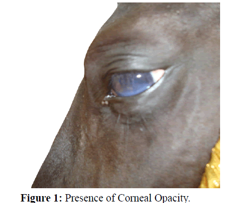
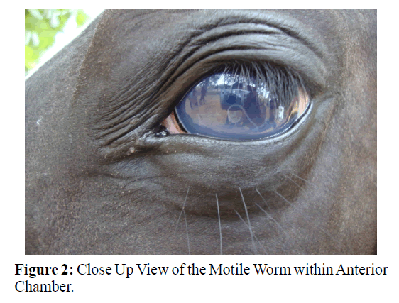
Twelve year old non descriptive (local breed) stallion with poor body condition was presented to the hospital nearer to the Ongole of Prakasam District in Andhra Pradesh, India. It had lacrimation, corneal opacity of left eye and abnormal gait for the past two weeks. Upon clinical examination, it showed congested conjunctival mucus membranes, elevated rectal temperature (102.6ºF), capillary refill time (3 s), respiratory rate (26 per minute), and heart rate (66 per minute). Shortened stride of fore limb was noticed while movement. Left eye showed corneal opacity with impaired vision. Examination of the left eye revealed motile white worm in the aqueous humor (Figures 1 and 2). Right eye was free from abnormalities. Ophthalmic examination was carried out by focal light source which found mild blepharospasm of left eye. Right eye was normal up on direct and consensual pupillary light reflexes but, left eye was not responded to these reflexes. Both eyes had hyperaemic and chemotic palpebral conjunctivae.
Condition was diagnosed as ocular setariosis and treated with inj. ivermectin@200 μg/kg body weight subcutaneously on the day of observation. Additionally it was treated with inj. ketoprofen@2.2 mg kg body weight I.M., inj. vitamin A-10,000 IU, I.M., for 3 days and ophthalmic drops (ofloxacin and prednisolone) were advised regularly up to clinical improvement (Soulsby, 1982., Sivajothi., Reddy, 2015).
Assessment of the clinical improvement and follow up was carried out by the local veterinarian. By the third day of treatment, movement of the worm was reduced and sluggish. By the 10th day, worm was active than the previous days. It might be due to insufficient concentration of ivermectin to kill the worm in the anterior chamber. Injection ivermectin (@200 μg/kg body weight subcutaneously) was given weekly twice for six doses along with intramuscular administration of inj.prednisolone@1 mg/kg body weight as tapering dose for seven days. By the end of 3rd dose of ivermectin, worm was not active and there is no movement of the worm. Horse attains its normal visibility after 26 days of therapy. Improvement in the condition was accordance with the previous reports of (Jaiswal et al. 2006) and (Sivajothi, 2015., Reddy 2015).
Previously similar conditions were managed by ivermectin administration in horses (Muhammad, 2007., Saqib, 2007), but in their study activeness of the worm was not noticed. Treatment with ivermectin is likely promising but, relapsing of condition may occur until the parasite completely dies. Sometimes delayed absorption of the dead parasites may cause inflammation of the anterior chamber (Basak et al. 2007) reported corneal edema due to dead filarial worm in the anterior chamber in their study.
Conclusion
Present communication reports about the successful management of ocular setariosis in stallion with repeated doses of ivermectin injection along with supportive therapy.
Acknowledgment
Authors are expressed their thankfulness to the late Dr. Dukki Anjaneya Reddy, Veterinary Assistant Surgeon who assisted and collected the information of the follow up.
References
- Basak, S.K., Hazra, T.K., Bhattacharya, D.(2007). Persistent corneal edema secondary to presumed dead adult filarial worm in the anteriorchamber. Indian J Ophthalmol,55, 67-69.
- Gangwar, A.K., Sangeetha, D., Singh, H.N., Singh, A. (2008).Ocular filariasis in equines, Indian Vet J,85, 547-548.
- Jaiswal, S., Singh, S.Y., Singh, B., Singh, H.N. (2006). Ocular setariosis in a horse in Intas Polivet, 7(1), 67-68.
- Mritunjay, K., Monsang, S.W., Pawde, A.M., Singh, S.K., Madhu, D.N., Zama, M.M.S. (2011). Post surgical healing effect of placentrex in equine (Equus cabalus) ocular setariasis: A review of 22 cases. The Indian J Am Vet Med Assoc, 6, 71-73.
- Muhammad, G., Saqib, M. (2007).Successful treatment of ocular equine microfilariasis (Setaria species) with ivermectin. Vet Rec, 160, 25-26.
- Sivajothi, S., Reddy, B.S. (2014).Brisket Oedema due to Microfilariosis in a buffalo. Research& Reviews: Journal of Veterinary Science and Technology,3(3), 17-19.
- Sivajothi, S., Reddy, B.S. (2015). Corneal opacity due to ocular setariosis in a buffalo. Inventi Impact: Veterinary Science,3, 93-94.
- Soulsby, E.J.L. (1982).Helminths, Arthropods and Protozoa of Domesticated Animals. 7th ed. Bailliere Tindall, London, 316-319.
- Tuntivanich, N., Sonthaya, T., Pranee, T. (2011). Success of Anterior Chamber Paracentesis as a treatment for Ocular Setariasis in Equine Eye: Case Report. J Equine Vet Sci,31,8-12.

