Case Report - Journal of Clinical Dentistry and Oral Health (2022) Volume 6, Issue 1
Management of mandibular first molar with three mesial canals and three distal canals-Two case reports.
Divyashree D Dandavati*, Madhu Pujar, Chetan Patil, Pallavi Gopeshetti, Veerendra Uppin
Department of Conservative and Endodontics, Maratha Mandal’s Nathajirao G. Halgekar Institute of Dental Sciences& Research Centre, Belagavi, Karnataka, India
- Corresponding Author:
- Dandavati DD
Department of Conservative and Endodontics
Maratha Mandal’s Nathajirao G. Halgekar Institute of Dental Sciences & Research Centre
Belagavi, Karnataka, India
E-mail: 3dddivyashri@gmail.com
Received: 03-Jan-2022, Manuscript No. AACDOH-22-51788; Editor assigned: 05-Jan-2022, PreQC No. AACDOH-22-51788(PQ); Reviewed: 17-Jan-2022, QC No. AACDOH-22-51788; Revised: 21-Jan-2022, Manuscript No. AACDOH-22-51788(R); Published: 28-Jan-2022, DOI: 10.35841/aacdoh-6.1.101
Citation: Dandavati DD, Pujar M, Patil C, et al. Management of mandibular first molar with three mesial canals and three distal canals–Two case reports. J Clin Dentistry Oral Health. 2022; 6(1):01-04.
Abstract
For the success of endodontic treatment, it is very critical for a clinician to have thorough knowledge of root canal morphology. There are a number of cases in literature wherein unusual anatomy have been discovered in mandibular first molars. Although the most common configuration is two roots and three root canals, mandibular molars might have many different combinations. There have been fewer reports regarding three mesial canals, three distal canals in mandibular first molars. The possibility of additional root canals should be considered even in teeth with a low frequency of abnormal root canal anatomy. This paper describes exploring of the unexplored, unusual anatomy of three mesial canals and three distal canals in mandibular first molars.
Keywords
Middle mesial canal, Three distal canal, Mandibular first molar, Extra canal, Distal root, Mesial root
Introduction
The success of root canal treatment depends on number of factors such as proper diagnosis, assessment of prognosis, complete debridement and obturation of the canals. If any of these protocols are not followed then it results in endodontic failures. These failures can result because of persistent infection, iatrogenic errors such as perforations, ledge formations, missed canals etc. In order to prevent such errors, it is very essential to understand the root canal morphology.
Mandibular first molars have different variations in the aspect of size, shape, number of roots/ canals and growth alterations. This paper describes about mandibular molars with three mesial canals and three distal canals [1].
Case 1
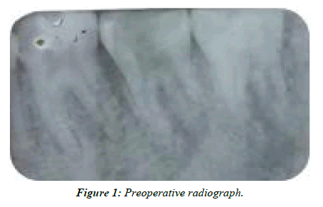
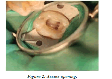
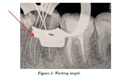
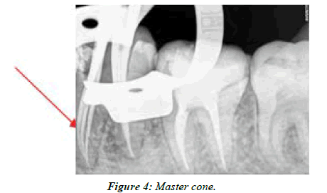
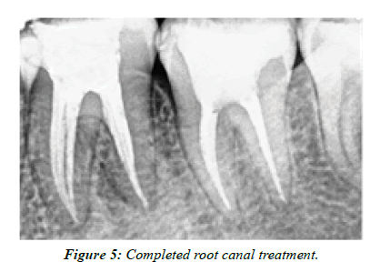
An 18 year old female patient reported with a chief complaint of pain in lower left back tooth region since 1 month. The pain aggravated on chewing food and during night. Clinical examination revealed a deep carious lesion and the tooth was tender to percussion. The tooth had no response to the vitality tests. The diagnosis was made as pulp necrosis with symptomatic apical periodontitis and endodontic treatment was planned. A 2% local anaesthesia was administered and access cavity was prepared under rubber damn isolation. During access opening a third canal was discovered between the mesiobuccal and the mesiolingual canals (Figure 1 and Figure 2). Working length determination was done using apex locator (NSK). Radiograph confirmed the presence of three mesial canals (mesiobuccal, mesiolingual and middle mesial) and two distal canals (Figure 3). Orifice enlargement was done using GG drills (MANI) and apical preparation up to 20 no K file (2%) was carried out in the mesial and distal canals. Cleaning and shaping was continued with Protaper gold files (Dentsply Maillefer) in crown down technique in mesiobuccal, mesiolingual and distal canals with lubricant. As the middle mesial canal was narrow, Hyflex 4% (Coltene) was used for cleaning and shaping. Irrigation was carried out using normal saline and 3% sodium hypochlorite. After drying the canals with paper points (Dentsply, India), master cones (Dentsply, India) were placed and radiograph was taken (Figure 4). The canals were obturated using AH Plus (DeTrey/Dentsply, Germany) sealer by warm lateral compaction technique (Figure 5 and Figure 6). Follow was done upto 1 year.
Case 2
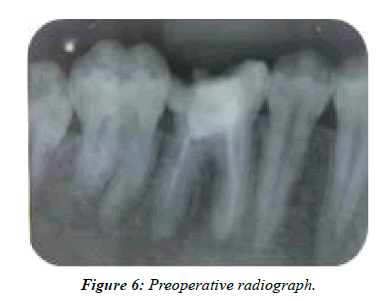
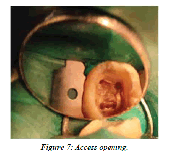
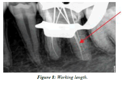
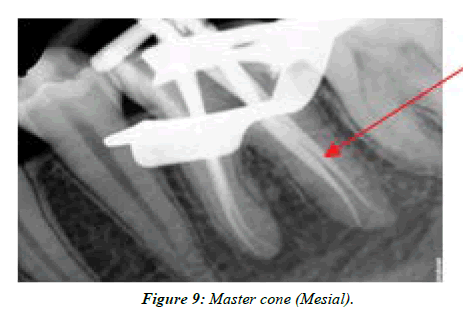
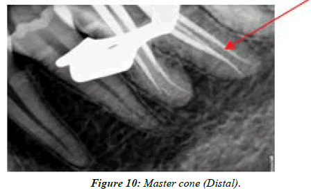
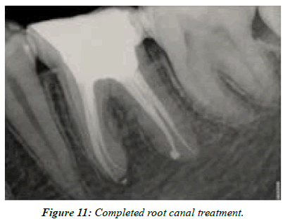
A 20 year old male patient reported with a chief complaint of pain in lower right back tooth region since 2 years. The patient gave history of root canal treatment 2 years back and was experiencing pain since then. Clinical examination revealed a fractured restoration and the tooth was tender to percussion. The tooth had no response to the vitality tests. The diagnosis was made as persistent symptomatic apical periodontitis and endodontic retreatment was planned. A 2% local anaesthesia was administered and previous restoration was removed rubber damn isolation. During modification of access cavity, a third canal was discovered in the distal root, which was missed and unobturated (Figure 7). Working length determination was done using apex locator (NSK). Radiograph confirmed the presence of three distal canals and two mesial canals (Figure 8). Orifice enlargement was done using GG drills (MANI) and apical preparation up to 20 no K file (2%) was carried out in the mesial and distal canals. Cleaning and shaping was continued with Protaper gold files (Dentsply Maillefer) in crown down technique in distobuccal, distolingual and mesial canals with lubricant. As the third distal canal was narrow, Hyflex 4% (Coltene) was used for cleaning and shaping. Irrigation was carried out using normal saline and 3% sodium hypochlorite. After drying the canals with paper points (Dentsply, India), master cones (Dentsply, India) were placed and radiograph was taken (Figures 9 and Figure 10). The canals were obturated using AH Plus (DeTrey/Dentsply, Germany) sealer by warm lateral compaction technique (Figure 11).
Discussion
Most of the times clinicians usually perceive that a particular tooth will have a predetermined number of roots and root canals. Extra roots/canals are those that show up more than average in different populations. Complicated and diverse root canal systems pose a challenge to successful diagnosis and treatment [1,2].The most common root canal morphology of mandibular first molars is the presence of 2 roots with either 3 or 4 root canals. The presence of a third canal in the mesial root is known as the middle mesial canal, which is found more towards mesiobuccal canal than the mesiolingual canal. Baugh and Wallace in a review, reported that the prevalence of a third canal in the mesial root of mandibular first molars was about 1-15%, but the existence of three canals in the distal root of mandibular first molars is uncommon [3].
Nosrat et al and Versiani et al found significant differences in the incidence of MMC between White (12.2%) and non-White (29.4%) patients. Table 1 shows the prevalence of configuration types of middle mesial canal [3].
| Author | Year | MMC (%) | Independent (%) | Fin (%) | Confluent (%) |
|---|---|---|---|---|---|
| Barker et al | 1969 | 1 | 1 | ||
| Vertucci and Williams | 1974 | 1 | 1 | ||
| Pomeranz et al | 1981 | 12 | 1 (I),1(II) | 5(I),3(II) | 1(I),1(II) |
| Walker | 1988 | 1 | 1 | ||
| Fabra Campos et al | 1989 | 2.6 | 0.1 | 2 | |
| Goel et al | 1991 | 15 | 6.6 | 8.3 | |
| Caliskan et al | 1995 | 3 | 3 | ||
| Gulabivala et al | 2001 | 10.8 | 1 | 9 | |
| Wasti et al | 2001 | 3 | 3 | ||
| Gulabivala et al | 2002 | 6.7 | 0.7 | 6 | |
| Sert et al | 2004 | 1.5 | 1.5 | ||
| Villegas et al | 2004 | 5 | 5 | ||
| Peiris et al | 2007 | 4.52 | 1.2 | 3.3 | |
| Shahi et al | 2008 | 0.95 | 0.95 | ||
| Chen et al | 2009 | 6 | 6 | ||
| Gu et al | 2009 | 0.82 | 0.82 | ||
| Karapinar | 2010 | 20 | 0 | ||
| Kazandag et al, Mukhaimer | 2014 | 3.1 | 0 | 0 | 3.1 |
| Azim et al | 2015 | 46.2 | 9.5 | 12 | 78.5 |
| Nosrat et al | 2015 | 20 | 20 | 33.3 | 46.7 |
| Chavda and Garg | 2016 | 50 | 0 | 0 | 96 |
Table 1: Prevalence of configuration types of middle mesial canal.
Pomeranz et al. in their study of 100 molars (61 first and 39 second molars) reported 12 cases of middle mesial canals. They classified middle mesial canal into three categories: (1) fin, when at any stage of debridement the instrument could pass freely between mesiobuccal or mesiolingual canal and the middle mesial canal, (2) confluent, when the prepared canal originated as a separate orifice but apically joined the mesiobuccal or mesiolingual canal, and (3) independent, when the prepared canal originated as a separate orifice and terminated as a separate foramen [4,5].
The incidence of a third canal in distal root is a rare occurrence with a prevalence rate of 0.2% to 3% in different ethnic groups [6-10].
There are only few comprehensive studies reporting the incidence of three distal canals in the mandibular first molars which have been summarized in Table 2 [2].
| Investigator | Year | Teeth | Method | Incidence (%) | Population Group |
|---|---|---|---|---|---|
| Caliskan et al | 1995 | 100 | Extracted | 1.7 | Turkish |
| Goel et al | 1996 | 60 | Extracted | 1.7 | Indian |
| Sperber | 1998 | 480 | Extracted | 0..2 | Senegalese |
| Gulabivala et al | 2001 | 139 | Extracted | 0.7 | Burmese |
| Gulabivala et al | 2002 | 118 | Extracted | 1.6 | Thai |
| Sert et al | 2004 | 20 | Extracted | 1 | Turkish |
| Ahmed et al | 2007 | 100 | Extracted | 3 | Sudanese |
| Al-qudah and Awawdeh | 2009 | 330 | Extracted | 0.3 | Jordanian |
Table 2:Incidence of three distal canals in the mandibular first molars.
Conclusion
It is very essential to understand when to suspect abnormality. An extra cusp (tuberculum paramolare), prominent occlusal distal/distal lingual lobe, cervical prominence, may indicate the presence of an additional root/canal. Additional off-angle radiographs, pulpal "footprint" can help to locate canals. Other aids such as micro-openers, magnification tools, red line test, white line test, perio probing can be used. The traditional “champagne bubble test,” still remains the gold standard for locating the root canal orifices. Additionally, CBCT and micro CT provide adequate information regarding the root canal morphology and are more superior to conventional radiographs.
As there is an axiom that states “You can treat, what you can see” [1] it is very essential for the clinician to carefully inspect the pulp chamber floor to locate possible accessory canal orifices. A thorough understanding of root canal morphology, angulated radiographs and exploring the root canals under magnification tools are the prerequisites for a successful treatment outcome [7-9].
References
- Weine FS. Case report: Three canals in the mesial root of a mandibular first molar (?). J Endo. 1982;8(11):517-20.
- Bansal P, Nikhil V, Shekhar S. Three distal root canals in mandibular first molar with different canal configurations: Report of two cases and literature review. Saudi Endo J. 2015;5(1):51.
- Bansal R, Hegde S, Astekar M. Morphology and prevalence of middle canals in the mandibular molars: A systematic review. J Oral Maxillofac Patho. 2018;22(2):216.
- Kottoor J, Sudha R, Velmurugan N. Middle distal canal of the mandibular first molar: a case report and literature review. Int Endo J. 2010;43(8):714-22.
- Ballullaya SV, Vemuri S, Kumar PR. Variable permanent mandibular first molar: Review of literature. J Conser Dent: JCD. 2013;16(2):99.
- Deepalakshmi M, Karumaran CS, Miglani R, et al. Independent and confluent middle mesial root canals in mandibular first molars: A report of four cases. Case Rep Dent. 2012 25;2012.
- Bhosale S, Balasubramanian A, Maroli R, et al. Middle Mesial Canal: A Common Finding-A Report of Three Cases. J Contemp Dent. 2014;4(3):152.
- Cohen S. Orofacial dental pain emergencies, endodontic diagnoses and management. Pathways Pulp. 2001:31-75.
- Pomeranz HH, Eidelman DL, Goldberg MG. Treatment considerations of the middle mesial canal of mandibular first and second molars. J Endo. 198;7(12):565-8.
Indexed at, Google Scholar, Cross Ref
Indexed at, Google Scholar, Cross Ref
Indexed at, Google Scholar, Cross Ref
Indexed at, Google Scholar, Cross Ref
Indexed at, Google Scholar, Cross Ref
