Case Report - Otolaryngology Online Journal (2016) Volume 6, Issue 3
Lipoma of Buccal Mucosa: Report of Two Cases and Literature Review
Abhay Sinha, Nitin Kumar Jain*, Megha Jain and Vaishali Gupta
Department of Otorhinolaryngology and Head and Neck surgery, Institute of Medical Sciences and Research, India
- *Corresponding Author:
- Nitin Kumar Jain
Department of Otorhinolaryngology and Head and Neck surgery
Institute of Medical Sciences and Research, Saifai, Etawah, (UP), India
Tel: 7534052763
E-mail: nitinjain70@gmail.com
Received date: April 09, 2016; Accepted date: May 02, 2016; Published date: May 05, 2016
Abstract
Lipomas are benign mesenchymal neoplasms composed of mature adipocytes, usually surrounded by a thin fibrous capsule. They are uncommon intraoral tumors with 1% to 4% occurring in this region. The literature is scanty on lipomas occurring in the buccal soft tissue. Here, we present two rare cases of lipoma occurring in the cheek of a 35-year old and a 45 year-old male. These were excised extra-orally. Histopathologically, these lesions were composed of the mature adipocytes with clear cytoplasm. There has been no evidence of tumor recurrence postoperatively.
Introduction
Initial description of Oral lipoma was provided by Roux in 1848 and he referred it as “yellow epulis” [1]. Lipomas of maxillofacial region are supposed to be neoplasms of adipocytes, occasionally associated with trauma [2]. Lipoma comprises 4-5% of all benign tumors in the body whereas oral lipoma constitutes 2.2% of all lipomas and 2.4% of all benign tumors of oral cavity [3]. Lipomas are usually asymptomatic unless they compress any neurovascular structure [4]. Commonest site of oral lipoma includes cheek, tongue, palate, mandible and lip [5,6]. Superficial lipomas impart yellow surface discolouration. Well capsulated tumors are freely movable beneath mucosa [2]. MRI is helpful in clinical diagnosis but CT and USG are unreliable. Multiple head and neck lipomas have been observed in neurofibromatosis, Gardner’s syndrome, Encephalocraniocutaneous lipomatosis, multiple familial lipomatosis and proteus syndrome [7,8].
Report of Two Cases
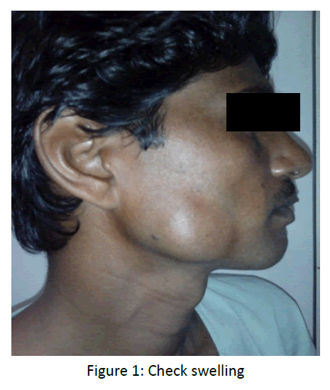
First case - A 35 years-old male patient reported to the Department of Otorhinolaryngology and Head and neck surgery, with the chief complain of swelling in the right cheek region. As per the patient, he was alright 4 year back then he developed a small swelling over right cheek which gradually increased to present size. Extraoral examination revealed a solitary swelling over right side of face approximately 4 cm × 4 cm in size. (Figure 1) On palpation, swelling was soft in consistency. On clenching the teeth, the swelling became firm and prominent; and soft and diffused on relaxation. There was no skin ulceration present. No abnormality detected on intraoral examination and there was no mucosal swelling.
Second case - A 45 years male patient was reported to the Department of otorhinolaryngology and head and neck surgery UPRIMS and R, Saifai, with the chief complain of swelling in the left cheek region since 2 years. Extra-orally, a single swelling over left side of face measuring approximately 3 cm × 2 cm was present. The swelling was soft in consistency on palpation.
A provisional diagnosis of lipoma of cheek was given for both cases.
For both cases, an excisional biopsy was done under local anaesthesia; specimens were fixed in formalin and then sent for histopathological examination.
Surgical Specimens and Histopathology
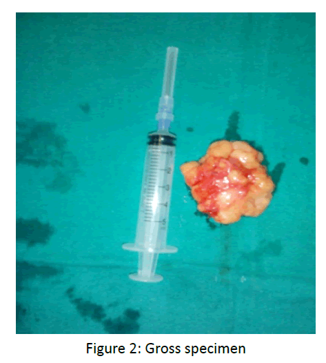
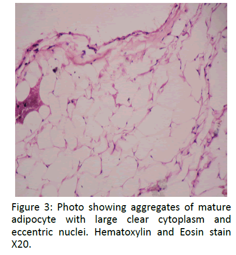
Grossly, lesional tissue appeared as capsulated fibro-fatty mass pale yellowish to greyish white in colour, soft in consistency and greasy to touch (Figure 2). As with all fatty tissue, a lipoma will float on the surface of formalin rather than to sink at the bottom of jar. Formalin fixed tissues were processed followed by sectioning and staining. Microscopic examination of the excised soft tissue mass revealed capsulated lesion composed of mature adipocytes containing large clear cytoplasm and eccentric nuclei. They were arranged in lobules separated by fibrous septa. Few dilated and congested blood vessels were also noted (Figure 3). These features are consistent with a classic diagnosis of a lipoma.
Discussion
Lipoma is predominantly composed of mature adipocytes admixed with collagen streaks and is often well demarcated from surrounding connective tissue. A thin, fibrous capsule may be seen and a distinct lobular pattern can be present [2]. Lipomas are the most common soft tissue mesenchymal neoplasms, with 15 to 20% of the cases involving the head and neck region and 1 to 4% affecting the oral cavity9. Several histological types of lipoma have been described and reported in literature containing glandular structure, cartilage, bone, vascular component [10-13]. Ocassionally, lipoma cannot be distinguished from a herniated buccal fat pad except by lack of history of sudden onset after trauma [2]. The peak age of incidence is usually in the 5th or 6th decade of life while the occurrence in children is very uncommon. Generally there is no gender predilection [14].
Adequate surgical excision is the treatment for oral lipomas. The surgical approach is dependent on the site of the tumor and the proposed cosmetic result [15].
Conclusion
Lipomas of buccal mucosa are uncommon and unusual tumors. Surgical excision is the ideal treatment with excellent outcome. Clinical course is usually slow and asymptomatic until they get larger in size and compress any neurovascular structures. Prognosis is considered good. Complete surgical excision is mandatory to avoid postoperative recurrence.
References
- Roux M (1848) On exostoses: Their character. Am J Dent Sc 9: 133-134.
- Gnepp DR, Diagnostic surgical Pathology, (2ndedn) Philadelphia, 2001. Saunders, Elseviers.
- Bataineh AB, Mansour MJ, Abalkhail A (1996) Oral Infiltrating lipomas. Br J Oral MaxillofacSurg 34: 520-523.
- Kaeser MA, Smith LW andKettner NW (2010) A case report of an intermuscular lipoma: presentation, pathophysiology, differential diagnosis. J Chiropr Med 9: 127-131.
- Adoga AA, Nimkur TL, Manasseh AN and GO (2008) Buccal soft tissue lipoma in an adult Nigerian: a case report and literature review. Journal of Medical Case Reports 2: 382
- Lawoyin JO,Akande OO, Kolude B andAgbaje JO (2001)Lipoma of the oral cavity: clinicopathological review of seven cases from Ibadan 10: 177-181.
- Barnes L, Surgical Pathology of Head and Neck, (2ndedn). Philadelphia PA, USA. Informa health care.
- Gorli RT, Cohen MM, Raoul CM and Hennekam RCM (2001) Syndromes of head and neck. 4thedn. Oxford: Oxford University press.
- Trandafir D, Gogalniceanu D, Trandafir V and Caruntu ID (2007) Lipomas ofthe oral cavity-a retrospective study. Rev Med ChirSoc MedNat Iasi 111: 754-758.
- Okada H, Yokoyama M, Hara M, Akimoto Y, Kaneda T, et al. (2009)Oral Surg Oral Med Oral Pathol Oral RadiolEndod 108: 571-576.
- Raj V, Dwivedi N, Sah K, and Chandra S (2014)Chondrolipoma: Report of a rare intra oral variant with review of histiogenetic concepts. J Oral MaxillofacPathol 18: 276-280.
- Kuyama K, Fifita SF, Komiya M, Sun Y, Akimoto Y, et al. (2009) “Rare LipomatousTumors with Osseous and/or Chondroid Differentiation in the Oral Cavity Report of Two Cases and Review of the Literature,” International Journal of Dentistry.
- Hemavathy S, Roy S and Kiresur A (2012)Intraosseousangiolipoma of the mandible 16: 83-287.
- Annibali S, Cristalli MP, Monaca GL, Giannone N, Testa NF, Russo LL, et al. (2009) Lipoma in soft tissue of the floor of mouth: A case report. Open Otorhinolaryngol J 3: 11-13.
- Furlong MA, Fanburg-Smith JC, Childers EL (2004) Lipoma of the oral and maxillofacial region: Site and subclassification of 125 cases. Oral Surg Oral Med Oral Pathol Oral RadiolEndod 98: 441-450.


