Review Article - Journal of Cancer Immunology & Therapy (2019) Volume 2, Issue 1
Irreversible electroporation of renal tumours: A review and an update.
Anthony Kodzo-Grey Venyo*
Department of Urology, North Manchester General Hospital, Manchester, United Kingdom
- *Corresponding Author:
- Anthony Kodzo-Grey Venyo
Department of Urology, North Manchester General Hospital, Crumpsall, Manchester, M8 5RB, United Kingdom
E-mail: akodzogrey@yahoo.co.uk
Accepted on January 21, 2019
Citation: Venyo AKG. Irreversible electroporation of renal tumours: A review and an update. J Cancer Immunol Ther. 2019;2(1):35-41.
Abstract
Background: Irreversible electroporation (IRE) for small localized renal carcinomas is a new treatment option for renal tumours than is being sporadically reported but majority of clinicians are not familiar with the procedure globally. Aim: To review the literature on IRE. Method: Various internet data bases were searched. Results: Irreversible electroporation (IRE) refers to a soft tissue ablation technique which utilizes ultra-short but strong electrical fields to create permanent and thus lethal nanopores within the cell membrane which tends to disrupt the cellular homeostasis. The ensuing cell death emanates from apoptosis and not necrosis as does result in all other types of thermal or radiation-based ablation techniques. The main idea behind the utilization of irreversible electroporation relates to the ablation of tumours within regions where there is importance or need for precision and conservation of the extracellular matrix, blood flow as well as nerves. The technique of irreversible electroporation, UN the form of NanoKnife ablation system, is, basically for the surgical ablation of soft tissue tumours. IRE of kidney tumours can be undertaken under radiological image guidance at open surgery, laparoscopic surgery or as a percutaneous procedure and generally the procedure tends to be associated with short-hospital stay, minimal Clavien Dindo grade 1 complication or higher. Successful renal tumour destruction tends to be high and greater than in 90% of patients and this can be detected by evidence of non-contrast enhancement of the lesion on radiological imaging following the procedure. Residual/recurrent lesions can be further treated by further IRE, radiofrequency ablation, cryotherapy or salvage partial nephrectomy. The short- and intermediate- term oncological outcome associated with IRE for small localized renal carcinomas is good but there are not many long-term follow-ups on patients with small renal tumours treated by IRE. IRE can be used to treat patients with significant comorbidities that would preclude them for partial or radical nephrectomy. A large global multi-centre study of IRE for small localized renal carcinomas would be recommended to ascertain if IRE should be recommended as a gold standard alternative to partial/radical nephrectomy. Conclusion: IRE for small renal carcinomas is an emerging treatment for small renal carcinomas associated with short hospital stay, minimal complications, reasonably good rate of destruction of tumour and good short-and medium term oncological outcome but possible residual tumour can be diagnosed by presence of persistent contrast enhanced renal lesion after the IRE which can be retreated by various techniques including re-IRE.Keywords
Irreversible electroporation, Renal cell carcinoma, Clavien-Dindo complications, Partial nephrectomy, Radical nephrectomy, Computed tomography scan, Magnetic resonance imaging scan, Ultrasound scan.
Introduction
Irreversible electroporation (IRE) is a terminology that refers to a non-thermal injury ablative modality of treatment which has been in clinical use since 2008 in the management of locally advanced soft-tissue tumours [1]. Irreversible electroporation has been reported to have been undertaken intraoperatively, laparoscopically and well as a percutaneous procedure [1]. It has been iterated that the mode of action of irreversible electroporation tends to rely upon a high voltage up to a maximum of 3,000 volts of small microseconds pulse lengths for 70 to 90 microseconds to induce cell membrane porosity that leads to slow/protracted cell death overtime [1]. Most localized renal carcinomas tend to be treated with curative intent by means of partial nephrectomy or by radical nephrectomy. Some very small renal carcinomas have just been observed and if there is evidence of increase in size of the tumours with time then these tumours would be treated with curative intent. Occasionally other minimal invasive treatment options have sporadically been undertaken in specialist centres and usually these minimally invasive procedures tend to be available in majority of hospitals globally. Some of these minimal non-invasive or minimally invasive procedures for renal carcinomas include: Cryotherapy, radiofrequency ablation, high intensity ultrasound, and irreversible electroporation of renal tumour. Because only very few renal tumours have been treated globally by irreversible electroporation, many clinicians would be unfamiliar with the use of the procedure in treating renal carcinomas as well as with the outcome and complications associated with the treatment. The ensuing article is divided into two parts (A) overview and (B) miscellaneous narrations and discussions related to irreversible electroporation of renal carcinomas.
Aim
To review the literature on irreversible electroporation of renal cancer.
Methods
Various internet data bases were searched including: PUBMED, google, google scholar, and Educus. The search words used included: Irreversible electroporation of carcinoma of kidney; Irreversible electroporation of renal cell carcinoma; irreversible electroporation of renal cancer.
Literature Review
Overview
General comments and definition: Irreversible electroporation (IRE) which is also called non-thermal irreversible electroporation refers to a soft tissue ablation technique which utilizes ultra-short but strong electrical fields to create permanent and thus lethal nanopores within the cell membrane which tends to disrupt the cellular homeostasis [2]. The ensuing cell death emanates from apoptosis and not necrosis as does result in all other types of thermal or radiation based ablation techniques [2].
The main idea behind the utilization of irreversible electroporation relates to the ablation of tumours within regions where there is importance or need for precision and conservation of the extracellular matrix, blood flow as well as nerves [2].
The technique of irreversible electroporation, in the form of Nano Knife ablation system, became available to be utilized for research commercially in 2009, basically for the surgical ablation of soft tissue tumours [2,3].
History: Fuller in 1898 was the first person to observe the irreversible electroporation effects [2,4].
Nollet was the first person to report the systematic findings of the appearance of red spots on human and animal skin when they were exposed to electric sparks [2,5].
Neumann et al. [6] reported their seminal work in 1982 which initiated the modern utilization of irreversible electroporation in modern times.
Pulsed electric fields had since then been utilized to temporarily permeabilize cell membranes to deliver foreign DNA into cells. In the ensuing decade, high-voltage pulsed electric fields had been combined with the chemotherapeutic medication as well as combined with DNA which yielded new clinical applications that are referred to as electrochemotherapy and gene electro-transfer, respectively [2,7-11].
Davalos et al. [12] in 2005 reported the first study of a potential use of irreversible electroporation. Medical Irreversible electroporation can be undertaken by means of (a) open surgical approach, (b) laparoscopic approach, and (c) percutaneous approach.
Mechanism: The mechanism of electroporation and irreversible electroporation have been summated as follows: [2]
Upon utilization of ultra-short pulsed but strong electrical fields, micropores as well as nanopores tend to be induced within the phospholipid bilayers that constitute the outer cell membranes and emanating from these two types of damage could occur which include reversible electroporation and irreversible electroporation.
• Reversible electroporation (RE) occurs when temporary and limited pathways for the transport of molecules are formed through nanopores, but pursuant to the end of the electric pulse, the transport tends to cease, and the cells tend to remain viable. Medical utilization of reversible electroporation does include local introduction of intracellular cytotoxic chemotherapeutic agent for example bleomycin (electroporation and electrochemotherapy.
• Irreversible electroporation (IRE) occurs when following a certain degree of damage to the membranes of the cells by electroporation, the leakage of the intracellular of intracellular contents has become too severe or the process of resealing of the cell membrane has become too slow, which does leave healthy and / cancerous cells damaged irreversibly. The cells tend to die by apoptosis, what has been said to be unique to irreversible electroporation, in comparison with all other techniques of ablation which tend to induce necrosis by heat or radiation [2]. It has been stated that although the method of ablation has been accepted generally to be apoptosis, some observations would seem to contradict a pure apoptosis cell death and because of this it had been suggested that the exact process by which irreversible electroporation induces cell death is uncertain [13].
• With regards to the mechanism of irreversible electroporation, it had been iterated that the mechanism had not been well understood; nevertheless, the conjectured theories include: [14]
It has been suggested that when an electric field greater than 0.5 V/nml [15] has been applied to the resting trans-membrane potential water tends to enter the cell during this di-electric breakdown and hydrophilic pores tend to be formed [16] [17]. A molecular dynamics simulation reported by Tarek [18] did illustrate the afore-mentioned proposed pore formation in two steps [14]:
1. Following the application of an electrical field, water molecules tend to line up in single file and tend to penetrate the hydrophobic centre of the bilayer lipid membrane.
2. The water channels tend to continue to grow in length and diameter and to expand into water-filled pores, and at that point, they tend to be stabilized by the lipid head groups which tend to move from the membrane-water interface into the middle of the bilayer.
• It had been suggested that as the applied electrical field increases, the greater tends to be the perturbation of the phospholipid head groups, and in turn tends to increase the number of water-filled pores [19]. The entire process could occur in a few nanoseconds [18]. It had also been stated that the average sizes of nanopores were likely cell-type specific and that in livers of Swine the nanopores average around 340 nm to 360 nm, as found in scanning electron microscope (SEM) [14].
Radiology: Diagnosis of a renal mass can be made by using ultrasound scan, magnetic resonance imaging (MRI) scan, as well as computed tomography (CT) scan.
Ultrasound scan: Ultrasound scan of the renal tract could use to establish the site, size, location, as well as type of renal tumour whether solid, cystic or mixed solid/cystic and the ultrasound scan could also determine whether there is any extra-renal mass.
• Ultrasound-scan guided irreversible electroporation of the renal lesion can be undertaken/
• Ultrasound scan of renal tract and abdomen and pelvis could be undertaken as follow-up surveillance.
• Contrast enhanced ultrasound scan (CEUS) can also be undertaken in the assessment of a renal mass.
Computed tomography (CT) scan: CT scan of renal tract / abdomen and pelvis can be undertaken in the diagnosis and initial assessment of the renal mass without and post contrast.
• CT scan-guided irreversible electroporation of the renal tumour can be undertaken.
• CT scan of renal tract could be undertaken following the procedure at intervals as part of the surveillance programme to ascertain whether the renal has recurred or not. Successful and complete irreversible electroporation of the renal tumour would be evidenced by non-contrast enhancement of the lesion after the irreversible electroporation.
Magnetic Resonance Imaging (MRI) scan: MRI scan of renal tract / abdomen and pelvis can be undertaken in the diagnosis and initial assessment of the renal mass without and post contrast.
• MRI scan-guided irreversible electroporation of the renal tumour can be undertaken.
• MRI scan of renal tract could be undertaken following the procedure at intervals as part of the surveillance programme to ascertain whether the renal has recurred or not. Successful and complete irreversible electroporation of the renal tumour would be evidenced by non-contrast enhancement of the lesion after the irreversible electroporation.
Some uses of Irreversible electroporation in the treatment of human tumours: Some of the lesions that have been treated recently by Irreverble electroporation include:
• Lung lesions [20]
• Pancreatic tumours [21]
• Renal tumours [20]
• Bone [20]
• Carcinoma of the prostate gland [22].
Discussion
Canvasser et al. [23] did report on the first short-term oncological outcomes of percutaneous irreversible electroporation for small kidney masses. Canvasser et al. [23] reviewed patients who had cT1a kidney masses which had been treated by means of irreversible electroporation from April 2013 to December 2016. With regards to methods, Canvasser et al. [23] reported that small, low complexity tumours had been generally selected for irreversible electroporation by the utilization of the NanoKnife R System (Angiodynamics, Latham, NY, USA). They had performed surveillance imaging and utilized Kaplan-Meier method to analyse surveillance analysis. Canvasser et al. [23] summated the results as follows:
• A total of 42 kidney tumours in 41 patients had been treated by irreversible electroporation.
• The man size of the kidney tumours was 2.0 cm and the median RENAL nephrometry score was 5.0.
• Twenty-nine patients which did amount to 71% of the patients had been discharged on the same day of undergoing the procedure and furthermore, there was no major Clavien grade II or higher intra-operative or postoperative complications.
• The success rate of the initial treatment of irreversible electroporation was 93%; there were 3 failures which accounted for 7% of the patients had undergone salvage radiofrequency ablation.
• Pursuant to a mean follow-up 22-months, the 2-year local recurrence-free survival amounted to 83% for the patients who did have biopsy proven renal cell carcinoma, 87% with biopsy proven or a history of renal cell carcinoma, and 92% with regards to the intent-to-treat cohort of patients.
Canvasser et al. [23] made the ensuing conclusions:
• Even though with low morbidity, as compared with surgical extirpation and conventional thermal ablation treatment options, irreversible electroporation is associated with suboptimal short-term local disease control outcomes in their series of small, low complexity tumours.
• Larger series of patients undergoing irreversible electroporation for small kidney tumours would, associated with longer follow-up would determine the durability of modality of treatment.
Diehl et al. [24] reported five patients including 2 females and 3 males, who had a mean age of 66 years, with 7 kidney lesions who had undergone irreversible electroporation of kidney tumour in a solitary kidney between August 2014 and August 2015. These patients were retrospectively wed. They did evaluate changes in signal intensity (SI) of the treated kidney lesions on contrast-enhanced magnetic resonance imaging (MRI) scan. To assess the functional outcomes of the patients following their procedures. Diehl et al. [24] compared the creatinine level, estimated glomerular filtration rate (eGFR) versus baseline after 1 day, 2 to 7 days, 3 to 6 weeks, and 6 to 12 weeks pursuant to the irreversible electroporation. Diehl et al. [24] summarized the results as follows:
• The diameter of the tumours had ranged between 15 mm and 38 mm and the mean diameter of the kidney tumours was 24.4 mm; the RENAL Criteria score had ranged between 4 and 9 and the average RENAL criteria score was 7.7 (radius, exophytic/endophytic, nearness to collecting system or renal sinus, anterior/posterior and location relative to polar lines).
• They observed progressive, significant decrease in treated tumour signal intensity (SI) upon the follow-up imaging with a mean of 70% to 82% which did suggest a treatment response rate of 100% at follow-up that ranged from 3 to 11 months and a mean follow-up of 6.4 months.
• Two minor acute complications based upon the Society of Interventional Radiology Class A criteria did occur and these included: visible haematuria, and stage I acute kidney failure.
• In whole. there was no significant decrease in the estimated glomerular filtration rate which had been between – 2.75 ml / min over 3 months, although 1 patient's estimated glomerular filtration rate had decreased from greater than 60 ml / min / 1.73 m(2) to 44 ml / min / 1.73 m(2).
Diehl et al. [24] made the following conclusions:
• Their data had suggested that percutaneous irreversible electroporation for kidney mass in patients who have a solitary kidney is safe and feasible.
• Irreversible electroporation for kidney mass could help in preserving kidney function and it does offer promising short-term oncological results.
De la Flor-Robledo et al. [25] reported a 66-year-old patient who had been diagnosed with an adenocarcinoma of the kidney and who had undergone without intention to cure irreversible electroporation of the tumour under general anaesthesia. They stated that their case represented the first case of renal tumour to be treated by means of irreversible electroporation in Spain. Details of the case are not available to the author.
Wendler et al. [26] reported their study of the first three patients who had undergone irreversible electroporation for small renal cell carcinomas (T1a tumours) to investigate the efficiency of ablation of irreversible electroporation to assess whether a complete ablation of T1a renal cell carcinoma and tissue preservation with the Nanoknife System was possible and to decide if the ablation parameters needed to be altered. The patients did undergo resection of their tumours 4 weeks pursuant to the irreversible electroporation. The success of the ablation as well as the detailed histopathological description of the specimens were utilized to check the parameters of the ablation. Wendler et al. [26] summarized the results as follows:
• The irreversible electroporation did lead to a high degree of damage to the kidney tumours of which 1 tumour was central, and 2 tumours were peripheral, and the size of the tumour had ranged between 15 mm and 17 mm.
• They had only been able to partly confirm the postulated homogeneous, isomorphic damage.
• They had found a zone; structuring of the ablation zone, negative margins, and enclosed within the zone of ablation, very small tumour residues of unclear malignancy.
Wendler et al. [26] made the following conclusion:
According to the preliminary results of the first three kidney cases, a new zonal distribution of irreversible electroporation damage had been described and the curative intended, kidney saving focal ablation of localized renal cell carcinoma less than 3 cm in size, by percutaneous irreversible electroporation by the NanoKnife system would appear to be possible; nevertheless, needs further, systematic evaluation for this therapeutic modality and treatment protocol.
Trimmer et al. [27] evaluated whether irreversible electroporation (IRE) could be used as an ablation technique for the treatment of small kidney tumours (T1a cancers or small benign tumours) as well as to describe features pursuant to ablation on computed tomography (CT) or magnetic resonance imaging (MRI). Trimmer et al. [27] undertook a retrospective study which had included 20 patients whose mean age was 65 years ±12.8 years who had undergone CTguided irreversible electroporation of T1a carcinoma of kidney (13, in total), or small benign or intermediate renal masses that measured less than 4 cm in size of which there were 7. The mean size of the tumours was 2.2 cm ± 0.7 cm. They verified the ablation area with contrast-enhanced imaging which was performed immediately pursuant to the procedure to determine the technical success. Radiological imaging was undertaken at 6 weeks in 20 of the 20 patients, 6 months in 15 of the 20 patients, and 12 months in 6 out of the 20 patients pursuant to the ablation. Trimmer et al. [27] reviewed the medical records and CT scan/MRI scan features of all the patients for recurrence of tumour, symptoms, and complications following treatment. Trimmer et al. [27] summarized the results as follows:
• They did achieve technical success in all patients (100%); they did not encounter any major procedure-related complications; minor complications did occur in 7 patients who included self-limiting perinephric haematomas, pain which was difficult to control, and retention of urine.
• The mean time of the procedure was 2.0 hours ± 0.7 hours.
• At 6 months 2 of the patients did require salvage therapy as a result of incomplete ablation.
• At 6 months, all 15 patients who had radiological imaging studies available did have no evidence of recurrence. At 1 year, 1 patient out of 6 patients was noted to have developed recurrence.
• Computed tomography (CT) scan / Magnetic resonance imaging (MRI) scan after irreversible electroporation ablation did demonstrate an area of non-enhancement within the treatment zone which involuted over about 6 months.
Trimmer et al. [27] made the following conclusions:
• Irreversible electroporation of the kidney would appear to be a safe treatment for small kidney tumours.
• Tumours that were treated with irreversible electroporation did demonstrate nonenhancement within the treatment zone with involution upon follow-up computed tomography (CT) / magnetic resonance imaging (MRI) scan.
Pech et al. assessed the feasibility and safety of ablating renal cell carcinoma tissue by irreversible electroporation. They included six patients who were scheduled to undergo curative resection of renal cell carcinoma who did undergo irreversible electroporation during anaesthesia immediately preceding the resection with electrographic synchronisation. They recorded central haemodynamics preceding and 5 minutes pursuant to the electroporation. Pech et al. utilized five-channel electrocardiography (ECG) for detailed analysis of ST waveforms. Pech et al. did blood sampling and did perform 12- lead ECG before, during, and at scheduled intervals following the intervention [28]. Pech et al. [28] summarized the results as follows:
• Analysis of ST waveforms and axis deviations did not show any relevant changes during the entire period of study.
• They did not notice any changes in central haemodynamics 5 min after irreversible electroporation.
• Likewise, during the investigation period haematological, serum biochemical, and ECG variables did not reveal any relevant differences.
• They did not find any changes in cardiac function following the irreversible electroporation treatment.
• However, there was one case of supraventricular extrasystole that was encountered.
• Initial histopathological examination did not show any immediate adverse effects of irreversible electroporation, but the observation of delayed effects would require a different study design.
Pech et al. concluded that irreversible electroporation would seem to offer a feasible and safe technique by which to treat patients with renal tumours and might offer some potential advantages over current thermal ablative techniques [28].
Tracy et al. did evaluate the effects of irreversible electroporation (IRE) on renal parenchyma and the renal collecting system in a porcine model. They reported that eight female Yorkshire pigs had undergone a series of laparoscopic renal ablations with the utilization of bipolar irreversible electroporation (IRE) which entailed the use of Angiodynamics, Queensbury, New York United States of America equipment. The pigs had been killed within 10 minutes and 14 days following the irreversible electroporation, and the kidneys had been removed for macroscopic and histopathological examination which included NADH staining for cellular viability. Tracy et al. [29] summated the results as follows:
• In all, 24 renal ablations had been undertaken and all the pigs had survived without developing any complications.
• The initial observed macroscopic lesions were diffusely haemorrhagic, which decreased progressively in in size by 30% to 40% to small white scars over 14th day period.
• Immediately pursuant to the irreversible electroporation, the ablated tissue had been characterized by diffuse tubular desquamation, eosinophilia, and nuclear pyknosis as well as absence of cellular viability by NADH.
• At day 7 following the irreversible electroporation (IRE), there was evidence of diffuse cellular necrosis with early peripheral granulation changes, and by the 14th day post irreversible electroporation there was evidence of marked tissue granulation, chronic inflammation, as well as dystrophic calcification associated with early fibrosis and cellular contraction. By the 14th day pursuant to the irreversible electroporation, the initial patchy urothelial injury and ulceration had shown signs of repair and viability.
Tracy et al. made the ensuing conclusions [29]:
• Irreversible electroporation of the kidney in porcine kidney does lead to predictable histological features of cellular death within one hour of ablation, with relative sparing of the urothelium.
• Further animal studies would be warranted to ascertain safety and efficacy of the novel ablation technique.
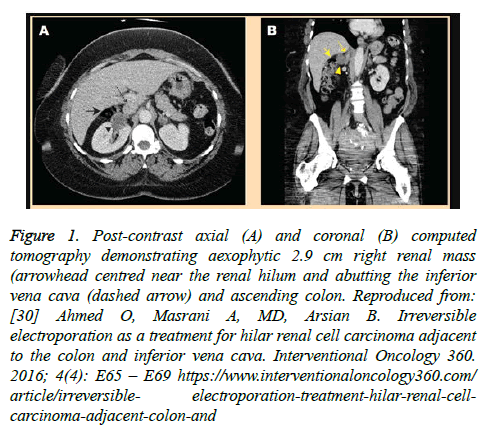
Ahmed et al. [30] reported a 54-year-old woman who was incidentally found to have a right renal mass which had grown 5mm on computed tomography (CT) scanning over 18 months that had been adjudged suspicious for renal cell carcinoma (RCC). The right renal tumour mass was contrast-enhancing and did measure 2.9 cm x 2.7 cm and had been located centrally near the renal hilum and it was abutting both the colon and inferior vena cava (IVC), (see Figure 1).
Figure 1. Post-contrast axial (A) and coronal (B) computed tomography demonstrating aexophytic 2.9 cm right renal mass (arrowhead centred near the renal hilum and abutting the inferior vena cava (dashed arrow) and ascending colon. Reproduced from: [30] Ahmed O, Masrani A, MD, Arsian B. Irreversible electroporation as a treatment for hilar renal cell carcinoma adjacent to the colon and inferior vena cava. Interventional Oncology 360. 2016; 4(4): E65 – E69 https://www.interventionaloncology360.com/article/irreversible- electroporation-treatment-hilar-renal-cellcarcinoma-adjacent-colon-and
She did have initially computed tomography (CT) scan-guided biopsy of the right renal mass and histology examination of the specimen was reported as being consistent with features that had confirmed the diagnosis of papillary type of renal cell carcinoma (RCC). Her treatment was strategized to include intraarterial injection of lipiodol with irreversible electroporation (IRE) the ensuing day after taking into consideration the closeness of the tumour to the renal vein, the colon and the IVC. Administration of lipiodol was undertaken in order to localise the tumour to enable accurate placement of probes for the IRE. The next day she had IRE under general anaesthesia. Five points of entry had been determined following a diagonal configuration with 3 central probes. Five separate 19-gauge, 15-cm length, Nanoknife (Angiodynamics) probes that had 2.5 cm exposure had been inserted (see Figure 2).
Figure 2. Intraprocedural computed tomography during irreversible electroporation demonstrates three 19 gauge probes placed approximately 1.5 cm apart and across the centre of the tumour. A single additional probe (not shown) was placed 11.5 cm was placed above and below the 3 probes respectively to complete the intended diamond configuration prior to initiation of treatment. Reproduced from: [30] Ahmed O, Masrani A, MD, Arsian B. Irreversible electroporation as a treatment for hilar renal cell carcinoma adjacent to the colon and inferior vena cava. Interventional Oncology 360. 2016; 4(4): E65 – E69 https://www.interventionaloncology360.com/article/irreversible- electroporation-treatment-hilar-renal-cellcarcinoma-adjacent-colon-and
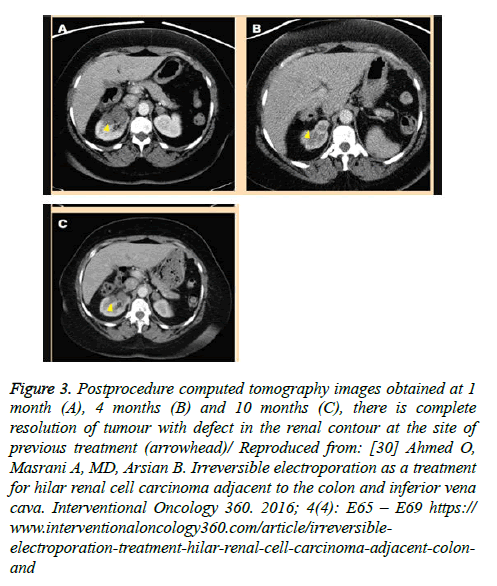
Two separate cycles of IRE had been undertaken distally and proximally. Completion computed tomography (CT) scan images did reveal iatrogenic gas collection in the lesion as was expected. No complications developed and the patient was discharged after 23 hours of observation. She did have followup CT scans at 1-month, 4- months, and 10-months pursuant to the IRE procedure and the scans did not reveal any residual, or recurrent tumour but the scans did show progressive decrease in size of the tumour at 4 months and complete resolution of the tumour at ten months (see Figure 3). During her follow-up her haematology and serum biochemistry profile remained similar to her pre-procedure values.
Figure 3. Postprocedure computed tomography images obtained at 1 month (A), 4 months (B) and 10 months (C), there is complete resolution of tumour with defect in the renal contour at the site of previous treatment (arrowhead)/ Reproduced from: [30] Ahmed O, Masrani A, MD, Arsian B. Irreversible electroporation as a treatment for hilar renal cell carcinoma adjacent to the colon and inferior vena cava. Interventional Oncology 360. 2016; 4(4): E65 – E69 https://www.interventionaloncology360.com/article/irreversibleelectroporation-treatment-hilar-renal-cell-carcinoma-adjacent-colonand
Conclusion
Irreversible electroporation of the kidney would appear to be a safe treatment for small kidney tumours.
Tumours that were treated with irreversible electroporation did demonstrate nonenhancement within the treatment zone with involution upon follow-up computed tomography (CT) / magnetic resonance imaging (MRI) scan.
A large multi-centre trial of irreversible electroporation of small renal carcinomas of the kidney with a long period of follow-up would be recommended in order to ascertain whether irreversible electroporation of small renal carcinomas should be strongly recommended as good alternative treatment of curative intent to be recommended for global first line mode of treatment.
Conflict of Interest
None
Acknowledgement
Ahmed O, Masrani A, Arsian B. Interventional Oncology 10 360 degrees for allowing figures from their journal to be reproduced provided the original source is properly cited.
References
- Martin RCG. Use of irreversible electroporation in unresectable pancreatic cancer. HepatobiliarySurgNutr. 2015; 4(3):211–21.
- https://en.wikipedia.org/wiki/Irreversible_electroporation
- Boutros C. Outcomes of Ablation of Unresectable Pancreatic Cancer Using the NanoKnife Irreversible Electroporation (IRE) System. ClinicalTrials.gov. 2017.
- Fuller GW. Purification of the Ohio River water at Louiseville Kentucky. University of Wisconsin, Madison, 1898.
- Nollet JA. Recherchessur les causes particulieres des phenomeneselectriques. Paris : H.-L. Guérin et L.-F. Delatour. 1754.
- Neumann E, Schaeffer-Ridder M, Wang Y, et al. Gene transfer into mouse lyoma cells by electroporation in high electric fields. EMBO J. 1982;1(7): 841–5.
- Mir LM, Belehradek M, Domenge C, et al. Electrochemotherapy, a new antitumor treatment: first clinical trial. C R AcadSci III. 1991; 313(13):613–8.
- Okino M, Mohri H. Effects of a high-voltage electrical impulse and an anticancer drug on in vivo growing tumors. JPN J Cancer Res. 1987;78(12):1319–21.
- Orlowski S, Belehradek J Jr, Paoletti C, et al. Transient electropermeabilization of cells in culture. Increase of the cytotoxicity of anticancer drugs. BiochemPharmacol. 1988;37(24):4727-33.
- Daud AI, DeConti RC, Andrews S, et al. Phase I trial of interleukin - 12 plasmid electroporation in patients with metastatic melanoma. J ClinOncol. 2008;26(36):5896–903.
- Titomirovav AV, Sukharev S, Kistanova E. In vivo electroporation and stable transformation of skin cells of newborn mice by plasmid DNA. BiochimBiophysActa. 1991;1088 (1):131–4.
- Davalos RV, Mir IL, Rubinsky B. Tissue ablation with irreversible electroporation. Ann Biomed Eng. 2005;33(2): 223–31.
- Golberg A, Yarmush ML. Nonthermal irreversible electroporation: fundamentals, applications, and challenges. IEEE. 2013;60(3):707 –714.
- Lee EW, Wong D, Prikhodko SV, et al. Electron microscopic demonstration and evaluation of irreversible electroporation-induced nanopores on hepatocyte membranes. J VascIntervRadiol. 2012;23(1): 107–13.
- Tieleman DP, Leontiadou H, Mark AE, et al. Simulation of pore formation in lipid bilayers by mechanical stress and electric fields. J Am Chem Soc. 2003;125(21):6382– 6383.
- Weaver JC. Molecular Basis for Cell Membrane Electroporation. Ann N Y Acad Sci. 1994; 720:141–52.
- Neumann E, Kakorin S, Toensing K. Fundamentals of electroporative delivery of drugs and genes. BioelectrochemBioenerg. 1999;48(1): 3–16.
- Tarek M. Membrane Electroporation: A Molecular Dynamics Simulation. Biophys J. 2005;88(6): 4045–53.
- Chen C, Smye SW, Robinson MP, et al. Membrane electroporation theories: a review. Med BiolEngComput. 2006; 44(1-2): 5–14.
- Vroomen LGPH, Petre EN, Cornelis FH, et al. Irreversible electroporation and thermal ablation of tumors in the liver, lung, kidney, and bone: What are the differences?. DiagnInterv Imaging. 2017; 98(9): 609–17.
- Philips P, Hays D, Martin RC. Irreversible Electroporation Ablation (IRE) of Unresectable Soft Tissue Tumors: Learning Curve Evaluation in the first 150 Patients treated. PloS One. 2013; 8(11): e76260.
- Ting F, Tran M, Bohm M, et al. Focal irreversible electroporation for prostate cancer: functional outcomes and short-term oncological control. Prostate Cancer Prostatic Dis. 2016;19(1): 46–52.
- Canvasser NE, Sorokin I, Lay AH, et al. Irreversible electroporation of small renal masses: suboptimal oncologic efficacy in an early series. World J Urol. 2017;35(10):1549-55.
- Diehl SJ, Rathmann N, Kostrzewa M, et al. Irreversible Electroporation for Surgical Renal Masses in Solitary Kidneys: Short-Term Interventional and Functional Outcome. J VascIntervRadiol. 2016;27(9):1407–13.
- de la Flor-Robledo M, Solis Munoz P, Sanjuan-Alvrez M, et al. Percutaneous irreversible electroporation of a renal tumour: Anesthetic management. Rev EspAnestesiolReanim. 2016;63(7):419-22.
- Wendler JJ, Ricke J, Pech M, et al. First Delayed Resection Findings after Irreversible Electroporation (IRE) of Human Localised Renal Cell Carcinoma (RCC) in the IRENE Pilot Phase 2a Trial. CardiovascInterventRadiol. 2016; 39(2):239–50.
- Trimmer CK, Khosla A, Morgan M, et al. Minimally Invasive Percutaneous Treatment of Small Renal Tumors with Irreversible Electoporation: A Single-Center Experience. J VascIntervRadiol. 2015;26(10):1465–71.
- Pech M, Janitzky A, Wendler JJ, et al. Irreversible electroporation of renal cell carcinoma: a first-in-man phase I clinical study. CardiovascInterventRadiol. 2011; 34(1): 132–8.
- Tracy CR, Kabbani W, Cadeddu JA. Irreversible electroporation (IRE): a novel method for renal tissue ablation. BJU Int. 2011;107(12): 1982–7.
- Ahmed O, MasraniA, Arslan B. Irreversible electroporation as a treatment for hilar renal cell carcinoma adjacent to the colon and inferior vena cava. IntervOncol 360. 2016;4(4): E6 –9.

