Case Blog - Journal of Neurology and Neurorehabilitation Research (2016) Volume 1, Issue 2
Intracranial wooden foreign body with uncertain route of entry.
Kyaw Linn*, Chawsu Hlaing, Hnin Wint Wint Aung, Cho Thair, Yi Yi Mar, Aye Mu Saan, Aye Mya Min Aye, Khin Thant Sin, Soe Moe, Khine Mi Mi Ko
Secretary General, Yangon Children Hospital, Myanmar
- *Corresponding Author:
- Kyaw Linn
Secretary General
Yangon Children Hospital
Myanmar
E-mail: linnkyaw.neuro@gmail.com
Accepted date: December 27, 2016
DOI: 10.35841/neurology-neurorehabilitation.1.2.55
Visit for more related articles at Journal of Neurology and Neurorehabilitation ResearchAbstract
Penetrating craniocerebral injuries are not uncommon in children, however, it can be obscured if history or examination is missed.
Introduction
Penetrating craniocerebral injuries are not uncommon in children, however, it can be obscured if history or examination is missed.
Case Blog
A boy aged 1 ½ year, from a village in middle Myanmar, had fever, irritability, vomiting and partially healed lacerated wound noted at the back (lower thoracic region) due to penetrating wooden stick injury 2 weeks ago. Apart from neck stiffness, the rest of neurological examinations were normal. CT head showed multiple brain abscesses with radiodensed linear foreign body extending from foramen magnum to left lateral ventricle (CT photo) (Figure 1).
Detailed history of injury revealed that his grandfather used to place him supine on a piece of cloth and pull across the ragged wooden floor. He was injured to the back when that cloth tore. Grandfather was not sure whether he got other injuries or not. He was re-examined for any other old injuries, however, nothing was found.
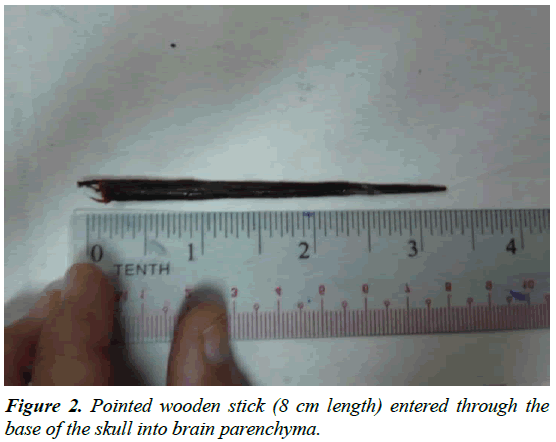
Surgical operation was done and found out that pointed wooden stick (8 cm length) entered through the base of the skull into brain parenchyma (wooden stick photo). He was treated with antibiotics for 4 weeks and well without neurological deficit at 3 months’ follow-up (Figure 2).
Discussion
Usually, the entry sites of foreign bodies include relatively vulnerable portion of the cranial bone, e.g. orbital roof, temporal squam and cribriform plate. The mystery of this case is lack of history and external entry site. Possible explanations might be previously missed history with completely healed entry site or migration form back due to contraction of the paraspinal muscles.

