Clinical Science - Case Reports in Surgery and Invasive Procedures (2017) Case Reports in Surgery and Invasive Procedures (Special Issue 2-2017)
Intestinal perforation caused by incidental ingestion of a fish bone.
Sagrario María Santos Seoane*
Hospital San Agustín, Health Service of the Principality of Asturias, Spain
- *Corresponding Author:
- Sagrario María Santos Seoane
Hospital San Agustín Health
Service of the Principality of Asturias, Spain
Tel: +34 902 430 960
E-mail: smsspulp@yahoo.es
Accepted date: July 12, 2017
Citation: Seoane SMS. Intestinal perforation caused by incidental ingestion of a fish bone. Case Rep Surg Invasive Proced. 2017;1(2):12-13.
Abstract
Intestinal perforations by fish bones are very rare. Most of the foreign bodies that are ingested advance through the intestinal tract without causing complications, especially if they pass the oesophagus. Only less than 1% causes intestinal perforation and is often long, sharp objects such as toothpicks, spines, chicken bones, or needles. They can occur at any point in the digestive tract, but are more common in segments with a closed angle such as pylorus, Treitz angle, distal ileum, and recto-sigmoid junction.
Keywords
Intestinal perforations, Digestive, Abdominal pain, Foreign body.
Introduction
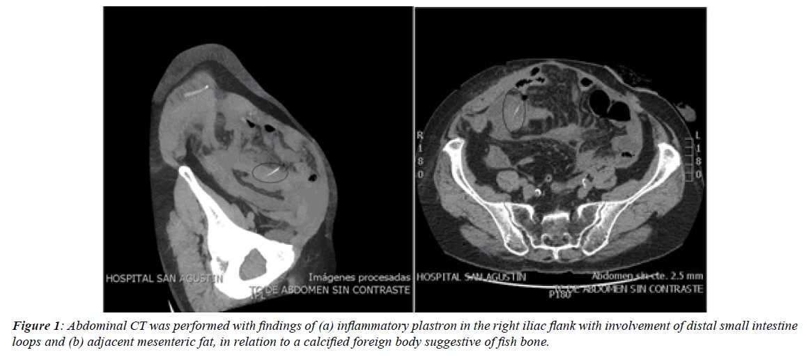
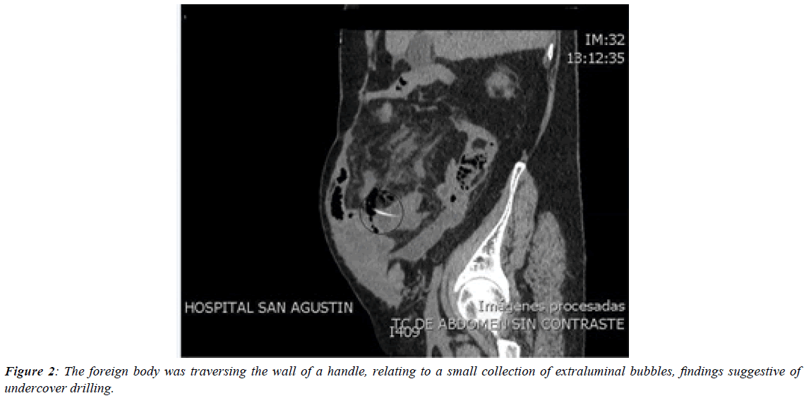
We present the case of an 88-year-old woman with a history two years before of left hemi-colectomy due to a colon neoplasia. She was admitted to the hospital for diffuse abdominal pain and oral intolerance of about 36 hours of evolution, with severe leukocytosis as well as pain and mass effect on right flank. Abdominal CT was performed with findings of inflammatory plastron in the right iliac flank with involvement of distal small intestine loops and adjacent mesenteric fat, in relation to a calcified foreign body suggestive of fish bone (Figures 1a and 1b). This foreign body was traversing the wall of a handle, relating to a small collection of extraluminal bubbles, findings suggestive of undercover drilling (Figure 2). Laparotomy and segmental resection of the small intestine were performed with extraction of the foreign body.

