Research Article - Virology Research Journal (2017) Virology Research Journal (Special Issue 1-2017)
Incorporation of antigens from whole cell lysates and purified virions from MP12 into fluorescence microsphere immunoassays for the detection of antibodies against Rift Valley fever virus
Mohammad M Hossain*, William C Wilson, Raymond R RowlandDepartment of Diagnostic Medicine/Pathobiology, College of Veterinary Medicine, Kansas State University, Manhattan, Kansas 66506 USA
- Corresponding Author:
- Mohammad M. Hossain, PhD
Department of Diagnostic Medicine/Pathobiology
College of Veterinary Medicine
Kansas State University
USA
Tel: 785-532-5660
E-mail: mofazzal@vet.k-state.edu
Accepted Date: March 15, 2017
Citation: Hossain MM, Wilson WC, Rowland RR. Incorporation of antigens from whole cell lysates and purified virions from MP12 into fluorescence microsphere immunoassays for the detection of antibodies against Rift Valley fever virus. Virol Res J. 2017;1(1):24-31.
Abstract
Background: The purpose of this study was the development of multiplex fluorescence microsphere immunoassay (FMIA) for the detection of Rift Valley fever virus (RVFV) IgG and IgM antibodies by incorporation of antigens from whole cell lysates and purified virions from MP12. Methods and Findings: Viral antigens were derived from whole cell material prepared from BHK cells infected with MP-12 (C-MP12). Virions from cell-free culture supernatant were purified on a sucrose cushion (V-MP12). The antigen material was treated with Triton X-100 or NP-40 prior to coating onto Luminex beads. V-MP12 and C-MP12 were assembled into a multiplex and tested in sheep and cattle infected with wild-type RVFV and the vaccine strain MP12. In these studies, V-MP12, C-MP12, and recombinant antigens (Gn, NSs, NSm, N) conjugated beads used in multiplex FMIA and the maximum level of IgG response to sheep in V-MP12 was recorded. IgG reactivity for the antigen targets varied with V-MP12>C-MP12. The positivity-negativity were evaluated by calculating S/P ratio using serum from RVFV-negative sheep (n=100) and negative cattle (n=100). The reaction of monoclonal anti-RVFV N antibody has been tested against N, V-MP12, and CMP12 by FMIA and all the antigens were reactive to antibody N>V-MP12>C-MP12. Conclusions: The experimental results demonstrate that the incorporation of V-MP12 and/or C-MP12 in the FMIA provides an improved sensitivity and specificity of FMIA with internal confirmation (i.e. multiple antigen targets) thus providing advantages over the traditional ELISA.
Keywords
Rift Valley fever virus-MP12, IgG, IgM antibody response, Cell-free viral lysates, Cell-associated viral lysates, Recombinant proteins, Fluorescence microsphere immunoassay, Luminex.
Introduction
Rift Valley fever virus (RVFV) is a zoonotic virus belonging to the genus Phlebovirus and one of the five genera in the family Bunyaviridae [1]. In ruminants, the disease is characterized by abortions, fetal deformities, and high mortality rates [2,3]. Humans are also readily infected through aerosols from infected animals or by exposure to infected animal tissues, aborted foetuses, and from infected mosquitoes [4,5]. RVFV is transmitted among ruminants and humans by direct contact with infectious tissues or by the bites of infected mosquito species of the Aedes and Culex genuses [6-8]. Global warming has certainly had a great impact on the transmission patterns of RVF [9,10]. Since the first outbreak in 1930, the geographical distribution of the virus has been expanded to several countries of Africa and the Arabian Peninsula [11-13]. Now, there are growing concerns of the continued spread of RVF to other parts of the world, including the United States and European Union [14-16].
Early detection of the virus is the key to initiating a rapid response and preventing the spread of RVFV. Several immunodiagnostic tests have been developed for the diagnosis of RVFV infection. The basic techniques, including hemagglutination-inhibition, complement fixation, indirect immunofluorescence, and virus neutralization test, have been used for detecting antibodies against RVFV [17]. The disadvantages of these techniques include the health risks to laboratory personnel, from manipulating live viruses, and the limitations in the number of sera sample that are able to be run in an assay [18]. Several ELISA-based tests have been developed to detect antibodies against various structural and non-structural components of RVFV [19-21]. Recently, inhibition/competition based ELISA assays were developed for the detection of antibodies against RVFV in several animal species, including domestic and wild ruminants [22,23]. These assays have proved to be safe alternatives to gold standard techniques, such as the virus neutralization test (VNT).
Fluorescent microsphere-based immunoassay (FMIA) is a highthroughput multiplex platform for the detection of antibodies to at least 100 target antigens in a small sample volume. Multiplex platforms which allow for the detection of antibodies to multiple pathogens in the same sample are a significant improvement in the diagnosis of numerous infectious diseases in humans and animals. A recent study described the development of a FMIA for the detection of antibody using Protein A, G, and A/G in place of species-specific secondary conjugate IgG [24]. In the previous studies, a FMIA have been developed for the detection of antibodies to RVFV proteins, including a surface glycoprotein, Gn, the non-structural proteins, NSs and NSm; and the nucleoprotein, N [25]. This assay is a diagnostic tool to detect cattle and sheep IgM and IgG antibodies to several
RVFV recombinant proteins. Also, the inclusion of bead sets coated with non-structural proteins provided differential infected from vaccinated animal (DIVA) compatibility. One limitation of the current assay is that it relies on recombinantly produced proteins that may not provide all the viral epitopes in the proper conformation. The objective of this study was to develop a FMIA based on whole cell and cell-free extracts from RVFV infected cells and determine if the incorporation of these antigens would increase test sensitivity and specificity.
Materials and Methods
Reagents and antibodies
Gelatin from cold water fish skin (Sigma-Aldrich, St. Louis, MO), phosphate buffered saline (PBS) (Sigma-Aldrich), sodium dihydrogen phosphate Tween-20 (Fisher Scientific, Fair Lawn, NJ), Tween-20 (Fisher Scientific), Nonidet P-40 (Sigma-Aldrich), TritonX 100 (Sigma-Aldrich), 1-Ethyl-3- (3-dimethylaminopropyl)-carbodiimide hydrochloride (EDC) (Thermo Scientific, Rockford, IL), N-hydroxy sulfosuccinimide (sulfo-NHS) (Thermo Scientific), Streptavidin-conjugated phycoerythrin (SA-PE) (Moss, Inc., Pasadena, MD), Carboxylated microspheres beads (Luminex Corporation, Austin, TX) were obtained from commercial sources. Antibody reagents were used: biotin-conjugated rabbit anti-sheep IgG (H+L) (Thermo Scientific), goat anti-bovine IgG (Thermo Scientific), rabbit anti-sheep IgM (Fc specific) (MyBioSource, Inc., San Diego, CA), sheep anti-bovine IgM (Bio-Rad AbD Serotec, Kidlington, Oxon, UK), rabbit anti-mouse IgG (H &L) (Rockland, Sunnyvale, CA), monoclonal anti-RVFV N antibodies and biotin-conjugated monoclonal antibody (Maine Biotechnology Services, Inc., Portland, ME).
Preparation of sucrose cushion purified MP12
Baby Hamster Kidney 21 (BHK-21) (American Type Culture Collection, Manassas, VA) cells were cultured in growth media, Dulbeco?s Eagle minimum essential media (MEM) (Invitrogen Corp., Carlsbad, CA), supplemented with 10% of fetal culf serum (FCS) (Atlanta Biologicals, Inc., Flowery, GA), L-alanyl-L-glutamine (Life Technologies, Grand Island, NY), anti-mycotic/antibiotic solution (Life Technologies) supplemented with 10,000 U/ml of penicillin, 10,000 µg/ ml of streptomycin, and 25 µg/ml of fungizone. The cells were incubated at 37°C, in a humidified incubator with 5% CO2. For large scale production, 4.6 à 106 BHK21 cells were seeded into cell culture flasks T-160 (Corning Incorporated, Corning, NY). On the day of infection, the cells were semiconfluent at a concentration of approximately 18.4 à 106 per flask. RVFV MP-12, at a multiplicity of infection (MOI) of 0.01 was seeded directly into the BHK21 cells. Flasks were incubated at 37°C in a 5% humidified incubator and observed for cytopathic effect (CPE). Cells were harvested when 60% to 80% of the cells were detached from the monolayer. The supernatant was harvested and centrifuged at 300 à g for 10 min at 4°C and then passaged through 0.45 µm filter to remove the cell debris and stored at -80°C. Cell-free supernatants were pelleted by ultracentrifugation at 24,000 à g for 2 h on a 40% sucrose cushion with a type 28 rotor (Beckman Coulter, Brea, CA). After centrifugation supernatant was discarded and the viral pellets were re-suspended in PBS containing 1% TritonX 100. Cell-free virus was disrupted by vortex and incubated at 37°C for 1 h. The concentration of protein in V-MP12 and C-MP12 were measured using a commercially available reagent (BioRad, Hercules, CA). V-MP12 was aliquoted and stored at -80°C.
Preparation of MP12 from infected cells
The infected cells were scraped from the flasks with a cell scraper and centrifuged at 300 à g for 10 min at 4°C. The supernatant was discarded and cells were washed in phosphate buffer saline (PBS) (Sigma-Aldrich). The final pellets were re-suspended in PBS containing 1% detergent (1% Nonidet P-40) (Sigma-Aldrich), vortexed, and ultrasonic disruption was performed. The disruption period was 30 s followed by 60 s intervals in an ice bath followed by incubation on ice for 1 h. The samples were kept in a salt-ice bath during the cell disruption to prevent overheating. The remaining cell debris was removed by centrifugation, known as C-MP12, and stored at -80°C.
Conjugation of antigens to microsphere magnetic beads
Conjugation of RVFV recombinant antigens (N, Gn, NSm and NSs) to carboxylated microsphere beads (Luminex Corporation) is described elsewhere [25]. Detergent-disrupted viral antigens were covalently coupled to magnetic beads (Luminex Corporation), as previously described, but with slight modification [26-28]. Briefly, microsphere beads in 80 µl of activation buffer (0.1 M sodium dihydrogen phosphate pH 6.2) was activated in the presence of 10 µl of 50 mg/mL N-hydroxy sulfosuccinimide (sulfo-NHS) (Thermo Scientific) and 10 µl of 50 mg/mL 1- Ethyl-3-(3-dimethylaminopropyl) carbodiimide hydrochloride (EDC) (Thermo Scientific). The activated beads were re-suspended in 100 µl coupling buffer (PBS, pH 7.4). To prepare a 500 µl reaction mixture for each bead set, 25 µg of protein and additional coupling buffer was added to each to a total volume of 500 µl bead set. For negative background controls, mock antigens were prepared from tissue supernatant and adherent cells using the protocol described above for virus infected cells. The reaction mixture was gently rotated on a shaker (Thermo Scientific) for two h at room temperature in the dark for the conjugation of antigen with the beads. The coupled beads were washed three times with washing buffer (0.1 M PBS, Tween 20 0.5 ml, sodium azide 0.5 g, pH 7.4, 1 litre). The beads were suspended in 1 ml of assay buffer (0.1 M PBS, Tween 20 0.5 ml, sodium azide 0.5 g, fish gelatin 10 g, pH 7.4, 1 litre) and stored at 2°C to 8°C in the dark until needed.
Source of serum samples
Serum samples from previous experimental RVFV MP-12 infection and vaccination trials (Kindly provided by Dr. Juergen A. Richt, Kansas State University, Manhattan, KS) were used in this study; specifically, seven cattle and four sheep infected with wild-type virus ZH501 (Kindly provided by Dr. Hana Weingartl, National Center for Foreign Animal Disease, Canadian Food Inspection Authority (CFIA), Winnipeg, Canada) were utilized. Prior to use, sera were heat-inactivated at 56ºC for 30 min in the presence of 0.25% Tween 20 and then safety tested to ensure the absence of infectious virus prior to removal from BSL3+ laboratories.
Fluorescence microsphere immunoassay (FMIA)
The different bead set mixtures and diluted test sera were prepared in an assay buffer separately. Prior to making the bead mixture, the number of beads was counted under a microscope. A 50 µl volume containing 1,250 beads from each bead set was added to each well. Fifty microliters of diluted serum was added to each well of a polystyrene white round bottom 96 well plate (Corning Incorporated, Corning, NY) along with 50 µl of premixed beads. Two replicates of each dilution were tested against RVFV positive and negative control serum. The plate was incubated in the dark at room temperature for 30 min on a plate shaker (Thermo Scientific). The beads were concentrated from the supernatant by placing the plate on the magnetic particle concentrator for 1 min and supernatant was discarded from each well. The beads were washed three times with 190 µl assay buffer.
The beads were suspended in 50 µl of biotin-conjugated secondary antibody appropriately diluted in assay buffer and incubated for 30 min followed by three washes, as described previously. Fifty microliters of streptavidin-conjugated phycoerythrin (SAPE) (Moss, Inc.), diluted 1:500 in assay buffer, was added to each well and incubated for 30 min at room temperature with shaking. The beads were washed three times with assay buffer and suspended in 100 µl of assay buffer. The plate was read (Luminex Corporation) and data analyzed using on-board software (Luminex Corporation). The results were reported as mean fluorescence intensity (MFI) obtained from the median value for at least 100 beads [25]. Results were also reported by the signal/positive (S/P) ratio.
S/P=(Mean MFI of test sample-Mean MFI of negative control)/ (Mean MFI of positive control-Mean MFI of negative control)
Estimation of N antigen in V-MP12 and C-MP12
A mouse monoclonal anti-RVFV N antibody was used to estimate the relative amount of MP12 antigen present on each bead sets coated with V-MP12 or C-MP12. The FMIA was performed as described above, with the exception that a 1:200 dilution of mAb was added to the beads instead of serum and a biotinylated mouse mAb (Maine Biotechnology Services) used for the detection of bound antibody.
Results
Preparation of V-MP12 and C-MP12 antigens
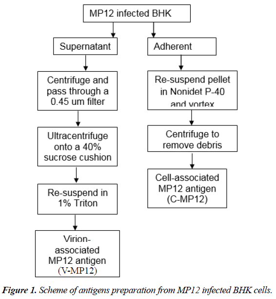
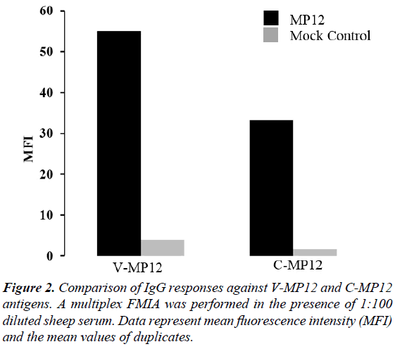
The peak level of MP12 infected BHK-21 cell infection was determined by cytopathic effect (CPE) under the microscope at day 3 after infection. Cell-free (V-MP12) and cell-associated (C-MP12) antigens were prepared from infected BHK-21 cells as described in Figure 1. Virus was purified from cell culture supernatant using ultracentrifugation on the sucrose cushion and lysed with 1% Triton-X. To prepare cell-associated viral antigens, the cells were lysed with 1% NP-40. Cell-free and cell associated viral lysates were conjugated to the beads, as described in the materials and methods. V-MP12 showed a higher, almost 2-fold higher signal to the RVFV positive serum than the C-MP12 (Figure 2).
Detection of N protein in V-MP12 and C-MP12
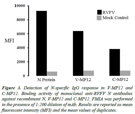
To detect the RVFV protein in V-MP12 and C-MP12, monoclonal anti-RVFV N antibody (N-mAb) were used to detect N protein in the V-MP12 and C-MP12 (Figure 3). Recombinant N, V-MP12 and C-MP12 were positive to anti-RVFV antibody. Each antigen showed different binding efficiencies despite the addition of a similar quantity of protein to protein-bead conjugation reactions. For example, recombinant N showed the highest reactivity with monoclonal anti-RVFV N antibody followed by beads conjugated with V-MP12 and C-MP12 respectively. The differences in monoclonal anti-RVFV N antibody binding may represent differential amount of N antigen in V-MP12 and C-MP12.
Figure 3: Detection of N-specific IgG response in V-MP12 and C-MP12. Binding activity of monoclonal anti-RVFV N antibodies against recombinant N, V-MP12 and C-MP12. FMIA was performed in the presence of 1:200 dilution of mAb. Results are reported as mean fluorescent intensity (MFI) and the mean values of duplicates.
Comparison of IgG responses to V-MP12, C-MP12 and recombinant RVFV antigens
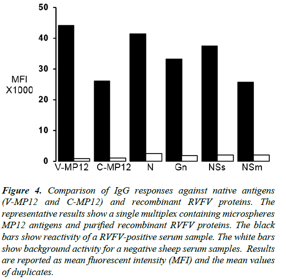
To compare the IgG responses between native cell culture produced antigens and recombinant RVFV antigens, surface glycoprotein Gn, non-structural proteins NSs, NSm and the nucleoprotein N were produced as described previously [25]. In these studies, out of six antigen conjugated beads used in multiplex FMIA, the highest level of IgG response to sheep in V-MP12 was recorded (Figure 4). V-MP12 showed strong antibody response compare to recombinant antigens of RVFV. On the other hand, IgG response to C-MP12 was moderate compare to recombinant antigens.
Figure 4: Comparison of IgG responses against native antigens (V-MP12 and C-MP12) and recombinant RVFV proteins. The representative results show a single multiplex containing microspheres MP12 antigens and purified recombinant RVFV proteins. The black bars show reactivity of a RVFV-positive serum sample. The white bars show background activity for a negative sheep serum samples. Results are reported as mean fluorescent intensity (MFI) and the mean values of duplicates.
IgG and IgM responses to native antigens (V-MP12, C-MP12) and recombinant N
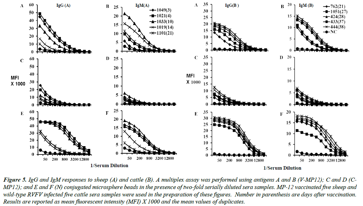
Serological responses were measured in sheep and cattle experimentally infected with the RVFV MP-12. Results for IgM and IgG from five sheep and five cattle are shown in Figure 5. In these studies, for sheep IgG, the greatest level of antibody response with the target was V-MP12, with the maximum values approaching 50,000 MFI (Figure 5A).
Figure 5: IgG and IgM responses to sheep (A) and cattle (B). A multiplex assay was performed using antigens A and B (V-MP12); C and D (CMP12); and E and F (N) conjugated microsphere beads in the presence of two-fold serially diluted sera samples. MP-12 vaccinated five sheep and wild-type RVFV infected five cattle sera samples were used in the preparation of these figures. Number in parenthesis are days after vaccination. Results are reported as mean fluorescent intensity (MFI) X 1000 and the mean values of duplicates.
This was followed by other native antigen C-MP12. Based on our previous findings, recombinant N antigen has been incorporated in this study. IgG antibody response in V-MP12 and RVFV recombinant N were similar and the maximum values of MFI between N and V-MP12 was 45,000-50,000 respectively (Figure 5A).
Similar results for V-MP12, C-MP12, and N antigens were obtained for cattle and sheep IgM, except that MFI values were decreased in cattle (Figure 5B). The V-MP12 was also immunodominant in infected sheep as shown in the Figure 5.
MFI values for sheep IgG approached 50,000, considerably higher than the MFI for cattle IgG. The increased reactivity of sheep IgG compared to cattle IgG was a consistent observation in our previous study [25]. The MFI values for sheep antibodies against C-MP12 were lower compare to V-MP12, but still easily detected by the assay.
Time course changes in anti-V-MP12 IgG and IgM
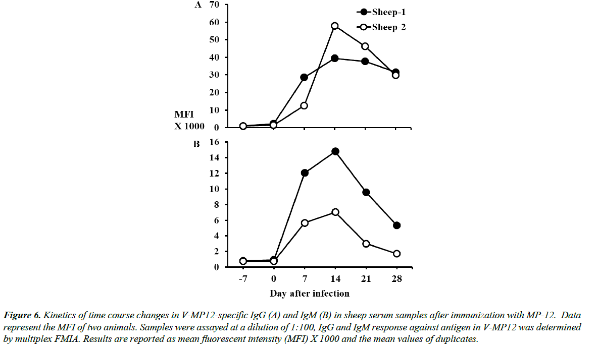
To determine changes in RVFV-specific IgG and IgM over time, two sheep were immunized with the RVFV modified live virus, MP12, and serum samples were collected at -7, 0, 7, 14, 21, and 28 days post-infection. IgG and IgM response against native antigen V-MP12 was determined by FMIA and reactivity of antibody was followed over time, as shown in Figure 6. sheep IgG was first detected at day 7 after immunization and was observed to remained elevated throughout the remainder of 28 days study period (Figure 6A). In contrast, sheep IgM peaked at 14 days post immunization and, by 28 days, had decreased to near background MFI values (Figure 6B).
Figure 6: Kinetics of time course changes in V-MP12-specific IgG (A) and IgM (B) in sheep serum samples after immunization with MP-12. Data represent the MFI of two animals. Samples were assayed at a dilution of 1:100, IgG and IgM response against antigen in V-MP12 was determined by multiplex FMIA. Results are reported as mean fluorescent intensity (MFI) X 1000 and the mean values of duplicates.
Signal/positive (S/P) ratio analysis in the V-MP12 and C-MP12
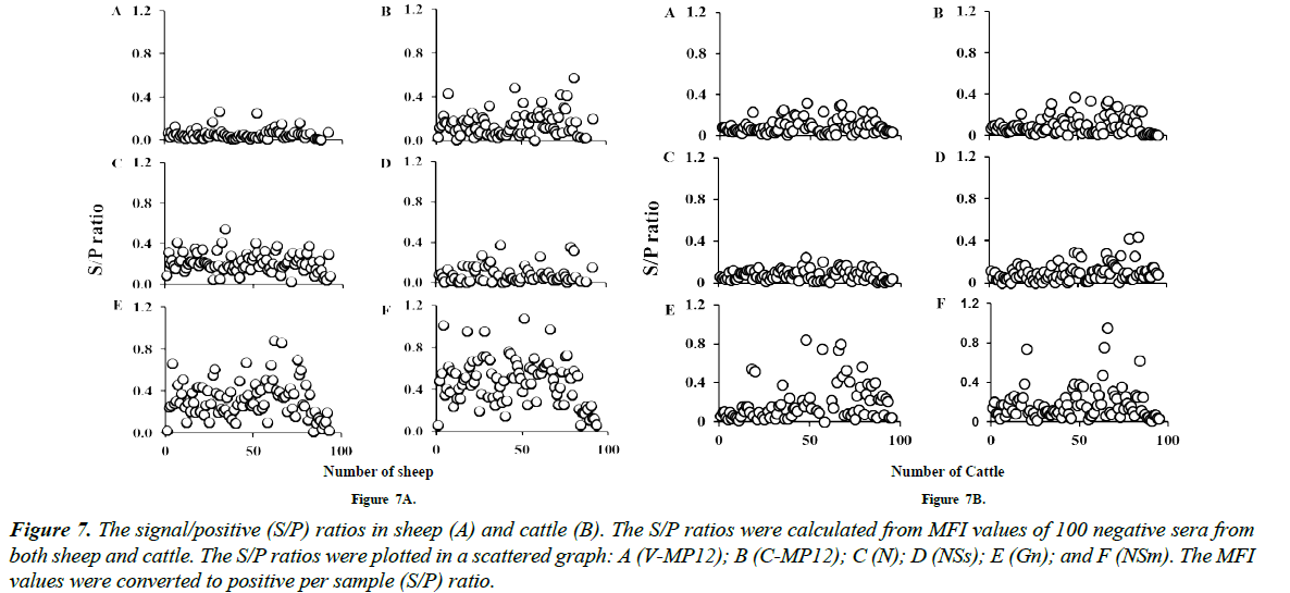
Serum dilution was optimized as 1:200 before screening all the negative sera. Diluted sera were incubated with native antigens (V-MP12 and C-MP12); RVFV recombinant N, Gn, NSm, NSs antigens coupled beads. The two native and four recombinant antigens conjugated bead sets were combined with an unconjugated bead set as a background control. Median MFI values were calculated as MFI test sample minus MFI of the background bead set. Signal/positive ratio was calculated as mentioned in materials and methods. All the negative samples were plotted in a scattered graph and cut-off values were determined based on the grouping of negative samples in comparison to positive. Data showed that sheep IgG were highly specific to native antigens (V-MP12 and C-MP12) compare to recombinantly produced antigens N, NSs, Gn, and NSm in sheep (Figure 7A). In the case of cattle, IgG responses were highly specific to native antigens (V-MP12 and C-MP12) and recombinantly produced N antigen in comparison to glycoprotein Gn and nonstructural proteins NSs and NSm (Figure 7B).
Figure 7: The signal/positive (S/P) ratios in sheep (A) and cattle (B). The S/P ratios were calculated from MFI values of 100 negative sera from both sheep and cattle. The S/P ratios were plotted in a scattered graph: A (V-MP12); B (C-MP12); C (N); D (NSs); E (Gn); and F (NSm). The MFI values were converted to positive per sample (S/P) ratio.
Discussion
A fluorescent microsphere-based immunoassay that uses virion-associated MP12 antigen (V-MP12) and cell-associated MP12 antigen (C-MP12) for the detection of IgG and IgM against RVFV in sheep and cattle were successfully developed. These studies describe a very simple and rapid method for the production of V-MP12 and C-MP12 and these antigens are useful for the diagnosis of RVFV infection in sheep and cattle. For the production of V-MP12 and C-MP12, BHK-21 cell line was infected with MP12 virus. MP12 is a mutant strain of RVFV that was generated by the serial passage of virus in the presence of mutagen 5-fluorouracil and, at the twelfth passage RVFV attenuated as a result of multiple mutation across the genome [29]. Mutated, a slow cytopathic strain of MP12 were used in this study than a highly cytopathic wild-type strain, which causes a faster destruction of cells after infection. In these studies, Triton X-100 and NP-40 disrupted cell-free or cell associated RVFV antigens were used in FMIA. The V-MP12 showed the highest antibody response in cattle and sheep with lower background compared to C-MP12 (Figures 2, 7A and 7B). Previous reports have also demonstrated a high level of antibody response using antigens produced from virus infected cultured cells in ELISA [30,31]. The data in the present study support the previously reported findings, which have shown reduced background in plasma sample [27]. Interestingly, when whole virus, as an antigen (no detergent lysis), is used to conjugate the beads, no signal was found (data not shown) which suggested that the proper lysis of the virus with detergent is a crucial step for the conjugation of viral antigen to the beads. Therefore, detergent disruption of virus is required for the use of V-MP12 and C-MP12 antigens in FMIA.
The FMIA described in the present study uses native antigens (V-MP12 and C-MP12), in contrast to using recombinant antigens (N, Gn, NSm, and NSs) for the detection RVFV antibody responses. A strong antibody response to NSs and NSm in FMIA suggested that NSs and NSm can be suitable candidate for Differentiate Infected from Vaccinated Animal (DIVA) (Figure 4). Recently, it has been suggested that the incorporation of NSs and/or NSm proteins into the FMIA can function as a companion DIVA test for gene-deleted MLV. The FMIA identified distinct differences in the IgG and IgM antibody responses in sheep when V-MP12 was used an antigen target. Anti-IgG activities in the sheep samples were elevated compared with IgM. In contrast, the IgM response in sheep followed the expected pattern for a primary antibody response, reaching an early peak and returning to background level (Figure 6), the previously reported results from this set of samples were similar when RVFV N as an antigen target [15].
In the present studies, V-MP12 showed the highest MFI signal in comparison to C-MP12 and recombinant antigens, suggested that V-MP12 might have the additional epitopes that increase the antigenicity of V-MP12. Also, our study showed that antibodies against V-MP12 were higher in both cattle and sheep; one animal showed MFI value up to 50,000 in sheep (Figure 5A). Consistantly, antibody response to native antigen (V-MP12 and C-MP12) showed strong MFI signals compared to recombinant antigens. The results showed that the IgG response to V-MP12 and N antigen are similar (Figure 5), suggested that antigen in V-MP12 might be the N protein. Recent studies [32-35] have identified recombinant N protein as a superior target for the diagnosis of infection, providing both sensitivity and specificity in ELISA-based diganostic assays. This conclusion is supported by our previous FMIA-based approaches [25].
In this study, detection of N protein in V-MP12 and C-MP12 using monoclonal anti-RVFV N antibodies (Figure 3) suggested that N protein in V-MP12 and C-MP12 produced from viral lysates and infected cell lysates might be N protein. In this respect, the fusion of the viral envelope and cell membrane causes disruption of the viral membrane through which viral ribonucleocapsid (RNP) and N protein are released into the cytoplasm; others supported the hypothesis [36-38].
FMIA for the detection of antibodies to native antigens (VMP12 and C-MP12) can be applied to the development of rapid diagnostic tests for the early detection of RVFV with high sensitivity and specificity in the diagnostic laboratory, especially in the case of urgent need during outbreaks of Rift Valley fever. The FMIA platform allows the generation of multiplex assays where a foreign animal disease could be included in a flag diagnostic submission to be sent more quickly to a federal lab. This potential would afford early detection of an introduced pathogen. This paper describes an improved sensitivity and rapid FMIA which would be an alternative diagnostic tool over the traditional ELISA.
References
- Elliott RM, Emerging viruses: the Bunyaviridae. Mol Med. 1997;3:572-7.
- Bird BH, Nichol ST. Breaking the chain: Rift Valley fever virus control via livestock vaccination. Curr Opin Virol. 2012;2:315-23.
- Ikegami T, Makino S. The pathogenesis of Rift Valley fever. Viruses. 2011;3:493-519.
- Amwayi SA, Gould LH, Sharif SK, et al. Risk factors for severe Rift Valley fever infection in Kenya, 2007. Am J Trop Med Hyg. 2010;83:14-21.
- Reed C, Lin K,Wilhelmsen C,et al. Aerosol exposure to Rift Valley fever virus causes earlier and more severe neuropathology in the murine model, which has important implications for therapeutic development. PLoS Negl Trop Dis. 2013;7:e2156.
- Jupp PG, Kemp A, Grobbelaar A, et al. The 2000 epidemic of Rift Valley fever in Saudi Arabia: mosquito vector studies. Med Vet Entomol. 2002;16:245-52.
- Nakouné E, Kamgang B,Berthet N, et al. Rift Valley fever virus circulating among ruminants, mosquitoes and humans in the Central African Republic. PLoS Negl Trop Dis. 2016;10:e0005082.
- Seufi AM, Galal FH. Role of Culex and Anopheles mosquito species as potential vectors of Rift Valley fever virus in Sudan outbreak, 2007. BMC Infect Dis. 2010;10:65.
- Gould EA, Higgs S. Impact of climate change and other factors on emerging arbovirus diseases. Trans R Soc Trop Med Hyg. 2009;103:109-121.
- Martin V, Chevalier V, Ceccato P, et al. The impact of climate change on the epidemiology and control of Rift Valley fever. Rev Sci Tech. 2008;27:413-26.
- Clements AC, Pfeiffer DU, Martin V, et al. A Rift valley fever atlas for Africa. Prev Vet Med. 2007;82:72-82.
- Jäckel S, Eiden M, EL Mamy BO, et al. Molecular and serological studies on the Rift Valley fever outbreak in Mauritania in 2010. Transbound Emerg Dis. 2013;60:31-9.
- Nanyingi MO, Munyua P, Kiama SG, et al. A systematic review of Rift Valley Fever epidemiology 1931-2014. InfecEcolEpidemiol.2015;5:28024.
- Gale P, Brouwer A, Ramnial V, et al. Assessing the impact of climate change on vector-borne viruses in the EU through the elicitation of expert opinion. EpidemiolInfec. 2010;138:214-25.
- Konrad SK, Miller SN. A temperature-limited assessment of the risk of Rift Valley fever transmission and establishment in the continental United States of America. Geospat Health. 2012;6:161-70
- Mansfield KL, Banyard AC, McElhinney L, et al. Rift Valley fever virus: A review of diagnosis and vaccination, and implications for emergence in Europe. Vaccine. 2015;33:5520-31.
- Swanepoel R, Struther JK, Erasmus MJ, et al. Comparison of techniques for demonstrating antibodies to Rift Valley fever virus. J Hyg (Lond.). 1986;97:317-29.
- Paweska JT, Burt FJ, Swanepoel R, et al. IgG-sandwich and IgM-capture enzyme-linked immunosorbent assay for the detection of antibody to Rift Valley fever virus in domestic ruminants. J Virol Methods. 2003;113:103-12.
- Jackel S, Eiden M, Balkema-Buschmann A, et al. A novel indirect ELISA based on glycoprotein Gn for the detection of IgG antibodies against Rift Valley fever virus in small ruminants. Res Vet Sci. 2013;95:725-30.
- Jansen van Vuren P, Potgieter AC, Paweska JT, et al. Preparation and evaluation of a recombinant Rift Valley fever virus N protein for the detection of IgG and IgM antibodies in humans and animals by indirect ELISA. J Virol Methods. 2007;140:106-14.
- McElroy AK, Albariño CG, Nicholet ST, al. Development of a RVFV ELISA that can distinguish infected from vaccinated animals. Virol J. 2009;6:125.
- Kim HJ, Nah JJ, Moon JS, et al. Competitive ELISA for the detection of antibodies to Rift Valley fever virus in goats and cattle. J Vet Med Sci.2012;74:321-7.
- Paweska JT, Mortimer E, Leman PA, et al. An inhibition enzyme-linked immunosorbent assay for the detection of antibody to Rift Valley fever virus in humans, domestic and wild ruminants. J Virol Methods. 2005;127:10-8.
- Hossain MM, Rowland RR. Fluorescence microsphere immunoassay for detection of antibodies to porcine reproductive and respiratory syndrome virus and porcine circovirus type 2 using protein A, protein G, and protein A/G. J Immunol Tech Infect Dis. 2017;6:1-6.
- Hossain MM, Wilson WC, Faburay B, et al. Multiplex detection of IgG and IgM to Rift Valley fever virus nucleoprotein, nonstructural proteins, and glycoprotein in ovine and bovine. Vector Borne Zoonotic Dis.2016;16:550-7.
- Anderson S, Wakeley P, Wibberley G, et al. Development and evaluation of a Luminex multiplex serology assay to detect antibodies to bovine herpes virus 1, parainfluenza 3 virus, bovine viral diarrhoea virus, and bovine respiratory syncytial virus, with comparison to existing ELISA detection methods. J Immunol Methods. 2011;366:79-88.
- Kuller L, Watanabe R, Anderson S, et al. Development of a whole-virus multiplex flow cytometric assay for antibody screening of a specific pathogen-free primate colony. Diagn Microbiol Infect Dis.2005; 53:185-93.
- Ryan JT, Rose TM. Development of whole-virus multiplex luminex-based serological assays for diagnosis of infections with kaposi's sarcoma-associated herpesvirus/human herpesvirus 8 homologs in macaques. Clin Vaccine Immunol. 2013;20:409-19.
- Caplen H, Peters CJ, Bishop DH, et al. Mutagen-directed attenuation of Rift Valley fever virus as a method for vaccine development. J Gen Virol. 1985;(Pt 10):2271-77.
- Ansari MZ, Shope RE, Malik S. Evaluation of vero cell lysate antigen for the ELISA of flaviviruses. J Clin Lab Anal. 1993;7:230-37.
- Johnson JE. Detection of human immunodeficiency virus type 1 antibody by using commercially available whole-cell viral lysate, synthetic peptide, and recombinant protein enzyme immunoassay systems. J Clin Microbiol.1992;30:216-18.
- Ellis CE, Mareledwane VE, Williams R, et al. Validation of an ELISA for the concurrent detection of total antibodies (IgM and IgG) to Rift Valley fever virus. Onderstepoort J Vet Res. 2014;81.
- Fafetine JM, Tijhaar E, Paweska JT, et al. Cloning and expression of Rift Valley fever virus nucleocapsid (N) protein and evaluation of a N-protein based indirect ELISA for the detection of specific IgG and IgM antibodies in domestic ruminants. Vet Microbiol. 2007;121:29-38.
- Fukushi S, Nakauchi M, Mizutani T, et al. Antigen-capture ELISA for the detection of Rift Valley fever virus nucleoprotein using new monoclonal antibodies. J Virol Methods.2012;180:68-74.
- Williams R, Ellis CE, Smith SJ, et al. Validation of an IgM antibody capture with Rift Valley fever virus. J Virol Methods.2011;177:140-46.
- Frese M, Kochs G, Feldmann H, et al. Inhibition of bunyaviruses, phleboviruses, and hantaviruses by human MxA protein. J Virol.1996;70:915-23.
- Piper ME, Sorenson DR,Gerrard SR, et al.Efficient cellular release of Rift Valley fever virus requires genomic RNA. PLOS ONE. 2011;6:e18070.
- Ribeiro D, Borst JW, Goldbach R, et al. Tomato spotted wilt virus nucleocapsid protein interacts with both viral glycoproteins Gn and Gc in planta. Virology.2009;383:121-30.
