Case Report - Biomedical Research (2017) Volume 28, Issue 22
Incarceration of a colonoscope within an inguinal hernia: An unusual complication
Xiaojun Wang and Yizhong Zhang*
Department of General surgery, the Affiliated Hospital of Medical College of Ningbo University, Ningbo, PR China
- *Corresponding Author:
- Yizhong Zhang
Department of General surgery
The Affiliated Hospital of Medical College of Ningbo University, PR China
Accepted date: October 13, 2017
Abstract
Colonoscopy, as a frequently performed diagnostic and therapeutic procedure, is fairly safe to patients with colorectal disorders generally. Incarceration of a colonoscope refers to the colonoscope is unable to be advanced or withdrawn due to some causes e.g. inguinal hernia, which is an unusual complication during colonoscope procedure. We present a case report of incarceration of the colonoscope in a left inguinal hernia. This case report reiterates the importance of this rare complication of colonoscopy and discusses its etiology and treatment. Clinicians and colonoscopists should be cautious of this possible complication to prevent unfavorable outcomes.
Keywords
Colonoscopy, Fluoroscopy, Hernia, Inguinal
Introduction
Colonoscopy, a frequently performed diagnostic and therapeutic procedure, is fairly safe. However, given the large volume of procedures performed, along with the increasing number of unusual complications [1], awareness of such complications is imperative. Here we present an unusual case of colonoscope incarceration in a left inguinal hernia.
Case Presentation
A 70 y old man was referred to the Department of Digestion Clinic with complaints of watery diarrhea for 1 year. Patient history indicated a left inguinal region moveable mass that had been present for 5 years. The mass could not be returned after it protruded without pain during the last 12 months. The colonoscopy was inserted without difficulty or significant abdominal discomfort until at approximately 60 cm, when it became impossible to advance or withdraw the colonoscope because of resistance and pain. Physical examination revealed a bulge in his left inguinal area consistent with incarceration of the colonoscope in the sac of the inguinal hernia.
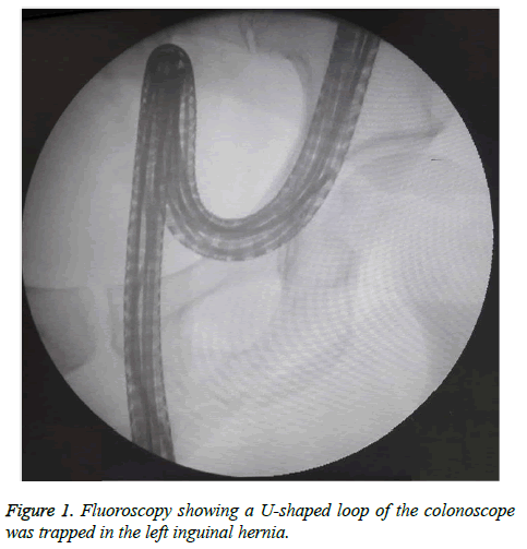
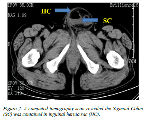
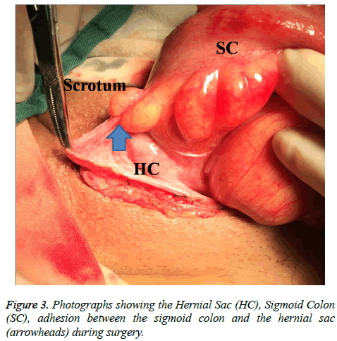
After unsuccessful attempts of manual reduction, the patient underwent fluoroscopy. A U-shaped loop of the colonoscope in the left inguinal hernial sac was found (Figure 1). Under radiographic guidance and general anesthesia, the incarcerated colonoscopy was attempted to be reduced by pushing and rotating the scope, combined with careful withdrawal through gentle traction. Eventually, the incarcerated colonoscope was reduced. The patient remained calm throughout the reduction of the incarcerated colonoscope. When presented with the option of hernia repair, the patient refused. He had watery diarrhea three times the night after the colonoscopy and complained of discomfort in the left inguinal region. Simultaneously, the left inguinal mass increased significantly. Immediate computed tomography showed a folded intestinal tract in the hernial sac (Figure 2). Therefore, emergency left inguinal hernia repair was performed. The sigmoid colon was identified as the contents of the hernia. Conspicuous adhesion was detected between the sigmoid colon and the hernial sac (Figure 3). The patient made an uneventful recovery.
Discussion
Only a limited number of colonoscope incarcerations have been reported in the English literature published to date [2]. It may cause failure of the colonoscopy or result in a surgical operation to avoid enterorrhexis [2-4], if it is not treated correctly. Hence, prompt recognition of this complication is important.
The stiffness and diameter of the colonoscope could lead to the occurrence of incarceration [5]. In addition, the sigmoid colon is long-winded, and a colonoscope can form a twist on this site. The adhesion becomes a pivot point, making it easier for the sigmoid colon to enter and form a loop in the hernial sac. Moreover, the hernial sac neck intensively spasms when the colonoscope enters the sac. All of the above factors can lead to the incarceration of the colonoscope.
How can this condition be prevented or treated? As prophylaxis in cases where inguinal hernia is detected, colonoscopy should be delayed until surgical repair of the hernia is performed. However, in cases where surgery would be inappropriate and colonoscopy is warranted, some techniques can be used to reduce the risk of colonoscope incarceration. For example, external manual pressure on the hernia ring could prevent the protrusion of the hernial content and incarceration of the colonoscope [6]. Applying a large patch of adhesive plaster over the hernia could maintain a good reduction during the procedure [6]. Moreover, computed tomography colonoscopy could also be used [2].
With regard to the treatment of colonoscope incarceration, a range of techniques have been suggested by various authors to reduce it, including manual reduction with deep sedation, the “pulley” method of manual reduction [5], reduction under direct fluoroscopic guidance [7], and surgical reduction [3]. A cap attached to the tip of the colonoscope can facilitate the passage of the colonoscope through the loop of bowel that has prolapsed into the hernia with small and moderate hernia sac neck [2]. In wide or large hernial sac neck, externally applied manual pressure may enable the completion of the colonoscopy without incarceration [8]. Direct fluoroscopic guidance helps in estimating the width of the orifice of the hernial sac and can minimize the colonoscope loop in the scrotal sac [7]. If all else procedures fail, surgical reduction should be performed to avoid enterorrhexis [3].
In conclusion, incarceration of a colonoscope within an inguinal hernia is an unusual but dangerous complication of colonoscopy. Despite its rarity, clinicians and colonoscopists should be cautious of this possibility to prevent unfavorable outcomes.
Informed Consent
Informed consent was obtained from the patient.
Disclosure of Potential Conflicts of Interest
The authors declare no conflict of interests for this article.
References
- Day LW, Kwon A, Inadomi JM, Walter LC, Somsouk M. Adverse events in older patients undergoing colonoscopy: a systematic review and meta-analysis. Gastrointest Endosc 2011; 74: 885-896.
- Tan PY, Lee YT, Poon JTC. Incarceration of a colonoscope in an inguinal hernia: Case report and literature review. World J Gastrointest Endosc 2013; 5: 304-307.
- Tas A, Olmez S, Demir M. Colonoscope incarceration in an inguinal hernia: a complication of colonoscopy. Endoscopy 2015; 47: 125-126.
- Lee YT, Hui AY. Failed colonoscopy due to hernia. Endoscopy 2004; 36: 758.
- Koltun WA, Coller JA. Incarceration of colonoscope in an inguinal hernia pulley technique of removal. Dis Colon Rectum 1991; 34: 191-193.
- Leichtmann GA, Feingelrent H, Pomeranz IS, Novis, BH. Colonoscopy in patients with large inguinal hernias. Gastrointest Endosc 1991; 37: 494.
- Fan CS, Soon MS. Colonoscope incarceration in an inguinal hernia. Endoscopy 2007; 39: 185.
- Yamamoto K, Kadakia SC. Incarceration of a colonoscope in an inguinal hernia. Gastrointest Endosc 1994; 40: 396-397.


