Case Report - Allied Journal of Medical Research (2021) Volume 5, Issue 9
Immunoglobulin (IgG4) sclerosing cholecystitis-camouflaging gall bladder cancer: A case report
Rashpal Singh*, Puneet Mahajan, Rizul Prasher, Vivek Rajdev, Jagwinder Singh, Cliffin Mathai Kattoor, Kavita Mardi, Jagdish Gupta
Department of General Surgery, Indira Gandhi Medical College, Shimla, Himachal Pradesh, India
- Corresponding Author:
- Rashpal Singh
Department of General Surgery
Indira Gandhi Medical College
Shimla,
Himachal Pradesh
India
E-mail: rashpalcallingrashpal@gmail.com
Accepted date: December 15, 2021
Citation: Singh R, Mahajan P, Prasher R, et al. Immunoglobulin (IgG4) sclerosing cholecystitis-camouflaging gall bladder cancer: A case report. Allied J Med Res. 2021;5(9): 38-41.
Abstract
Background: IgG4 related disease is a rare systemic disorder having an underlying autoimmune cause. These disorders mainly affects pancreatico biliary tree pancreas, gall bladder and biliary tree (extra hepatic as well intrahepatic), but can also affect other part of body. Majority of disorders involving biliary tree are associated with autoimmune pancreatitis component. These disorders are difficult to diagnose clinically as they can mimic inflammatory as well malignancy and poses a real diagnostic challenge to manage and treat.
Case Presentation: 64 years old female known diabetic evaluated for pain in right hypochondrium. Gall bladder cancer was suspected clinically as well on radiologically basis. Patient underwent extended cholecystectomy as it was a resectable disease. Final histopathology revealed immunoglobulin G4 (IgG4) related cholecystitis which was confirmed after immunohistochemistry for CD 138 and IgG4.This disease could be managed conservatively by giving oral steroids, if it has been picked up preoperatively and major surgical intervention have been avoided. No defined blood test or tumour markers are currently available to diagnose this entity except serum immunoglobulin G4 which is costly and not feasible to get done in all patients especially in developing nations like India.
Conclusion: IgG4 cholecystitis is an immune mediated disease whose pathophysiology is still not completely understood. Every clinician should keep possibility of IgG4 cholecystitis in mind whenever any patient with abnormal gall bladder thickening or gall bladder mass is encountered in their clinical practice, as both these entities have completely different options of treatment. We should not rely solely upon clinical and radiological picture.
Keywords
Health lifestyle, Girl-children, Healthy behaviour, Northern nigeria.
Introduction
IgG4 related disease is a rare systemic disorder which can involve any organ of our body. To club these different organs involvement by this rare similar auto-immune phenomenon, Kamisawa Proposed a new common term of systemic IgG4-related autoimmune diseases in 2003 as IgG4 Related Disorder (IgG4-RD). These disorders are difficult to diagnose clinically as well radio logically due to the overlapping of clinical and radiological imaging features. These disorders mimics inflammatory as well malignancy and poses a real difficulty in diagnosis, treatment and predicting the natural course of disease [1].
These disorders are immune mediated and mainly affects pancreatic biliary tree i.e. pancreas, gall bladder and biliary tree (extra hepatic as well intrahepatic). Apart from these organs, it can involve head and neck region, retroperitoneal organs, genito-urinary organs, salivary glands and thorax [2,3]. But the incidence of involvement of all these organs/sites is less as compared to pancreas. Autoimmune pancreatitis is a well-studied entity and along with it involvement of gall bladder and biliary tree cholecystitis and cholangitis respectively is common. Approximately 20%-30% of gall bladder involvement is there in autoimmune pancreatitis [4,5]. All these immune related entities are covered under one umbrella and termed as-IgG4 Related Diseases (IgG-RD).
Isolated gall bladder involvement and its clinical manifestations are rare and still evolving entity and literature also has paucity of data related to it. It is imperative to differentiate this autoimmune entity of gall bladder IgG 4 sclerosing choleystitits from other causes especially malignancy of gall bladder in order to avoid overtreatment as both of these have different treatment and completely different prognosis. Diagnosis of this entity is really a head scratching task as it has a totally similar presentation and most of the time it is misdiagnosed and wrongly treated [6].
Isolated IgG4 cholecystitis mimicking as cancer is not much reported in literature and here we are reporting a case report of IgG4 cholecystitis masquerading as gall bladder mass/cancer. Most of the case report of IgG4 cholecystitis are associated with Autoimmune Pancreatitis (AIP) and reporting of isolated involvement of IgG4 cholecystitis are very few. Our case report will help in suspecting IgG4 cholecystitis whenever we encounter gall bladder mass on imaging and also adds up to the existing literature of isolated involvement of gall bladder in IgG4 cholecystitis [7].
Case Presentation
64 year lady, post-menopausal, non-smoker and nonalcoholic residing in rural area of the state with known history of diabetes mellitus with poor glycaemic control (HbA1C-10.4) as patient was not taking oral hypoglycemics in a regular manner. She was evaluated for pain in the right upper abdomen along with dyspeptic symptoms lasting for one year. Patient was referred to us in a tertiary care hospital. On examination, her vitals were stable and performance status was 1 as per ECOG (Eastern Cooperative Oncology Group) and her GPE (General Physical Examination) was within normal limits and abdomen was tender at right hypochondriac and epigastric region [8,9].
Laboratory investigations: Total leucocyte count 9800/mm3(Normal value 4-11/mm3), total bilirubin 1.07 mg/dL(normal range 0.1-0.8 mg/dl), aspartate aminotransferase 21 U/L(10-40 U/L), alanine aminotransferase 27 U/L(10-40 U/L) alkaline phosphatase 169 U/L (44-147 IU/L),albumin 4.2 g/Dl (3.5-5.5) and PT-11.8 (11-13.5 sec),INR 0.64 (0.8-1.1).Carbohydrate antigen 19–9 (CA 19–9) levels were 70 U/mL (0-37 U/ ml) and Serum Carcino-Embryonic Antigen (SCEA) level was within normal range. (0-2.5 mg/ml) Ultrasound abdomen showed focal gall bladder thickening at fundal approximately 9 mm along with gall bladder stones.
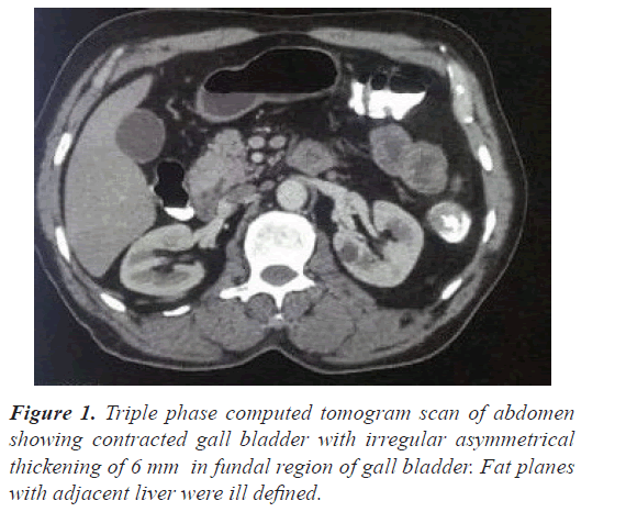
Patient was planned for triple phase CECT scan abdomen which showed in Figure 1 contracted gall bladder with irregular asymmetrical thickening of 6 mm along in fundal region of gall bladder. Fat planes with adjacent liver were ill defined. Subcentimetric lymphadenopathy, in interaortocaval region. Pancreas and bile duct and rest of the organ were normal. First possibility of gall bladder cancer was kept under the background of porcelain gall bladder and irregular thickness of 6 mm. Due to suspicion of gall bladder cancer, patient underwent ultrasound guided Fine Needle Aspiration Cytology (FNAC) from gall bladder which was inconclusive. In view of abnormal thickening, patient planned for surgery [10].
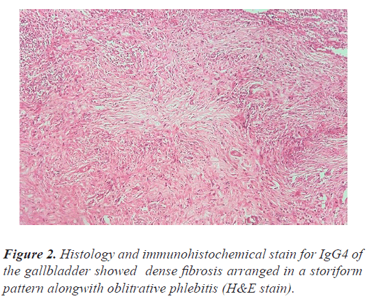
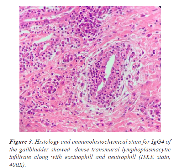
Patient underwent extended cholecystectomy after 3 weeks of acute attack of pain and good glycemic control was obtained. On opening the abdomen, there was no ascites and metastasis. Gall bladder was thickened at fundal region along with multiple stones in lumen. Centimetric lymph nodes were found along hepato duodenal ligament and inter-aortocaval region. Patient had uneventful postoperative recovery and discharged on postoperative day. Final histopathology report showed dense fibrosis (Figure 2) arranged in a storiform pattern alongwith oblitrative phlebitis and dense transmural lymphoplasmacytic infiltrate along with eosinophill and neutrophil (Figure 3). Lymph nodes were harvested and all showed sinus histocytic changes only. All these features were suggestive of IgG4 sclerosing cholecystitis. For confirmation we performed Immunohistochemistry (IHC) for IgG4 and CD138 which showed IgG4 positive 80 plasma cell and CD138 was positive in plasma cells. IgG was inconclusive. Serum IgG4 was 0.972 (reference range 0.03-2.01)
Results and Discussion
IgG4-cholecystitis is a total masquerader having a similar constellation of signs and symptoms as of gall bladder cancer. Etiopathogenesis of this entity is still not clear, but mostly it is stated that this is manifestation of autoimmune process driven by lymphoplasmacytic interaction in the organ involved. Depending upon the organ or system involved, it really poses difficulty in making final diagnosis based on clinical and radiological features. To circumvent this dilemma, we need a complete and indepth understanding of the clinical presentations of IgG4 related diseases. This will eventually lead to decrease unnecessary invasive forms of surgical interventions and ultimately benefits the patient and helps in reducing the futile exercise. It is prudent to put this rare entity in the list of differential diagnosis along with gall bladder cancer whenever any abnormal or suspicious features are there on clinically/ radiologically [11].
As per Zhang, the incidence of misdiagnosis of IgG4-C is 9.63% 8. This entity is more common in older people having more predilections towards male [9]. Patients with IgG4 choleycstitis on sonography shows hypoechoic, diffuse, circumferential thickening of the gallbladder Wall. As in our case, sonography showed gall stones along with, asymmetrical thickening of 6 mm in gall bladder. On CECT scan; it is mainly the delayed enhancement in IgG4 related cholecystitis which helps in differentiating it from gall bladder cancer. In our case, there was asymmetrical thickening of 6 mm along with porcelain bladder.
On MRI, it showed low-signal smooth diffuse gallbladder wall thickening on T2 weighted MR images, and delayed enhancement post-contrast. This thickening or enlargement of organ on imaging is generally due to infiltration of organs by lymphocytes and plasma cells along with coexisting fibrosis. As per Deshpande [11,12],the diagnostic criteria for diagnosing IgG4 related diseases are as mentioned in (Table 1).
| S.No. | Major histopathological features associated with IgG4-RD | Other histopathological features associated with IgG4-RD are: | Minimal criteria for IgG4-RD in a new organ/site |
|---|---|---|---|
| 1. | Dense lymphoplasmacytic infiltrate | Phlebitis without obliteration of the lumen | Characteristic histopathological findings with an elevated IgG4 plasma cells and IgG4-to-IgG ratio |
| 2. | Fibrosis, arranged at least focally in a storiform pattern | Increased numbers of eosinophils | High serum IgG4 concentrations |
| 3. | Obliterative phlebitis | Effective response to glucocorticoid therapy | |
| 4. | Reports of other organ involvement that is consistent with IgG4-RD |
Table 1: Three major histopathological features associated with igg4-related disease and the minimal criteria in a new organ/site in the international pathological consensus
In order to diagnose IgG4 cholecystitis, there should be at least 2 out of 3 major criteria as mentioned above and also presence of more than 50 percent plasma cell per high power field. In our case, all the three major criteria were present. Among minimal criteria as mentioned above, IgG4 positive in 80 plasma cells, but Serum IgG was within normal limit but Shin reported only 15% plasma cell per high power field in their case study in the background of IgG4 cholecystitis.
Role of serum IgG4 is also advocated by many studies, but routinely it is not practiced as it is not elevated in all cases of IgG4 related disease and also its accuracy only fits between 40%-50% and moreover in developing countries like India, it is difficult to get done due to financial and cost constraints. To differentiate IgG4 or other benign conditions from ca gall bladder, role of Carbohydrate Antigen (CA 19-9), Carcino-Embryonic Antigen (CEA) and Carbohydrate Antigen (CA-125) are more established [12]. Increased ratio >40% of IgG4/IgG also favours IgG4 related disease and is considered as a salient feature to diagnose IgG4 related disease.
Treatment decision is of paramount importance in suspecting IgG4 related cholecystitis. The main diagnostic ambiguity is with malignancy of gall bladder, as the treatment options for both these diseases are completely different. IgG4 cholecystits is mainly treated by steroids and showed a dramatic response, duration and relapse rate are yet not documented as no randomised controlled trial has not been yet performed. Role of immune modulators also has been tried in Mayo clinic [13]. Even if patient requires surgery after steroid treatment, the extent of surgery is definitely less as compared to surgery for gall bladder cancer.
Conclusion
Radical surgery in the form of extended cholecystectomy and regional lymphadenectomy is mainly done for gall bladder cancer. Therefore, a firm and confident diagnosis is required before embarking upon any modality of treatment. Any misdiagnosis will over treat IgG4 cholecystitis at the cost of other post-operative surgical complications which can be completely avoided if we have ruled out IgG4 cholecystits in case suspected to have gall bladder cancer.
Declarations
Ethics approval and consent to participate
Not applicable' for this section
Consent for publication
Consent for publication has been obtained from the patient in a proper informed consent form.
Availability of data and materials
Not applicable
Competing interest
The authors declare that they have no competing interests
Funding
None
Authors' Contribution
RS drafted the case manuscript and was the main operating surgeon.
PM-part of surgical team and helped in drafting the manuscript
RP,VR, JS,CK-all were part of surgical team and helped in citing references for the case report.
JG-helped in framing discussion part
KM-reported the final histopathology report
All authors read and approved the final manuscript
Acknowledgement
Not applicable
Authors' Information
Not applicable
References
- Kamisawa T, Funata N, Hayashi Y, et al. A new clinicopathological entity of IgG4-related autoimmune disease. J Gastroenterol. 2003;38: 982-984.
- Finkelberg DL, Sahani D, Deshpande V, et al. Autoimmune pancreatitis. N Engl J. 2006; 355: 2670–2676.
- Lopes J, Hochwald SN, Lancia N, et al. Autoimmune esophagitis: IgG4-related tumors of the esophagus. J Gastrointest Surg. 2010;14: 1031–1034.
- Abraham SC, Cruz-Correa M, Argani P, et al. Lymphoplasmacytic chronic cholecystitis and biliary tract disease in patients with lymphoplasmacytic sclerosing pancreatitis. Am J Surg Pathol. 2003; 27(4): 441–451.
- Umehara H, Okazaki K, Masaki Y, et al. A novel clinical entity, IgG4-related disease (IgG4RD): general concept and details. Mod Rheumatol. 2012;22: 1-14.
- Erdogan D, Kloek JJ, ten Kate FJ, et al. Immunoglobulin G4-related sclerosing cholangitis in patients resected for presumed malignant bile duct strictures. Br J Surg. 2008;95: 727-734.
- Zhang, R, Lin H.M, Cai Z.X, et al. Clinical strategies for differentiating IgG4-related cholecystitis from gallbladder carcinoma to avoid unnecessary surgical resection. Sci China Life Sci. 2019.
- Kamisawa T, Okamoto A. Autoimmune pancreatitis: proposal of IgG4-related sclerosing disease. J Gastroenterol. 2006;41(7): 613–625.
- Kurowecki D, Patlas MN, Haider EA, et al. Cross-sectional pictorial review of IgG4-related disease. Br J Radiol. 2019;92: 20190448.
- Vlachou PA, Khalili K, Jang HJ, et al. IgG4-related sclerosing disease: autoimmune pancreatitis and extrapancreatic manifestations. Radiographics. 2011; 31(5):1379–402.
- Deshpande V, Zen Y, Chan JK, et al. Consensus statement on the pathology of IgG4-related disease. Mod Pathol. 2012; 25: 1181-1192.
- Shin SW, Kim Y, Jeong WK, et al. Isolated cholecystitis mimicking gallbladder cancer: A case report. Clin Imaging. 2013;37: 969–971.
- Wang Y.F, Feng F.L, Zhao X.H, et al. Combined detection tumor markers for diagnosis and prognosis of gallbladder cancer. World J Gastroenterol. 2014; 20: 4085


