- Biomedical Research (2010) Volume 21, Issue 4
Helicobacter pylori in gall bladder disease
Jafri Deeba1*, Singhal Sanjay1, Malik Abida1, Khan Athar21Department of Microbiology and Department of Surgery, J. N. Medical College, Aligarh Muslim University, Aligarh (U.P) India
2Department of Surgery, J. N. Medical College, Aligarh Muslim University, Aligarh (U.P) India
Accepted August 24 2013
Abstract
In recent years, H. pylori has been detected in bile, liver and biliary epithelium obtained from patients with hepatobiliary diseases and is thought to be associated with these diseases. We, therefore, planned the present case control study to find the role of H. pylori in gall bladder disease through culture and serological studies. Serum and gall bladder tissue were obtained from 75 patients and only serum from 40 controls. Tissue was processed for smear and culture and sera were tested for presence of antibodies quantitatively by ELISA. H. py-lori was not detected by smear or culture from any sample, but statistically significant dif-ference was observed between patients and controls in positivity (80 % vs. 47.5%, p value < .05) as well as titres for antibodies against H. pylori (p value < .001). Statistically significant difference was observed between antibody levels in patients and controls. Further studies are required, to elucidate the role of Helicobacter in hepatobiliary diseases, especially with regard to the effective culture of the organisms from the biliary tree.
Keywords
Helicobacter pylori, Gall bladder, ELISA
Introduction
Helicobacter pylori is a newly discovered gram negative bacteria which causes chronic active gastric inflamma-tion. Infection seems to be life-long, unless eradicated by antibiotics, and is strongly associated with duodenal ul-ceration, gastric ulceration and gastric cancer. Recently, the bacterium has been implicated as a risk factor for various extraintestinal diseases including hepatobiliary diseases ranging from chronic cholecystitis and primary biliary sclerosing cholangitis to gall bladder cancer and primary hepatic carcinomas. In recent years, H. pylori has been detected in bile, liver and biliary epithelium obtained from patients with hepatobiliary diseases [1-4]. Many researchers have demonstrated the presence of Helico-bacter DNA in gall bladder of patients with gall stone disease [5-8], though none were able to isolate H. pylori. No such studies appear to have been done in India. We, therefore, planned the present case control study to find the role of H. pylori in gall bladder disease through cul-ture and serological studies.
Material and Methods
1. Patients and controls: seventy five patients undergo-ing cholecystectomy because of cholelithiasis or cholecystitis and 40 controls which were healthy vol-unteers without any complaints or history of dyspep- sia or gastritis were included in the study. A detailed relevant clinical history was recorded, with special emphasis on the presence of symptoms associated with H. pylori infection on a proforma from both the groups.
2. Specimens :
a) 3-4 ml of venous blood was collected from each patient a day before operation, and from controls. The serum was separated and sera were stored at -20ºC in sterile vials till further testing.
b) A small piece of gall bladder sample was collected from the operation theatre with sterile precautions in a vial containing five ml normal saline and rest of the gall bladder tissue put in 10% formalin and sent for histopathological examination by the surgery team, and not included in the study.
3. Specimen processing :
a) Gall bladder sample was crushed in a sterile mor-tar and pestle in one ml saline and the tissue homoge-nate used for rapid urease test, gram smear and cul-ture as described earlier [9,10].
b) Quantitative ELISA for IgG antibodies was per-formed in duplicate by using the kit manufactured by NovaTec Immunodiagnostica GmbH, Germany. The instructions given in the Users’ Manual with the kit were strictly followed. Titre of 20 NTU/ ml was given as cut-off, thus any titre > 20 was considered positive.
4. Quality control :
a) Each batch of urease test and culture media was tested with previously isolated H. pylori and were also simultaneously used for gastric biopsies.
b) Controls supplied with the kit were used for ELISA and the runs which were not giving defined values were rejected and repeated. Mismatching pairs of sera were also repeated again in duplicate.
5. Statistical Analysis: Statistical analysis was done us-ing Z – test for proportions to find the significance of differences.
Observations
Maximum number of patients (34.7%) was in the age group 21-30 years, followed by 29.3% patients in the age group 31-40 years. Females and males constituted 88.0% and 12.0% of the study group, respectively. The most fre-quent complaint was pain in right hypochondrium in 72 (96%), followed by vomiting in 59 (78.7%), dyspepsia in 41 (54.7%) and epigastric discomfort in 35 (46.7%) pa-tients.
All 75 (100%) patients in the study group had taken broad-spectrum antibiotics, 30 (40%) took metronidazole, 50 (66.7%) took NSAIDS and 25 (33.3%) antacids.
Rapid urease test, though meticulously prepared, was found to immediately turn alkaline upon addition of tissue homogenate due to bile, and therefore discarded and not done after twenty specimens.
No specimen was found to be positive in direct micros-copy or culture. The same batches of media were giving good growth of previously isolated H. pylori and from fresh gastric biopsies.
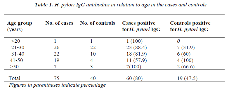
Serology for IgG antibody was done on all 75 samples, and this was found to be positive in 60 (80%) samples, while 19 controls were positive (Table 1).
Statistically significant difference was observed between positive cases and controls in all the age groups (p-value < .05) except in age < 20, where sample size was very low. A reverse positivity (statistically significant) was seen in age group 41-50 yrs.
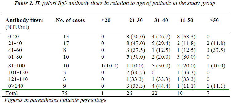
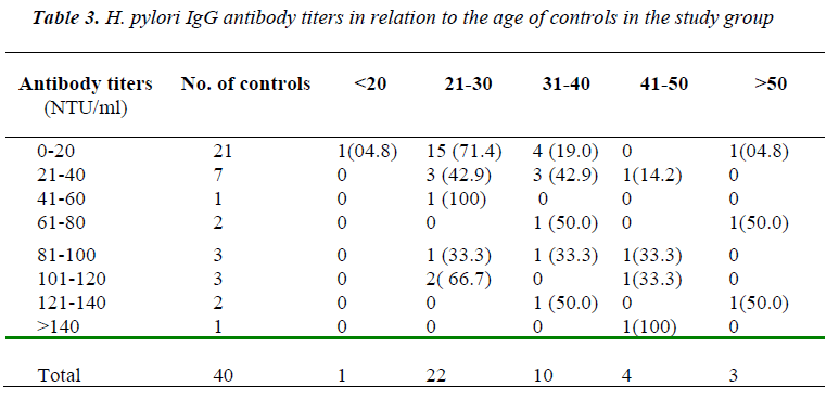
Table 2 and 3 show H. pylori IgG antibody titers in rela-tion to age of patients and controls in the study group re-spectively. The patients were having higher titers than the respective control groups, specially in lower age groups. These differences in titers were statistically significant (p value < .001) for most of the ranges.
Discussion
Detection of Helicobacter species in human bile has prompted a growing interest as to whether these organ-isms truly colonize the biliary tract of humans and cause hepatobiliary diseases. Bile-resistant Helicobacter species such as H. hepaticus, H. bilis and H. pullorum, have been discovered in man as well as animals. However, research in this area has been limited by the lack of gold standard in the diagnosis of these organisms in bile. A meta-analysis of some published work has shown strong asso-ciation between Helicobacter and gall bladder disease [11]. We, therefore, attempted to find a relation between Helicobacter pylori and gall bladder disease.
Most of the patients in our study were females with chole-lithiasis (88%). It is said that a fat, fertile, female of forty is more at risk of developing cholelithiasis. However, we found that a majority of females (34.7%) were in the age group of 21-30 years showing a decreasing trend in the age for development of cholelithiasis. This could proba-bly be attributed to the changes in lifestyle.
Culture remains the definitive investigation to prove the viability of H. pylori in bile and gall bladder tissue. De-spite prolonged incubation, we were not able to isolate H. pylori from any of the patients. This could be due to the fact that all the patients have taken broad spectrum antibi-otics and, moreover, 40% of them took metronidazole as well, which constitute a part of eradication therapy for H. pylori. It is possible that the number of bacteria remains very low and they may have been partially inhibited by adverse conditions in the biliary milieu. Other possibility is that there is patchy colonization of the biliary epithe-lium, thus culturing a small piece may not give positive results. Most other workers were also not successful in getting positive cultures, but could demonstrate DNA by PCR [1,5-8]. The lack of recovery has been attributed to the prolonged freezer storage of tissue and bile without glycerol or other preservatives or that the DNA detected was from non-viable organisms. However, in a recent study, H. pylori was isolated from 6 gall bladder samples by culture [12].
Direct microscopy was also found to be negative. A cor-ollary to the same finding is apparent in the work of oth-ers [1,13,14]. However, some workers [1,12] detected curved bacteria suggestive, but not diagnostic of Helico-bacter species, by different histopathological methods (Warthin Starry, Haematoxylin Eosin and Gram staining).
In the present study, statistically significant difference was found between the prevalence of anti H. pylori IgG in the study group and the controls. In contrast, some other workers [13,15] found no significant difference in the prevalence of H. pylori IgG antibodies in patients with cholelithiasis and controls. However, it has to be empha-sized that in most of the studies there was either no con-trol group or very few controls.
Various workers have studied antibodies in healthy indi-viduals [16-18] as well as in patients of dyspepsia [10] and have reported positivity ranging between 49 and 79%. Most of these workers [16,17] have observed increase in positivity for H. pylori IgG antibody with increasing age, and the same was observed in the present study among controls. However, we observed that among the patients, positivity was slightly more in the age group of 21-50 years which is also the most common age groups for cholelithiasis.
Some of our patients had symptoms like dyspepsia (54.7%) and epigastric discomfort (46.7%) and it may be suspected that they were suffering with acid-peptic dis-ease as well as with cholecystitis, but as both these symp-toms can be present in cholecystitis, they were not ex-plored for the presence of acid-peptic disease
In the present study, it was observed that 57.3% of the patients had titers of >40 NTU/ml while only 30% of in-dividuals in the control group had titers above 40 NTU/ml. This difference was found to be statistically sig-nificant (p<0.001). This is an important information sug-gesting that majority of patients of cholelithiasis in the study group were positive with higher titers.
These finding highlight the need to improve the condi-tions for the growth of Helicobacter species from the bil-iary tree and to develop experimental models for studying the role of Helicobacter in the genesis of biliary diseases. Further studies are required to elucidate the role of Helicobacter in hepatobiliary diseases, especially with regard to the effective culture of the organisms from the biliary tree, the development of accurate tests to identify these bacteria, large-scale autopsy studies and experimen-tal infection of the biliary tract in animal models.
References
- Fox JG, Dewhirst FE, Shen Z, Feng Y, Taylor NS, Pas- ter BJ, et al. Hepatic Helicobacter species identified in bile and gall bladder tissue from Chileans with chronic cholecystitis. Gastroenterology 1998; 114: 755-763.
- Nilsson HO, Mulchandani R, Tranberg KG, Taneera J, Caastedal M, Glatz E, et al. Helicobacter species iden- tified in liver from patients with cholangiocarcinoma and hepatocellular carcinoma. Gastroenterology 2001; 120: 323-324.
- Nilsson HO, Taneera J, Caastedal M, Glatz E, Olsson R, Wadstrom T. Identification of Helicobacter pylori and other Helicobacter species by PCR, hybridization and partial DNA sequencing in human liver samples from patients with primary sclerosing cholangitis or primary biliary cirrhosis. J Clin Microbiol 2000; 38: 1072-1076.
- Queiroz DMM, Santos A, Oliveira AG, Rocha GA, Moura SB, Camargo ERS, et al. Isolation of a Helico- bacter strain from the human liver. Gastroenterology 2001; 121: 1023-1024.
- Silva CP, Pereira-Lima JC, Oliveira AG, Guerra JB, Marques DL, Sarmanho L, et al. Association of the presence of Helicobacter in gall bladder tissue with cholelithiasis and cholecystitis. J Clin Microbiol. 2003; 41: 5615-5618.
- Mendez-Sanchez N, Pichardo R, Gonzalez J, Sanchez H, Moreno M, Barquera F, et al. Lack of association between Helicobacter species colonization and gallstone disease. J Clin Gastroenterol 2001; 32: 138-41.
- Kawaguchi M, Saito T, Ohno H, Midorokawa S, Sanji T, Handa Y, et al. Bacteria closely resembling Helico- bacter pylori detected immunohistologically and ge-netically in resected gall bladder mucosa. J Gastroen- terol 1996; 31: 294-298.
- Apostolov E, Al Soud WA, Nilsson I, Kornilovska I, Usenko V, Lyzogubov V, et al. Helicobacter pylori and other Helicobacter species in gall bladder and liver of patients with chronic cholecystitis detected by immu- nological and molecular methods. Scand J Gastroen- terol 2005; 40(1): 96-102.
- Prasad KN, Singhal S, Ayyagari A, Sharma S, Dhole TN. Association of Helicobacter pylori in patients with non-ulcer dyspepsia. Ind J Microbiol 1991; 31(4): 415-418.
- Malik A, Singhal S and Mukharji U. Evaluation of ELISA based antibody test against H. pyloriin cases of non-ulcer dyspepsia. J Med Microbiol 1999; 17 (3):137-139.
- Manoj P. Helicobacter species are associated with possible increase in risk of biliary lithiasis and benign biliary diseases World J Surgical Oncology 2007, 5:94
- Abayli B, Colakoglu S, Serin M, Erdogan S, Isikal YF, Tuncer I, et al. H. pyloriin the etiology of cholesterol gall stones. J Clin Gastroenterol. 2005; 39(2): 134-137.
- Myung SJ, Kim MH, Shim KN, Kim YS, Kim EO, Kim HJ, et al. Detection of Helicobacter pylori DNA in human biliary tree and its association with hepatolithi- asis. Dig Dis Sci 2000; 45: 1405-1412.
- Lin TT, Yeh CT, Wu CS and Liaw YF. Detection and partial sequence analysis of Helicobacter pylori DNA in the bile samples. Dig Dis Sci 1995; 40: 2214-2219.
- Figura N, Cetta F, Angelico M, Montalto G, Cetta D,Pacenti L, et al. Most Helicobacter pylori infected pa- tients have specific antibodies and some also have H. pyloriantigens and genomic material in bile: is it a risk factor for gall stone formation? Dig Dis Sci 1998; 43: 854-862.
- Alaganantham TP, Pai M, Vaidehi T, Thomas J. Se- roepidemiology of Helicobacter pylori infection in an urban, upper class population in Chennai. Ind J Gastro-enterol 1999; 18 (2) : 66-68.
- Graham DY, Adam E, Reddy GT, Agarwal JP, Agar- wal R, Evans DJ, Jr., et al. (1991). Seroepidemiology of Helicobacter pylori infection in India: Comparison of developing and developed centuries. Dig Dis Sci 1991; 36 (8):1084-1088.
- Sitas F, Forman D, Yarnell JWG, Burr ML, Elwood PC and Pedley S. Helicobacter pylori infection rates in relation to age and social class in a population of Welsh men. Gut 1991; 32: 25-28.


