Research Article - Biomedical Research (2017) Volume 28, Issue 15
Fiberoptic bronchoscopy in turban pin aspiration
Suat Konuk*
Chest Disease Specialist, Private Practice, Düzce, Turkey
Accepted date: July 13, 2017
Abstract
Purpose: To explore respiratory allergen profile and the relation between the month of birth and allergen type in Duzce Province in Turkey.
Method: The study was performed retrospectively. Between October 2011 and January 2017, 302 cases underwent in fiberoptic bronchoscopy were examined retrospectively. Among these, the characteristics of 7 female patients who applied to our clinic with turban pin aspiration were examined.
Results: The ages of our patients ranged from 19 to 34 y. Except one patient, the other patients applied within the first 12 h on the day of aspiration. The aspirated pin was localized in the bronchial system in the right lung in 6 patients and in the bronchial system in the left lung in 1 patient. In all of these patients the pin could be removed with a fiberoptic bronchoscope.
Conclusion: In patients who applied with turban pin aspiration, the fiberoptic bronchoscopy with a local anesthesia is a very successful method.
Keywords
Fiberoptic bronchoscopy, Rigid bronchoscopy, Pin
Introduction
The foreign body aspirations are frequently observed in the childhood age group and in children under 2 y old [1]. In adults, this is particularly the result of the neurological disorders that disrupt the defense mechanisms, the loss of consciousness due to trauma, or the use of sedative drugs or alcohol [2]. In elderly persons, in the cases of senility and debility, this is caused by the deep inspiration of the object which is swallowed. In Turkey, it is noteworthy that the number of the incidences due to the aspiration of the pins used during the attachment of the headscarves called “turban”, are growing steadily [3,4].
The treatment of the foreign body aspiration is possible with the bronchoscopic removal of the foreign body. For the bronchoscopy procedure, while previously the Rigid Bronchoscope (RB) was recommended, in recent years, it is reported that the Flexible Fiberoptic Bronchoscope (FOB) can be used [5-9].
In this study, the characteristics of 7 patients were discussed who applied to our clinic with turban pin aspiration used for the veiling and the success rate of the treatment with FOB are presented.
Methods
This retrospective case series study was performed at the Department of Chest Diseases in Bolu Abant Izzet Baysal University. The hospital archieve for patient files was investigated for the patients who applied with foreign body aspiration between October 2011 and January 2017. The patients with turban pin aspiration were selected among patients including all kinds of foreign body aspiration. The demographic (age, gender) and the clinical characteristics (the duration between the aspiration and the application to the clinic, the type of treatment, the localization of the aspirated pin and the treatment method for the removal of the foreign body) were noted.
Results
Seven patients with turban pin aspiration were detected during the hospital archive review. All the patients were female. The ages of the patients ranged between 19-34 y. Mean age was 19.5 ± 8.3 y. All the patients declared that they aspirated the pins as a result of sudden speech or breathing while the pin was between the lips.
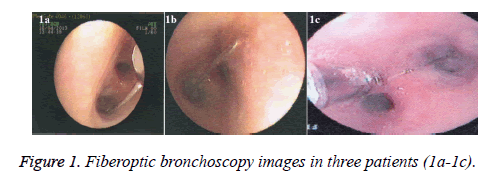
Except one patient, all are applied 2 to 12 h after the aspiration. The scopi method was used to localize the pins just before the procedure. Aspirated pins were localized in the bronchial system in the right lung in 6 patients (85.7%) and in the bronchial system in the left lung in 1 patient (14.3%). In all of the patients the pins were successfully removed by FOB. In one patient, the pin was in the in the left main bronchus on the lung graphy however, the pin was removed from the right lower lob bronchus with FOB. Some images detected by FOB in three patients are shown in Figure 1.
Discussion
Foreign body aspiration in adults is relatively rare compared to childhood age group. The nature of foreign body varies according to age, regions, eating habits and even clothing [10-13]. In one study on sixty patients with tracheobronchial foreign body, it was seen that the most frequently aspired materials were food particles; it was followed secondly by dental and medical devices [2]. In that study, the neurological disorders were the leading cause of the aspiration in adult patients. It is noticed that the tracheobronchial foreign body aspiration happens more easily (i) during dental procedures performed in the supine position, (ii) during medical procedures such as the cleaning, replacement or manipulation of the tracheostomy or the endotracheal tube, (iii) after the traumatic events that result in cervicofacial damages or unconsciousness, and (iv) in individuals using alcohol or sedative drugs [2]. However, foreign body aspiration can also occur in the absence of any predisposing factor [14]. In our study, there was no predisposing factor in all of the cases with turban pin aspiration. The aspiration has occurred as a result of sudden speech or breathing while the pin was between the lips in our patients.
Concerning the aspiration of the turban pins, it was noticed that between the years of 1988 and 1994, there were 47 women patients who applied to 5 different centers in Turkey with foreign body aspiration [3]. The proportion of turban pins among the foreign bodies removed in those centers has been noted as 24-86%. In that study, it was also indicated that in all patients, the pin aspiration has occurred as a result of a smile or speech while the pin was hold between the teeth or lips of the patients.
The placement of foreign bodies in the tracheobronchial tree is related to the posture of the patient at the moment of the aspiration as well as the anatomical feature. The most common aspiration site is in the right lower lobe and it is followed secondly by the left lower lobe [2]. In this study, the aspirated pins were localized in the bronchial system in the right lung in 6 patients (85.7%) and in the bronchial system in the left lung in 1 patient (14.3%). Ucan et al. reported that the placement rate of the pin in the right or left lower lobe was 51% [3].
The diagnosis can be difficult when the foreign body is aspirated by a person who has a predisposing factor. Sometimes such patients remain unidentified for years; the most common symptom for this type of patients is cough. The diagnosis can be easily made on the basis of the history in patients who were conscious at the moment of the aspiration as seen in our patients. The inhalation of pins is never asphyxiating because of the slender shape of the pins [10]. Initially, the symptoms are coughing, suffocation and dyspnea. These symptoms fade after a few minutes. The rate of asymptomatic patients was reported between 10% and 20% [10,11,15]. The postero-anterior and lateral lung graphs are adequate and enough in localizing the aspirated material, especially for radioactive materials such as pins. Thus, in all of our cases, the possible location of the pin was determined by radiological methods before the bronchoscopy procedure. In two of our cases, one of the pins which were seen on the lung graphy in the left main bronchus was removed from the right lower lob bronchus in the bronchoscopy. In their study including sixty-six patients, Weissberg et al. reported a 8 y old child with a pin in the right bronchus in the first graphy. However, the pin was in the left bronchus in the graphy retaken after a few hours of waiting [16]. The authors stated that this phenomenon is an example for "foreign body traveling" and emphasized the importance of taking the lung graphs just before bronchoscopy. In our cases, for the fact that the localization may not be compatible with the postero-anterior lung graphy, we used the scopi method to re-evaluate the case during the procedure.
RB was used largely before the 1970s, in the following years the FOB has gained popularity [5,6,9]. In 1978, Cunanan has published his experiences with the use of FOB for the foreign body removal in 300 cases with acute foreign body aspiration where the most of the patients were mentally or physically disabled. He emphasized that the mortality and morbidity rates reduced from 12% to 1% as a result of using a flexible FOB instead of RB [5]. In the study carried out by Lan et al. the foreign body was successfully removed with FOB in all of 33 adult patients but one [6]. The success rates of FOB have been reported as 56%, 62.5% and 80.6% in three studies [11,17,18]. Fenane et al. reported that the average number of attempts of FOB removal was 2.5 times per patient [10]. Kaptanoglu et al. reported that they could extract all but six of a total of 63 patients in the first attempt [19].
In the study published in 1997 by Chen et al. while the first FOB was successful on 25 adult patients (58%) over 43 patients; the aspired material could be removed by FOB intervention applied one or more times in 34 patients (74%). The authors suggested that FOB should be the first step in the treatment of foreign body aspiration [9]. It was suggested that FOB should be performed urgently before complications arise [20,21]. The reasons for unsuccessful endoscopy were reported as distal localization and pins embedded in bronchus wall with inflammatory reaction [10].
In the series of 60 patients studied by Limper et al. despite 60% success of FOB, it has been obtained a 98% of success with RB and the authors have expressed that they do not agree with the opinions arguing that the FOB should prevail compared to RB. But, the authors have indicated that the FOB will provide an advantage, for the removal of foreign bodies located distal to the rigid bronchoscope; in patients where the use of a rigid bronchoscopy is not possible due to the lack of cervical stabilization and in cases where the mechanical ventilation is applied [2].
Due to the fact that the pin can travel down to the segmental bronchi, the FOB may be more successful in pin removal than removal of other foreign bodies. In a study from Lebanon, the FOB performed with a general anesthesia has produced successful results in 5 patients [22]. In a case presented by Smith et al. the pin was removed with FOB after two unsuccessful RB attempts [23]. With a study in Turkey, Gürsu et al. [24] emphasized that the 77.4% of the cases aspirated an inorganic body and the 64.5% of them were beaded pins. In another study in Turkey, Kolbakır et al. [25] studied on 152 cases where children were also involved. The authors determined that the bean speckles were mostly aspired and this was followed on the second rank (19%) by pins.
In pin aspiration, the intervention made without loss of time with FOB shows successful results. In the literature, there are a low number of studies about the use of FOB in turban pin aspiration. This study differs from other studies and becomes more of an issue. It is revealed by our study that FOB is a successful method in foreign body aspiration cases, even in difficult cases.
References
- Black RE, Choi KJ, Syme WC, Johnson DG, Matlak ME. Bronchoscopic removal of aspirated foreign bodies in children. Am J surg 1984; 148: 778-781.
- Ucan ES, Tahaoglu K, Mogolkoc N, Dereli S, Basozdemir N, Basok O, Turktas H, Akkoclu A, Ates M. Turban pin aspiration syndrome: a new form of foreign body aspiration. Resp Med 1996; 90: 427-428.
- Dayioglu E, Rahimi M, Toker A, Akaslan I, Barlas S, Tireli E, Borteçen KH, Bostanci K, Barlas C. Brons Içi Yabanci Cisimler: Türban Ignesi Komplikasyonlari. GKD Cer Derg 1995; 3: 82-85.
- Gokirmak M, Hasanoglu HC, Koksal N, Yildirim Z, Hacievliyagil SS, Soysal O. Retrieving Aspirated pins by flexible bronchoscopy. J Bronchol 2002; 9: 10-14.
- Lan RS, Lee CH, Chiang YC, Wang WJ. Use of fiberoptic bronchoscopy to retrieve bronchial foreign bodies in adults. Am Rev Respir Dis 1989; 140: 1734-1737.
- Tong M, Kang X, Sakakibara H, Suetsugu S. Successful removal of a 12 year long intrabronchial fishbone through fibreoptic bronchoscopy. Respirol 1997; 2: 291-293.
- Chen CH, Lai CL, Tsai TT, Lee YC, Perng RP. Foreign body aspiration into the lower airway in Chinese adults. Chest 1997; 112: 129-133.
- Baharloo F, Veyckemans F, Francis C, Biettlot MP, Rodenstein DO. Tracheobronchial foreign bodies: presentation and management in children and adults. Chest 1999; 115: 1357-1362.
- Weissberg D. Foreign bodies in the tracheobronchial tree. Chest 1992; 102: 656.
- Fenane H, Bouchikh M, Bouti K, EL Maidi M, Ouchen F, Mbola TO, Damessane L, Achir A, Benosman A. Scarf pin inhalation: clinical characteristics and surgical treatment. J Cardiothorac Surg 2015; 10: 61.
- Hebbazi A, Afif H, El Khattabi W, Aichane A, Bouayad Z. Scarf pin: a new intrabronchial foreign body. Rev Mal Respir 2010; 27: 724-728.
- Koraichi A, Mokhtari M, Haddoury M, Kettani SE. Rigid bronchoscopy for pin extraction in children at the Childrens Hospital in Rabat, Morocco. Rev Pneumol Clin 2011; 67.
- Arsalane A, Zidane A, Atoini F, Traibi A, Kabiri EH. The surgical extraction of foreign bodies after the inhalation of a scarf pin: two cases. Rev Pneumol Clin 2009; 65: 293-296.
- Shabb B, Taha AM, Hamada F, Kanj N. Straight pin aspiration in young women. J Trauma 1996; 40: 827-828.
- Kaptanoglu M, Nadir A, Dogan K, Sahin E. The heterodox nature of Turban Pins in foreign body aspiration; the central anatolian experience. Int J Pediatr Otorhinolaryngol 2007; 71: 553-558.
- Smith LJ, Khan MA. Role of fiberoptic bronchoscopy in removal of a foreign body. Chest 1977; 72: 264-265.
- Zaghba N, Benjelloun H, Bakhatar A, Yassine N, Bahlaoui A. Scarf pin: an intrabronchial foreign body who is not unusual. Rev Pneumol Clin 2013; 69: 65-69.
- Al-Ali MAK, Khassawneh B, Alzoubi F. Utility of fiberoptic bronchoscopy for retrieval of aspirated headscarf pins. Resp Int Rev Thorac Dis 2007; 74.
- Kaptanoglu M, Dogan K, Onen A, Kunt N. Turban pin aspiration; a potential risk for young Islamic girls. Int J Pediatr Otorhinolaryngol 1999; 48.
- Uskul TB, Turker H, Arslan S, Selvi A, Kant A. Use of fiberoptic bronchcoscopy in endobronchial foreign body removal in adults. Turk Respir J 2007; 8.
- Al-Sarraf N, Jamal-Eddine H, Khaja F, Ayed AK. Headscarf pin tracheobronchial aspiration: a distinct clinical entity. Interact Cardiovasc Thorac Surg 2009; 9: 187-190.
- Rohde FC, Celis ME, Fernandez S. The removal of an endobronchial foreign body with the fiberoptic bronchoscope and image intensifier. Chest 1977; 72: 265.
- Debeljak A, Sorli J, Music E, Kecelj P. Bronchoscopic removal of foreign bodies in adults: experience with 62 patients from 1974-1998. Eur Resp J 1999; 14: 792-795.
- Gursu S, Sirmali M, Gezer S, Findik G, Türüt H, Aydin E, Kaya S, Tastepe I. Yetiskinlerde trakeobronsiyal yabanci cisim aspirasyonlari. Türk Gögüs Kalp Damar Cerrahisi Dergisi 2006; 14: 38-41.
- Kolbakir F, Keceligil HT, Ankan A, Erk MK. Yabanci Cisim Aspirasyonlari Bronkoskopi Yapilan 152 Olgunun Analizi. Türk Gögüs Kalp Damar Cerrahisi Dergisi 1995; 3: 117-120.
