Research Article - Biomedical Research (2017) Volume 28, Issue 19
Expression of Reg IV in inflammatory bowel disease and the predictive effect on lesion activity
Junwei Zhang*, Jing Bai, Limin Zhang and Aibin Cheng
North China University of Science Technology Affiliated Hospital, PR China
- *Corresponding Author:
- Junwei Zhang
North China University of Science Technology Affiliated Hospital, PR China
Accepted date: August 30, 2017
Abstract
Purpose: This article aims to observe the expression of the Regenerating gene (Reg) IV in intestinal tissues of patients of Inflammatory Bowel Disease (IBD) and explores the correlation between Reg IV expression level and the activity of IBD.
Method: 65 cases of IBD patients confirmed during January 2013-October 2013 as well as 30 cases of healthy people tested by colonoscopy inspection during corresponding period are taken for comparisons, including 30 cases of Ulcerative Colitis (UC) and 35 cases of Crohn’s Disease (CD). The immunohistochemistry method is adopted to detect the protein expression level of Reg IV. The RT-PCR method is adopted to inspect the mRNA expression level of Reg IV. In addition, the correlation with IBD is further analysed.
\Results: According to the quantitative results of immunohistochemistry staining, Reg IV has weak protein expression in health people, and Reg IV protein level of UC during active stage is significantly higher than that of remission stage (P<0.01); there is no significant difference on Reg IV protein expression level of CD during active stage and remission stage (P>0.05). The RT-PCR result is consistent with that of the immunohistochemistry.
Conclusion: Reg IV has a significantly higher expression in IBD, illustrating that Reg IV may be involved in the occurrence and development of IBD.
Keywords
Regenerating gene, Inflammatory bowel disease, Ulcerative colitis, Crohn’s disease
Introduction
With common symptoms of diarrhea and stomach-ache, some patients of Inflammatory Bowel Disease (IBD) even have bloody stools. IBD includes Ulcerative Colitis (UC) and Crohn’s Disease (CD) [1]. There has been no clear definition on the etiology and pathogenesis on IBD at present, which may be involved in environmental factors, genetic factors and microorganism infection, with changes on body immunity state [2-4]. The Regenerating gene (Reg) IV belongs to the calciumdependent phytohemagglutinin superfamily, and the protein coded belongs to the secreted protein, with expressions mainly in gastrointestinal tract and pancreas. According to existing researches, there is a close correlation between Reg IV and the occurrence and development of digestive system neoplasms [5]. However, little research on roles of Reg IV in the occurrence and development of IBD has been reported in literatures. Therefore, the immunohistochemistry method and the RT-PCR method are adopted in this research to detect the protein and mRNA expressions of Reg IV protein in intestinal tissues. In addition, analysis on the correlation between the expression level of Reg IV protein and the activity of diseases is conducted, so as to find out new indexes for assessment of IBD activity.
Materials and Methods
Clinical data
65 cases of IBD patients confirmed during January 2013- October 2013 were collected, including 30 cases of Ulcerative Colitis (UC) and 35 cases of Crohn’s Disease (CD). The diagnosis of all the patients was in accordance with the diagnostic criteria for IBD of 2007 [6]. At the same time, 30 healthy people with colonoscopy inspection were collected for contrast.
Immunohistochemistry staining
The samples were baked at the temperature of 65-70°C for 45 min, followed by gradient dewaxing and dehydration as well as washing for 3-5 times with distilled water. Then citrate buffer solution was added into the samples which are then put into microwave oven at 95-98°C for 10-15 min for antigen retrieval. The samples were then taken out and cooled to indoor temperature, followed by washing. Then 0.6% H2O2 (H2O2 diluted with absolute methanol) was added. Ten minutes later, the samples were washed with distilled water for 2 times and then with PBS for 2 times.
The samples were then sealed (with 5% Bovine Serum Albumin) at indoor temperature (18-20°C) for 10 min. They were then placed for a night at 4°C after addition of primary antibodies. They were cleaned with PBS thoroughly for 3 times after taking out, and 5 min for each time. They were then added with second antibodies at 37°C for 25 min. They were cleaned with PBS for 3 times after taking out, and 5 min for each time. Then they were added with tertiary antibodies (streptomycin) at 37°C for 30 min. They were cleaned for 3 times after taking out, and 5 min for each time. Then substrate was added for coloration.
RT-PCR detection method
The tissues were grinded to powders in liquid nitrogen. Then the Trizol method was adopted to extract the total RNA, and the RT-PCR method was utilized to measure the tissue expression quantity of the chemotactic factor. Please refer to the literature for the calculating method for the expression quantity of the chemotactic factor [7].
Quantitative method of the immunohistochemistry result
Please refer to the literature for specific methods [1].
Statistical method
The SPSS 16.0 statistical software was adopted for data analysis. T-test was adopted for comparisons between the data of 2 groups, and One-Way ANOVA test was adopted for comparisons among the data of multiple groups. P<0.05 meant the statistical significance of the difference.
Results
Reg IV protein expression in the detection with the immunohistochemistry method
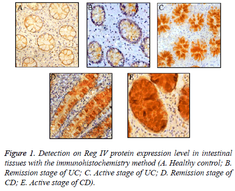
As shown in Table 1, Reg IV protein expression levels in comparison: no expression (0 cases), low expression (23 cases), moderate expression (5 cases), and high expression (2 cases). Reg IV protein expression levels in UC patients: no expression (0 cases), low expression (5 cases), moderate expression (9 cases), and high expression (16 cases). Reg IV protein expression levels in CD patients: no expression (0 cases), low expression (7 cases), moderate expression (12 cases), and high expression (16 cases). According to the representative immunohistochemistry staining results in Figure 1, healthy people had only low Reg IV protein expression; Reg IV protein level of UC during active stage was significantly higher than that of remission stage (P<0.01), and there was no statistical significance on Reg IV protein level of UC during remission stage than that of healthy people (P>0.05). There was no significant difference between Reg IV protein expression level of CD during active stage and that of remission stage (P>0.05); however, Reg IV protein expression levels of both of them were significantly higher than that of healthy people (P<0.01). There was certain correlation between the CRP content and Reg IV protein expression level of UC patients, and the CRP concentration increased with the strengthening of Reg IV protein expression (P<0.01). At the same time, the Mayo index of UC patients also increased with the strengthening of Reg IV protein expression.
| Group | Reg IV expression | |||
|---|---|---|---|---|
| - | + | ++ | +++ | |
| Healthy control group | 0 | 23 | 5 | 2 |
| UC group | 0 | 5 | 9 | 16 |
| CD group | 0 | 7 | 12 | 16 |
Table 1. Reg IV protein expression levels in comparison.
Detection on Reg IV mRNA expression level with the PT-PCR method
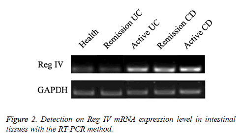
As shown in Figure 2, according to the RT-PCR results, healthy people had relatively low Reg IV mRNA expression level and Reg IV mRNA level of UC during active stage was significantly higher than that of the emission stage. There was no significant difference on Reg IV mRNA level of CD during active stage and emission stage, but Reg IV mRNA level of both of them was significantly higher than that of the healthy people.
Correlation between Reg IV expression and the serum CRP concentration of UC patients
As shown in Table 2, the CRP concentration of the low expression group of Reg IV was (5.83 ± 4.67 mg/L); the CRP concentration of the moderate expression group of Reg IV was (14.83 ± 10.94 mg/L); the CRP concentration of the high expression group of Reg IV was (81.74 ± 39.82 mg/L). The concentration of CRP gradually increased with the strengthening of Reg IV protein expression level (P<0.01).
| Reg IV expression concentration | Number of cases | CRP concentration |
|---|---|---|
| + | 5 | 5.83 ± 4.67 |
| ++ | 9 | 14.83 ± 10.94 |
| +++ | 16 | 81.74 ± 39.82 |
| F value | 27.826 | |
| P value | 0.001 |
Table 2. Correlation between Reg IV protein expression level and serum CRP concentration of UC patients.
Correlation between Reg IV expression level and Mayo index of UC patients
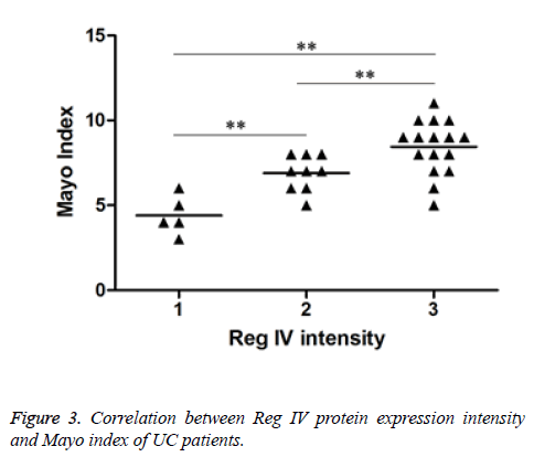
As shown in Figure 3, the Mayo index of the low expression group of Reg IV was 4.40 ± 1.14; the value of the moderate expression group was 6.89 ± 1.05; the value of the high expression group was 8.44 ± 1.59. Mayo index gradually increased with the strengthening of Reg IV protein expression level (P<0.01).
Discussion
With common symptoms of diarrhea and bellyache, some patients of Inflammatory Bowel Disease (IBD) even have bloody stools. Generally speaking, the diseases involved in ileum, rectum and colon are known as Inflammatory Bowel Disease (IBD). IBD includes Ulcerative Colitis (UC) and Crohn’s Disease (CD) [1]. There has been no clear definition on the etiology and pathogenesis on IBD at present, which may be related to environmental factors, genetic factors and microorganism infection, with changes on body immunity state [2-4]. Clinically, the diagnosis on IBD is mainly made according to the clinical presentations of patients, the endoscopy results and the laboratory inspection results. In addition, in order to make a definite diagnosis on IBD, it is needed to exclude other diseases of immune system and intestinal tract. Therefore, the IBD auxiliary diagnosis and indexes related to the state of IBD are helpful to clinical diagnosis and judgment on disease state of IBD.
The Regenerating gene (Reg) IV belongs to the calciumdependent phytohemagglutinin superfamily, and the coded protein belongs to the secreted protein, mainly expressed in gastrointestinal tract and pancreas [8-10]. Little research has been published on the roles of Reg IV in the occurrence and development of IBD. According to the findings of this research, Reg IV has low expression in intestinal tissues of healthy people, which significantly increases in that of IBD patients; in addition, Reg IV protein level of UC patients during active stage is significantly higher than that of remission stage (P<0.01). The subsequent RT-PCR experiment further verifies that Reg IV protein has low expressions in healthy people, but has high ones in patients of IBD.
According to the above experiment, there is difference on Reg IV expression levels only between the UC patients during active stage and that during remission stage, and there is no significant difference on Reg IV protein expression levels between the CD patients during active stage and that during remission stage (P>0.05). It illustrates that Reg IV expression level may be related to the disease activity of UC patients. In order to work out the correlation between Reg IV expression level and UC disease activity, the correlation between Reg IV expression level and the serum CRP and Mayo index of UC patients is observed in this article. According to research findings, there is certain correlation between CRP level and UC disease state [11-15]. In addition, with the rise on Reg IV expression level, the serum CRP level of the patients gradually increases; besides, Mayo index, the disease activity index, also increases with the rise on Reg IV expression level. It illustrates that Reg IV expression can illustrate the activity of the disease of UC patients to certain extent.
In conclusion, according to the findings of this research, Reg IV has significantly high expressions in IBD patients. In addition, Reg IV protein level of UC patients during active stage is significantly higher than that of remission stage; there is no significant difference on Reg IV protein expression level between CD patients of active stage and that of remission stage. According to further analysis on the correlation between Reg IV expression level and the disease activity of UC patients, the serum CRP level and the Mayo index of the patients gradually increase with the rise on Reg IV expression level. It illustrates that Reg IV may be involved in the occurrence and development of IBD, especially that of UC.
References
- Lu H, Tu H, Hu X. Expression and significance of CX3CR1 in Inflammatory Bowel Disease (IBD). Guide of China Med 2013; 11: 495-496.
- Xing Y, Yang D, Liu C. Expression and significance of triggering receptor-1 and epoxidase-2 of soluble myeloid cells of patients of inflammatory bowel disease. Chinese J Gerontol 2014; 34: 3341-3342.
- Zheng H, Wu Z, Yang F. Expression and clinical significance of S100A12 antibody in inflammatory bowel disease. J Anhui Med Univ 2012; 47: 295-298.
- Beltran CJ, Nunez LE, Diaz-Jimenez D. Characterization of the novel ST2/IL-33 system in patients with inflammatory Bowel disease. Inflamm Bowel Dis 2010; 16: 1097-1107.
- Zhou H, Xu L, Cui H. Preliminary study on REG IV gene functions of prostate glands cells. Prog Mod Biomed 2010; 10: 4407-4410.
- Cooperative Group of Inflammatory Bowel Disease of Gastroenterology Branch of Chinese Medical Association. Opinion Consensus on Norms on Diagnosis and Treatment on Inflammatory Bowel Disease in China. Gastroenterology 2007; 12: 488-495.
- Zou R, Hu Z, Chen H. Expression and significance of miR-199a in cervical cancer and intraepithelial neoplasia. J Med Res 2011; 40: 55-59.
- Fagan EA, Dyck RF, Maton PN. Serum levels of C-reactive protein in Crohns disease and ulcerative colitis. Eur J Clin Invest 1982; l 2: 35l-359.
- Zilberman L, Maharshak N, Arbel Y. Correlated expression of high-sensitivity C-reactive protein in relation to disease activity in inflammatory bowel disease: lack of differences between CrohnS disease and ulcerative colitis. Digestion 2006; 73: 205-209.
- Solem CA, Jr Loftus E, Tremaine WJ. Correlation of C-reactive protein with clinical, endoscopic, histologic, and radiographic activity in inflammatory bowel disease . Inflamm Bowel Dis, 2005; 11: 707-712.
- Xiong L, Xia B, Jiang L. Polymorphism of TLR4 gene Asp299Gly and TLR2 gene Arg753Glu and Arg677Trp as well as no correlation with inflammatory bowel disease of Han nationality in Hubei Province of China. World Chinese J Digestol 2006; 14: 212-215.
- Sheng J, Duan L, Miao Y. Progress in research on roles played by signal transduction and transcriptional activation factor family in pathogenesis of inflammatory bowel disease. Chinese J Pract Int Med 2007; 27: 1470-1472.
- Zhang T, Chen Y, Wang Z. Changes on intestinal microbial community structure of patients of inflammatory bowel disease and the correlation with inflammatory indexes. J South Med Univ 2013; 10: 1474-1498.
- Lv X, Zhan L, Jiang H. Influences of metalloprotein kinase of enterocyte matrix of inflammatory bowel disease on NAP-2. World Chinese J Digestol 2006; 14: 2510-2515.
- Xue H, Ni P, Wu J. Research on IL-1β gene polymorphism and linkage disequilibrium. J Shanghai Jiaotong Univ 2006; 26: 912-915.


