Research Article - Biomedical Research (2017) Volume 28, Issue 19
Evaluation of mandibular notch, coronoid process, and mandibular condyle configurations with cone beam computed tomography
Melek Tassoker1*, Anil Didem Aydin Kabakci2, Duygu Akin2 and Sevgi Sener1
1Department of Oral and Maxillofacial Radiology, Faculty of Dentistry, Necmettin Erbakan University, Konya, Turkey
2Department of Anatomy, Meram Medicine Faculty, Necmettin Erbakan University, Konya, Turkey
- *Corresponding Author:
- Melek Tassoker
Department of Oral and Maxillofacial Radiology
Faculty of Dentistry
Necmettin Erbakan University, Turkey
Accepted on September 18, 2017
Abstract
Objective: Having detailed knowledge about the anatomical structure of mandible is important for anthropologists, forensic scientists and reconstructive surgeons. There are several imaging modalities visualizing the mandibular morphology. Cone Beam Computed Tomography (CBCT) is a developing technique that is being increasingly used in dento-maxillo-facial imaging due to its relatively low dose and high spatial resolution features. It has created a revolution in maxillofacial imaging by providing the transition of dental diagnosis from two dimensional (2D) to three dimensional (3D) images. This study was designed to evaluate mandibular notch, coronoid process, and mandibular condyle morphology by using CBCT.
Methods: The study was conducted based on archived records of CBCT images of a total of 108 patients. Configurations of coronoid process, mandibular notch and condylar process were reviewed on axial, coronal and sagittal CBCT sections. 216 (108 mandibles) coronoid processes, mandibular notches and mandibular condyles from 108 mandibles were evaluated.
Results: There was statistically significant relation between age and condyle shapes on coronal sections (p<0.001). Gender had no effect on the condyle shapes on both coronal and sagittal CBCT sections (p>0.05).
Conclusion: The data were obtained from this study can be used as anthropological markers to assess different races. CBCT can be a preferred 3D imaging method to detect possible morphological modifications on the mandibular bone and coronal CBCT sections can be useful in forensic sciences based on the influence of aging on mandibular condyle morphology.
Keywords
CBCT, Coronoid process, Mandibular notch, Mandibular condyle.
Introduction
Bones have great importance in identification of a person and they also help in establishing the process of evolution, race and demographic profile [1]. Mandible is the most durable and sexually dimorphic bone of the skull and also resists post mortem changes [2,3]. The mandible bears a curve shaped body with two rami. Each ramus consists of two processes: coronoid and condylar [4,5]. Coronoid or condylar process (mandibular condyle) cannot be distinguished in the early stage of mandibular development [6].
The Coronoid Process (CRP) derived from a Greek word korone (meaning crow’s beak) [7]. The CRP of the mandible which giving attachments to temporalis muscle from its margins and medial surface is a thin flat triangular process from mandibular ramus projecting upwards and slightly forwards [8,9]. Its lateral surface provides attachment to anterior fibres of masseter. The temporalis and masseter are both important muscles of mastication which show morphofunctional dependence [5]. The CRP varies in size and shape [9]. The size and shape of CRP is influenced by dietary habit, hormones, genetic constitution, and mainly by temporalis muscle activity [4,5]. Muscle and bone may dynamically affect the function of each other and lead to change in the morphology of the bone involved [10]. CRP enlargement may be seen in some pathological condition like osteochondroma, exostosis, osteoma and other developmental anomalies [5]. The CRP is of clinical importance to the maxillofacial surgeon for reconstructive purposes and also helps in age, sex, race and species determination [4,8,9,11]. The CRP is favorable donor site which has the advantage of biocompatibility and availability and provides less operative time for harvesting [9].
The Mandibular Condyle (MC) or condiloid process is the part of the mandible that articulates with the articular fossa of the temporal bone [12]. The appearance of the MC varies greatly among different age groups and individuals. Morphologic changes may occur depending on the developmental variations, malocclusion, trauma, remodelling, endocrine disturbances, and various diseases [7,13,14]. Several studies have carried out to evaluate the morphology of the MC. Variation in the MC shapes was noted by previous researchers [14]. The most common morphologic changes are detected in the MC of elderly people due to the onset of joint degeneration [15].
The Mandibular Notch (MN) or the mandibular incisure of the mandible [16] is the gap between the CRP and MC [10]. It allows the passage of the masseteric nerve, masseteric artery and masseteric vein to and from masseter muscle [17]. The shape of MN depends on the shape of CRP and MC [10].
MC morphology has been studied on dry and autopsy human skulls, histology, radiographic exams, Magnetic Resonance Imaging (MRI), Computed Tomography (CT) and Cone Beam Computed Tomography (CBCT) methods [15]. Radiographic analysis by means of two dimensional (2D) conventional methods such as panoramic radiography has a high degree of difficulty for the examination of the mandibular morphology [14]. They suffer of the same limitations of all planar 2D projections: magnification, distortion, superimposition, and misrepresentation of structures [18].
Biomedical engineering is the application of the principles and problem solving techniques of engineering to biology and medicine [19-21]. Medical imaging is one of biomedical engineering fields. It provides novel tumor detection approaches such as melanoma [22] and develops new imaging systems. In dental imaging field, along with the advances in biomedical engineering, CBCT has been introduced and a transition from 2D to 3D imaging was provided for the maxillofacial region [18]. The images are reconstructed in a 3D data set using a modification of the original cone beam algorithm developed by Feldkampet al. [23] in 1984. This technique is widely used in different industrial and biomedical applications such as microCT. Among the first clinical applications were Single Photon Emission Computerized Tomography (SPECT), angiography and image-guided radiotherapy [24].
Various morphological features of mandible show changes in reference to age and sex which act as an anthropological marker for detection of races [4,5]. CBCT is superior over other imaging modalities for the bony morphology of MC by providing 3D images with minimal distortion [14]. The present study was undertaken to determine the different forms of CRP, MC and MN existing in a Turkish population by using CBCT images on both sides in relation to age and sex and compare it with other population studies.
Materials and Methods
The study protocol approved by the Necmettin Erbakan University Institutional Review Board and the study conformed to the guidelines laid out in the Declaration of Helsinki (decision no: 2016/011). The study was carried out with the archived records of CBCT images of patients who came for diagnostic or treatment purposes to Necmettin Erbakan University, Oral and Maxillofacial Radiology Department.
A total of 108 patients with a mean age of 35.7 (age range 17-79) consisting of 45 males and 63 females were included in the study. The patient groups were formed according to sex and age (young, adult and old group). The history of dental and maxillofacial fracture, Temporomandibular Joint (TMJ) dysfunction, developmental anomaly and pathological condition around TMJ was excluding criteria for the patients.
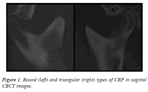
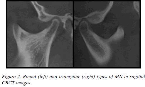
Confıgurations of CRP, MC and MN were reviewed on CBCT images. CRP and MN types were classified into triangular (type 1) and round (type 2) shaped by using sagittal CBCT sections (Figures 1 and 2).
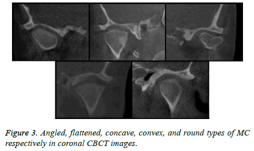
The MC types were categorized as convex (type 1), rounded (type 2), flat (type 3), angled (typed 4), and concave (type 5) shaped in the coronal CBCT sections using the revised classification of Yale et al. [25] (Figure 3).
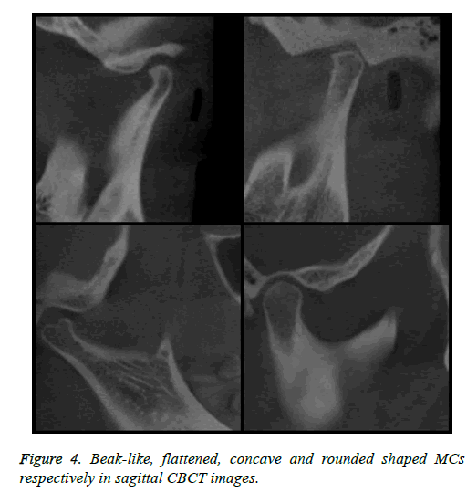
Rounded (type 1), flat (type 2), beak-like (type 3), and concave (type 4) shaped types were used for classification of MC in the sagittal sections using the revised classification of Koyamaet al. [26] and Shubhasini et al. [27] (Figure 4).
CBCT scanning was performed using Morita 3D Accuitomo machine (J. Morita Mfg. Corp. Kyoto. Japan). All CBCT images were evaluated by the same oral and maxillofacial radiologist with 5 year of experience. All CBCT images were examined in a dark room and in the same computer (Intel® Xeon® E5-2620. 2.0 GHz; NVIDIA quadro 2000; 32" Dell T7600 workstation with a resolution of 1280 × 1024 pixels. 8 GB memory. Windows 7 operating system) with the use of the i-Dixel software Ver. 2.0 (J. Morita MFG. Co.).
Statistical analysis was performed by SPSS version 21.0 (Statistical Package for Social Science Inc. Chicago. IL). Data set was analysed using descriptive statistics and chi-squared test.
Results
In the present study, 216 (of 108 mandibles) CRPs, MNs and MCs were evaluated. The most common shape for CRP observed was triangle (68.1%) and this was followed by round shape (31.9%). Round MN was found most (79.2%) among 216 sides. Shape of MC in the coronal section was classified as convex (42.6%), round (10.6%), flat (20.8%), angular (19.4%) and concave (6.5%). Similarly, round MC was the most common shape by 41.2% and this was followed by flat, beaklike and concave shapes by 41.2%, 37%, 20.4%, respectively in the sagittal plane (Table 1).
| Coronoid process | Shape | Sex | Total | Chi-square | p-value | ||||
|---|---|---|---|---|---|---|---|---|---|
| Male | Female | ||||||||
| n | % | n | % | n | % | ||||
| Triangular | 61 | 67.8 | 86 | 68.3 | 147 | 68.1 | 0.005 | 0.941 | |
| Round | 29 | 32.2 | 40 | 31.7 | 69 | 31.9 | |||
| Total | 90 | 100 | 126 | 100 | 216 | 100 | |||
| Mandibular notch | Triangular | 19 | 21.1 | 26 | 20.6 | 45 | 20.8 | 0.007 | 0.932 |
| Round | 71 | 78.9 | 100 | 79.4 | 171 | 79.2 | |||
| Total | 90 | 100 | 126 | 100 | 216 | 100 | |||
| Mandibular condyle-coronal section | Convex | 36 | 40 | 56 | 44.4 | 92 | 42.6 | 0.12 | 0.913 |
| Round | 12 | 13.3 | 11 | 8.7 | 23 | 10.6 | |||
| Flat | 16 | 17.8 | 29 | 23 | 45 | 20.8 | |||
| Angled | 24 | 26.7 | 18 | 14.3 | 42 | 19.4 | |||
| Concave | 2 | 2.2 | 12 | 9.5 | 14 | 6.5 | |||
| Total | 90 | 100 | 126 | 100 | 216 | 100 | |||
| Mandibular condyle -sagittal section | Round | 39 | 43.3 | 50 | 39.7 | 89 | 41.2 | 1.49 | 0.221 |
| Flat | 35 | 38.9 | 45 | 35.7 | 80 | 37 | |||
| Beak-like | 16 | 17.8 | 28 | 22.2 | 44 | 20.4 | |||
| Concave | 0 | 0 | 3 | 2.4 | 3 | 1.4 | |||
| Total | 90 | 100 | 126 | 100 | 216 | 100 | |||
Table 1. Shape prevalence of coronoid process, mandibular notch and mandibular condyle according to sex.
The most common shape for CRP observed was triangle both in males (67.8%) and females (68.3%). Type of MN and MC on the sagittal plane was mostly observed as round in both female and male individuals (Table 1). There was not any statistical association between mandibular parameters observed during the study and gender (Table 1).
The most common shape of the CRP observed in young individuals was triangle (n=85, 70.8%) when prevalence of mandibular parameters were compared with the age groups. Such higher rates of triangular shape were observed on 41 (78.8%) males and 44 (64.7%) females (Tables 2 and 3). MN was detected round in 94 (78.3%) young individuals consisted of 42 (80.8%) males and 52 (76.5%) females (Tables 2-4).
| Coronoid process | Shape | Age groups | Total | Chi-square | p-value | ||||||
|---|---|---|---|---|---|---|---|---|---|---|---|
| Young (18-35) | Adult (36-55) | Old (56-80) | |||||||||
| n | % | n | % | n | % | n | % | ||||
| Triangular | 85 | 70.8 | 32 | 64 | 30 | 65.2 | 147 | 68.1 | 0.701 | 0.403 | |
| Round | 35 | 29.2 | 18 | 36 | 16 | 34.8 | 69 | 31.9 | |||
| Total | 120 | 100 | 50 | 100 | 46 | 100 | 216 | 100 | |||
| Mandibular notch | Triangular | 26 | 21.7 | 12 | 24 | 7 | 15.2 | 45 | 20.8 | 0.551 | 0.458 |
| Round | 94 | 78.3 | 38 | 76 | 39 | 84.8 | 171 | 79.2 | |||
| Total | 120 | 100 | 50 | 100 | 46 | 100 | 216 | 100 | |||
| Mandibular condyle-coronal section | Convex | 62 | 51.7 | 14 | 28 | 16 | 34.8 | 92 | 42.6 | 13.262 | 0.000* |
| Round | 3 | 6 | 3 | 6 | 0 | 0 | 23 | 10.6 | |||
| Flat | 12 | 24 | 12 | 24 | 15 | 32.6 | 45 | 20.8 | |||
| Angled | 12 | 24 | 12 | 24 | 13 | 28.3 | 42 | 19.4 | |||
| Concave | 9 | 18 | 9 | 8 | 2 | 4.3 | 14 | 6.5 | |||
| Total | 120 | 100 | 50 | 100 | 46 | 100 | 216 | 100 | |||
| Mandibular condyle -sagittal section | Round | 66 | 55 | 13 | 26 | 10 | 21.7 | 89 | 41.2 | 2.477 | 0.116 |
| Flat | 26 | 21.7 | 28 | 56 | 26 | 56.5 | 80 | 37 | |||
| Beak-like | 27 | 22.5 | 7 | 14 | 10 | 21.7 | 44 | 20.4 | |||
| Concave | 1 | 0.8 | 2 | 4 | 0 | 0 | 3 | 1.6 | |||
| Total | 120 | 100 | 50 | 100 | 46 | 100 | 216 | 100 | |||
*The significance level is 0.001
Table 2. Shape prevalence of coronoid process, mandibular notch and mandibular condyle according to age groups.
| Age groups | Triangular | Round | |||||
|---|---|---|---|---|---|---|---|
| Right | Left | Total | Right | Left | Total | ||
| Male | Young | 20 (76.9%) | 21 (80.8%) | 41 (78.8%) | 6 (23.1%) | 5 (19.2%) | 11 (21.2%) |
| Adult | 5 (55.6%) | 4 (44.4%) | 9 (50%) | 4 (44.4%) | 5 (55.6%) | 9 (50%) | |
| Old | 5 (50%) | 6 (60%) | 11 (55%) | 5 (50%) | 4 (40%) | 9 (45%) | |
| Total | 30 (66.7%) | 31 (68.9%) | 61 (67.8%) | 15 (33.3%) | 14 (31.1%) | 29 (32.2%) | |
| Female | Young | 24 (70.6%) | 20 (58.8%) | 44 (64.7%) | 10 (29.4%) | 14 (41.2%) | 24 (35.3%) |
| Adult | 11 (68.8%) | 12 (75%) | 23 (71.9%) | 5 (31.3%) | 4 (25%) | 9 (28.1%) | |
| Old | 9 (69.2%) | 10 (76.9%) | 19 (73.1%) | 4 (30.8%) | 3 (23.1%) | 7 (26.9%) | |
| Total | 44 (69.8%) | 42 (66.7%) | 86 (68.3%) | 19 (30.2%) | 21 (33.3%) | 40 (31.7%) | |
Table 3. Prevalence of coronoid process shapes according to sex and age groups.
| Age groups | Triangular | Round | |||||
|---|---|---|---|---|---|---|---|
| Right | Left | Total | Right | Left | Total | ||
| Male | Young | 4 (15.4%) | 6 (23.1%) | 10 (19.2%) | 22 (84.6%) | 20 (76.9%) | 42 (80.8%) |
| Adult | 1 (11.1%) | 3 (33.3%) | 4 (22.2%) | 8 (88.9%) | 6 (66.7%) | 14 (77.8%) | |
| Old | 3 (30%) | 2 (20%) | 5 (25%) | 7 (70%) | 8 (80%) | 15 (75%) | |
| Total | 8 (17.8%) | 11 (24.4%) | 19 (21.1%) | 37 (82.2%) | 34 (75.6%) | 71 (78.9%) | |
| Female | Young | 10 (29.4%) | 6 (17.6%) | 16 (23.5%) | 24 (70.6%) | 28 (82.4%) | 52 (76.5%) |
| Adult | 2 (12.5%) | 6 (37.5%) | 8 (25%) | 14 (87.5%) | 10 (62.5%) | 24 (75%) | |
| Old | 0 (0%) | 2 (15.4%) | 2 (7.7%) | 13 (100%) | 11 (84.6%) | 24 (92.3%) | |
| Total | 12 (19%) | 14 (22.2%) | 26 (20.6%) | 51 (81%) | 49 (77.8%) | 100 (79.4%) | |
Table 4. Prevalence of mandibular notch shapes according to sex and age groups.
When prevalence of the MC was compared with the age groups on coronal section, the most common form observed was the convex one (42.6%). Such convex form was detected more among young females (54.4%) than young males (48.1%) (Tables 2 and 5). The most common form observed in young individuals on the sagittal section was round (41.2%) (Tables 2 and 6).
| Male | Female | ||||||||
|---|---|---|---|---|---|---|---|---|---|
| Young | Adult | Old | Total | Young | Adult | Old | Total | ||
| Convex | Right | 12 (46.2%) | 3 (33.3%) | 3 (30%) | 18 (40%) | 19 (55.9%) | 5 (31.3%) | 5 (38.5%) | 29 (46%) |
| Left | 13 (50%) | 2 (22.2%) | 3 (30%) | 18 (40%) | 18 (52.9%) | 4 (25%) | 5 (38.5%) | 27 (42.9%) | |
| Total | 25 (48.1%) | 5 (27.8%) | 6 (30%) | 36 (40%) | 37 (54.4%) | 9 (28.1%) | 10 (38.5%) | 56 (44.4%) | |
| Round | Right | 6 (23.1%) | 0 (0%) | 0 (0%) | 6 (13.3%) | 4 (11.8%) | 1 (6.3%) | 0 (0%) | 5 (7.9%) |
| Left | 5 (19.2%) | 1 (11.1%) | 0 (0%) | 6 (13.3%) | 5 (14.7%) | 1 (6.3%) | 0 (0%) | 6 (9.5%) | |
| Total | 11 (21.2%) | 1 (5.6%) | 0 (0%) | 12 (13.3%) | 9 (13.2%) | 2 (6.3%) | 0 (0%) | 11 (8.7%) | |
| Flat | Right | 4 (15.4%) | 3 (33.3%) | 3 (30%) | 10 (22.2%) | 5 (14.7%) | 3 (18.8%) | 5 (38.5%) | 13 (20.6%) |
| Left | 3 (11.5%) | 1 (11.1%) | 2 (20%) | 6 (13.3%) | 6 (17.6%) | 5 (31.3%) | 5 (38.5%) | 16 (25.4%) | |
| Total | 7 (13.5%) | 4 (22.2%) | 5 (25%) | 16 (17.8%) | 11 (16.2%) | 8 (25%) | 10 (38.5%) | 29 (23%) | |
| Angled | Right | 4 (15.4%) | 3 (33.3%) | 4 (40%) | 11 (24.4%) | 4 (11.8%) | 3 (18.8%) | 2 (15.4%) | 9 (14.3%) |
| Left | 5 (19.2%) | 3 (33.3%) | 5 (50%) | 13 (28.9%) | 4 (11.8%) | 3 (18.8%) | 2 (15.4%) | 9 (14.3%) | |
| Total | 9 (17.3%) | 6 (33.3%) | 9 (45%) | 24 (26.7%) | 8 (11.8%) | 6 (18%) | 4 (15.4%) | 18 (14.3%) | |
| Concave | Right | 0 (0%) | 0 (0%) | 0 (0%) | 0 (0%) | 2 (5.9%) | 4 (25%) | 1 (7.7%) | 7 (11.1%) |
| Left | 0 (0%) | 2 (22.2%) | 0 (0%) | 2 (4.4%) | 1 (2.9%) | 3 (18.8%) | 1 (7.7%) | 5 (7.9%) | |
| Total | 0 (0%) | 2 (11.1%) | 0 (0%) | 2 (2.2%) | 3 (4.4%) | 7 (21.9%) | 2 (7.7%) | 12 (9.5%) | |
Table 5. Prevalence of mandibular condyle according to sex and age groups in the coronal section.
| Male | Female | |||||||
|---|---|---|---|---|---|---|---|---|
| Young | Adult | Old | Total | Young | Adult | Old | Total | |
| Right | 17 (65.4%) | 3 (33.3%) | 3 (30%) | 23 (51.1%) | 19 (55.9%) | 5 (31.3%) | 2 (15.4%) | 26 (41.3%) |
| Left | 13 (50%) | 1 (11.1%) | 2 (20%) | 16 (35.6%) | 17 (50%) | 4 (25%) | 3 (23.1%) | 24 (38.1%) |
| Total | 30 (57.7%) | 4 (22.2%) | 5 (25%) | 39 (43.3%) | 36 (52.9%) | 9 (28.1%) | 5 (19.2%) | 50 (39.7%) |
| Right | 5 (19.2%) | 5 (55.6%) | 7 (70%) | 17 (37.8%) | 7 (20.6%) | 8 (50%) | 8 (61.5%) | 23 (36.5%) |
| Left | 6 (23.1%) | 6 (66.7%) | 6 (60%) | 18 (40%) | 8 (23.5%) | 9 (56.3%) | 5 (38.5%) | 22 (34.9%) |
| Total | 11 (21.2%) | 11 (61.1%) | 13 (65%) | 35 (38.9%) | 15 (22.1%) | 17 (53.1%) | 13 (50%) | 45 (35.7%) |
| Right | 4 (15.4%) | 1 (11.1%) | 0 (0%) | 5 (11.1%) | 7 (20.6%) | 2 (12.5%) | 3 (23.1%) | 12 (19%) |
| Left | 7 (26.9%) | 2 (22.2%) | 2 (20%) | 11 (24.4%) | 9 (26.5%) | 2 (12.5%) | 5 (38.5%) | 16 (25.4%) |
| Total | 11 (21.2%) | 3 (16.7%) | 2 (10%) | 16 (17.8%) | 16 (23.5%) | 4 (12.5%) | 8 (30.8%) | 28 (22.2%) |
| Right | 0 (0%) | 0 (0%) | 0 (0%) | 0 (0%) | 1 (2.9%) | 1 (6.3%) | 0 (0%) | 2 (3.2%) |
| Left | 0 (0%) | 0 (0%) | 0 (0%) | 0 (0%) | 0 (0%) | 1 (6.3%) | 0 (0%) | 1 (1.6%) |
| Total | 0 (0%) | 0 (0%) | 0 (0%) | 0 (0%) | 1 (1.5%) | 2 (6.3%) | 0 (0%) | 3 (2.4%) |
Table 6. Prevalence of mandibular condyle shapes according to sex and age groups in the sagittal section.
In the sagittal section, no significant association was between shapes of CRP, MN and MC and the age groups. However, the shape of the MC in the coronal section was statistically associated with age groups (p<0.001) (Table 2). The prevalence of convex, concave and round type condylar morphology decreased with increasing age, whereas flat and angled typed increased.
Discussion
CBCT is becoming an important tool in modern dental practice and provides excellent imaging of the mandibular bony structures with less radiation exposure compared to other techniques [15]. CBCT was used in previous studies [15,28,29] for detection of changes in condylar morphology. This study was performed to reveal the differences of MC, CRP and MC shapes in comparison with age, sex and other population studies. To the best of our knowledge this is the first CBCT study evaluating all of these parameters.
CBCT images of 108 individuals were reviewed in the present study. Our results revealed that aging had an effect on condylar shape on coronal sections. The number of convex, concave and round type condylar morphology decreased with age increase, whereas flat and angled type increased. Along with presenting 3D images of the bone, CBCT is commonly preferred method to detect possible morphological modifications on the bone when compared with 2D panoramic radiograph [27].
2D panoramic radiograph [10] and 3D medical CT [30] were also used for assessing the MC morphology in the literature. Panoramic radiograph has disadvantages of magnification, distortion, superimposition, and misrepresentation of structures [18]. These problems can be result in unreliable measurements [31]. Medical CT is another 3D imaging system but its application is limited in dentistry because of cost, accessibility, and dose considerations. CBCT is a recent technology and has created a revolution in maxillo-facial imaging providing accurate measurements in maxillofacial region. The early studies revealed that CBCT measurements of human dry skull are highly accurate and reproducible [32]. It uses a coneshaped X-ray beam and only one rotational sequence of the gantry is necessary to acquire enough data for image reconstruction, which result in reduced patient radiation dose. On the other hand, a traditional medical CT uses a fan-shaped x-ray beam and each slice requires a separate scan and separate 2D reconstruction which result in higher radiation dose [18].
Mandible (submaxilla) is a U-shaped bone which forms the jaw joint through articulation with the temporal bone. It consists of two parts including mandibular corpus and two mandibular rami. Each ramus has two processes called coronoid and condylar processes and a notch called mandibular notch [5,6]. The mandible is one of the strongest bone of the body used for determination of sociodemographic structure for anthropology and used widely in forensic medicine for age, gender and racial determination. In consideration of close relation of the mandible with surrounding formations, importance of morphological differences appeared in its structure has been revealed in different researches conducted by anatomists and surgeons [5,6].
Configurations of the CRP, MC and MN are developmentally connected to each other [17]. Contours of the coronoid and condylar processes are particularly important for formation of the mandibular notch. However, such formations may morphologically differ depending on genetic factors, hormones, nutritional habits and activity of the temporalis muscle. There are studies where different configurations of such formations were detected in the literature [5,6,10,17].
Maxillofacial surgeons consider the region due to its clinical importance for reconstructive purposes, surgical approach and management of the surgery [17]. The information about morphological forms of the MN obtained would allow a maxillofacial surgeon to treat chronic mandibular dislocations properly through a novel mini-plate introduced by Cavalcanti and Vasconcelos [17]. Mandibular CRP becomes increasingly important to be used as a graft for all types of reconstructive cranio-maxillofacial surgical procedures such as orbital floor reconstruction, paranasal augmentation, TMJ ankylosis, trauma, tumors, facial paralysis, alveolar defects, non-union fracture of mandible, osseous defects reconstruction [5,6,33]. Expansion of the CRP may be observed in some disorders including osteochondroma, exostosis, osteoma and some developmental abnormalities. Furthermore, science of anthropology interests in coronoid morphology well including clinical and other aspects through studies in the literature [6]. Morphological differences suggest that the measures of the condyle may not be generalized to the populations for manufacturing purposes of condylar prosthesis; and such measurements may be implemented for local populations even with differences for males and females [12].
Shakya et al. [10] classified MN and CRP shapes with 200 panoramic radiographs (102 males, 98 females). They reported the most common MN shape of both males (44.11%) and females (45.09%) as sloping form. Gender is not found to be an affecting factor on MN and CRP morphologies and triangular CRP was mostly seen type in their study similar to our results. Our study indicated that with aging process, condylar morphology can be changed and this can be seen on coronal CBCT section. Panoramic radiograph has the advantages of low cost and easy access in dentistry but cannot provide 3D nature of the bony structure [34].
Saad et al.[17] identified MN of 100 cadavers as triangular, round and truncated and detected prevalence of these types as 46% (12% for female, 34% for male), 34 (24% for female, 10% for male) and 20% (6% for female, 14% for male), respectively. MN was divided into two groups as triangular and round in the present study. Prevalence of the foresaid types were 20.8% (20.6% for female, 21.1% for male) and 79.2% (79.4% for female, 78.9% for male), respectively. Saad et al. [17] detected the most common form of MN as triangular (46%) whereas sloping form was the most common form in the study conducted by Shakya et al. [10]. The most common form observed in the present study was round (20.8%).
Recent studies indicated that different CRP forms including triangular, round, hook and miscellaneous are common in human mandibles [6,8,35-38] (Table 7). The most common form of CRP detected in the present study as well as in the studies carried out by Isaac and Holla [8], Prajapati et al. [24], Nirmale et al. [23], Pradhan et al. [5], Desai et al. [35], Quadri and Tanveer [38] was triangular, whereas Subbaramaiah et al. [6] stated the most common shape of the CRP as round (Table 7).
| Researcher | Samples | Type | Total | Male | Female |
|---|---|---|---|---|---|
| Isaac and Holla [8] | 157 dry human mandibles | Triangular shaped | 154 (49%) | 93 (46.5%) | 61 (53.5%) |
| 314 sides (100 male, 57 female) | Round shaped | 74 (23.6%) | 47 (23.5%) | 27 (23.6%) | |
| Hook shaped | 86 (27.4%) | 60 (30%) | 26 (22.8%) | ||
| Prajapati et al. [37] | 120 dry human mandibles | Triangular shaped | 130 (54.17%) | 84 (56%) | 46 (51.11%) |
| 240 sides (75 male, 45 female) | Round shaped | 59 (24.58%) | 34 (22.66%) | 25 (27.77%) | |
| Hook shaped | 51 (21.25%) | 32 (21.33%) | 19 (21.11%) | ||
| Nirmale et al. [36] | 84 dry human mandibles | Triangular shaped | 109 (65%) | 70 (41.66%) | 39 (23.21%) |
| 168 sides (62 male, 22 female) | Round shaped | 47 (28%) | 7 (4.16%) | 5 (2.97%) | |
| Hook shaped | 12 (7%) | 30 (17.85%) | 17 (10.11%) | ||
| Pradhan et al. [5] | 92 dry human mandibles | Triangular shaped | 86 (46.73%) | 44 (45.83%) | 42 (47.72%) |
| 184 sides (48 male, 44 female) | Round shaped | 33 (17.93%) | 31 (32.29%) | 21 (21.87%) | |
| Hook shaped | 65 (35.3%) | 21 (21.87%) | 12 (13.63%) | ||
| Desai et al. [35] | 100 dry human mandibles | Triangular shaped | 136 (68%) | 74 (37%) | 62 (31%) |
| 200 sides (56 male, 44 female) | Round shaped | 16 (8%) | 10 (5%) | 6 (3%) | |
| Hook shaped | 48 (24%) | 28 (14%) | 20 (10%) | ||
| Subbaramaiah et al. [6] | 100 dry human mandibles | Triangular shaped | 28 (14%) | 18 (18%) | 10 (10%) |
| 200 sides (50 male, 50 female) | Round shaped | 25 (12.5%) | 52 (52%) | 71 (71%) | |
| Hook shaped | 123 (61.5%) | 17 (17%) | 8 (8%) | ||
| Miscellaneous shaped | 24 (12%) | 13 (13%) | 11 (11%) | ||
| Quadri and Tanveer [38] | 200 dry human mandibles | Triangular shaped | 134 (67%) | 109 (72.2%) | 25 (51%) |
| 400 sides (135 male, 45 female) | Round shaped | 6 (3%) | 4 (2.6%) | 2 (4.1%) | |
| Hook shaped | 60 (30%) | 38 (25.2%) | 22 (44.9%) | ||
| Our study | 108 CBCT images | Triangular shaped | 147 (68.1%) | 61 (67.8%) | 86 (68.3%) |
| 216 sides (45 male and 63 female) | Round shaped | 69 (31.9%) | 29 (32.2%) | 40 (31.7%) |
Table 7. Coronoid process shapes prevalence according to researchers.
Yale et al. [25] reviewed 3008 MC at coronal section from 1560 skulls. They reported 1753 (58.3%) convex, 90 (3%) round, 759 (25.2%) flat and 348 (11.6%) angled shapes of MC. MC was classified as convex, round, flat, angled and concave at coronal section in our study. MC prevalence was determined as 92 (42.6%), 23 (6%), 45 (20.8%), 42 (19.4%) and 14 (6.5%) respectively. The obtained data from the present study are in line with the study conducted by Yale et al. [25].
Shubhasini et al. [27] detected the round form (71.9%) of MC as the most common shape at sagittal section with the sample size of 32 CBCT images. Similarly, the most common MC form was detected as round (41.2%) in the present study. The most commonly observed MC morphology on coronal sections was the angled followed by the convex. This difference can be related with racial diversities between Indian and Turkish populations. Sample size and study design are other affecting factors.
The present study conducted for anatomic differences of the CRP indicated the most common form as triangular both in males and females, followed by round shape. Round form of the MN was the most common type. The most common form of the MC was convex and round at coronal and sagittal sections, respectively. The data was obtained from a Turkish population and these results can be used as anthropological markers to assess different races. Our results indicated that aging had an effect on MC shapes on coronal sections. The presence of convex, concave and round type MC morphology decreased with age increase, whereas flat and angled typed increased. CBCT can be a preferred 3D imaging method to detect possible morphological modifications on the mandibular bone and coronal CBCT sections can be used in forensic sciences based on the influence of aging on MC morphology and would help forensic doctors to predict if someone is younger or older. This result requires verification with further investigations.
References
- Jembulingam S, Thenmozhi M. Sexual dimorphism of adult mandibles: a forensic tool. JIAFP 2016; 3: 3-5.
- Saini V. Metric study of fragmentary mandibles in a North Indian population. Bull Int Assoc Paleodont 2013; 7: 157-162.
- Singh R, Mishra SR, S, Passey J, Kumar P, Singh S, Sinha P, Gupta S. Sexual dimorphism in adult human mandible of North Indian origin. Forens Med Anat Res 2015; 3: 82-88.
- Kayalvili S. A study of morphological variaiton of lingula and coronoid process of adult human dry mandibles. J Pharm Sci Res 2015; 7: 1017-1020.
- Pradhan S, Bara D, Patra S, Nayak S, Mohapatra C. Anatomical study of various shapes of mandibular coronoid process in relation to gender and age. J Dent Med Sci 2014; 13: 9-14.
- Subbaramaiah M, Bajpe R, Jagannatha S, Jayanthi K. A study of various forms of mandibular coronoid process in determination of sex. Ind J Clin Anat Physiol 2015; 2: 199-203.
- Standring S, Ellis H, Healy J, Johnson D, Williams A, Collins P, Wigley C. Grays Anatomy: The anatomical basis of clinical practice. Am J Neuroradiol 2005; 26: 2703.
- Isaac B, Holla S. Variations in the shape of the coronoid process in the adult human mandible. J Anat Soc India 2001; 50: 137-39.
- Kadam SD, Roy PP, Ambali M, Doshi M. Variation in the shape of coronoid process in dry mandible of Maharashtra population. Int J Anat Res 2015; 3: 895-898.
- Shakya S, Ongole R, Nagraj SK. Morphology of coronoid process and sigmoid notch in orthopantomograms of South Indian population. World J Dent 2013; 4: 1-3.
- Rak Y, Ginzburg A, Geffen E. Gorilla-like anatomy on Australopithecus afarensis mandibles suggests Au. afarensis link to robust australopiths. Proc Natl Acad Sci USA 2007; 104: 6568-6572.
- Wangai L, Mandela P, Butt F, Ongeti K. Morphology of the mandibular condyle in a Kenyan population. Anat J Africa 2013; 2: 71-79.
- Alomar X, Medrano J, Cabratosa J, Clavero J, Lorente M, Serra I, Monill JM, Salvador A. Anatomy of the temporomandibular joint. Semin ultrasound CT MR 2007; 28: 170-183.
- Hegde S, Praveen B, Shetty S. Morphological and radiological variations of mandibular condyles in health and diseases: a systematic review. Dentistry 2013; 3: 2161-1122.
- Valladares Neto J, Estrela C, Bueno MR, Guedes OA, Porto OCL, Pecora JD. Mandibular condyle dimensional changes in subjects from 3 to 20 years of age using cone-beam computed tomography: a preliminary study. Dental Press J Orthod 2010; 15: 172-181.
- Field EJ, Harrison RJ. Anatomical terms: their origin and derivation. Anatomical terms: their origin and derivation. Br J Surg1947; 35: 111: 10.
- Saad AM, Abd-alla MAJM. Mandibular notch configuration in Iraqi adults. Tikrit J Dent Sci 2012; 2: 175-178.
- Scarfe WC, Farman AG. What is cone-beam CT and how does it work? Dent Clin N Am 2008; 52: 707-730.
- Ebrahimian H, Ojaroudi M, Ojaroudi N, Ghadimi N. Distributed diode single-balanced mixer using defected and protruded structures for Doppler radar applications. Appl Comp Electromagn Soc J 2015; 30: 313-319.
- Razmjooy N, Ramezani M, Ghadimi N. Imperialist competitive algorithm-based optimization of neuro-fuzzy system parameters for automatic red-eye removal. Int J Fuzzy Syst 2017; 19: 1144-1156.
- Ghadimi N, Ojaroudi M. A novel design of low power rectenna for wireless sensor and RFID applications. Wireless Persl Commun 2014; 78: 1177-1186.
- Parsian A, Ramezani M, Ghadimi N. A hybrid neural network-gray wolf optimization algorithm for melanoma detection. Biomed Res 2017; 28: 3408-3411.
- Feldkamp LA, Davis LC, Kress JW. Practical cone-beam algorithm. J Opt Soc Am 1994; 1: 612-619.
- De Vos W, Casselman J, Swennen GRJ. Cone-beam computerized tomography (CBCT) imaging of the oral and maxillofacial region: a systematic review of the literature. Int J Oral Maxillofac Surg 2009; 38: 609-625.
- Yale SH, Allison BD, Hauptfuehrer J. An epidemiological assessment of mandibular condyle morphology. Oral Surg Oral Med Oral Pathol 1966; 21: 169-177.
- Koyama J, Nishiyama H, Hayashi T. Follow-up study of condylar bony changes using helical computed tomography in patients with temporomandibular disorder. Dentomaxillofac Radiol 2014; 36: 472-477.
- Shubhasini A, Birur P, Shubha G, Keerthi G, Sunny SP, Nayak DS. Study of three dimensional morphology of mandibular condyle using cone beam computed tomography. MJDS 2016; 1: 7-12.
- Ejima K, Schulze D, Stippig A, Matsumoto K, Rottke D, Honda K. Relationship between the thickness of the roof of glenoid fossa, condyle morphology and remaining teeth in asymptomatic European patients based on cone beam CT data sets. Dentomaxillofac Radiol 2013; 42: 90929410.
- Al-koshab M, Nambiar P, John J. Assessment of condyle and glenoid fossa morphology using CBCT in South-East Asians. PloS One 2015; 10: 0121682.
- Gomes LR, Gomes MR, Gonçalves JR, Ruellas ACO, Wolford LM, Paniagua B, Cevidanes LHS. CBCT versus MSCT-based models on assessing condylar morphology. Oral Surg Oral Med Oral Pathol Oral Radiol 2016; 121: 96-105.
- Serman NJ. Pitfalls of panoramic radiology in implant surgery. Ann Dent 1989; 48: 13-16.
- Kamburoglu K, Kolsuz E, Kurt H, Kilic C, Ozen T, Paksoy CS. Accurracy of CBCT measurements of a human skull. J Digit Imaging 2011; 24: 787-793.
- Mintz SM, Ettinger A, Schmakel T, Gleason MJ. Contralateral coronoid process bone grafts for orbital floor reconstruction: an anatomic and clinical study. J Oral Maxillofac Surg 1998; 56: 1140-1144.
- Tang Z, Liu X, Chen K. Comparison of digital panoramic radiography versus cone beam computerized tomography for measuring alveolar bone. Head Face Med 2017; 13: 2.
- Desai VC, Desai S, Hussain S. Morphological study of mandible. J Pharm Sci Res 2014; 6: 175-177.
- Nirmale V, Mane U, Sukre S, Diwan C. Morphological features of human mandible. Int J Rec Trends Sci Technol 2012; 3: 33-43.
- Prajapati VP, Malukar O, Nagar S. Variations in the morphological appearance of the coronoid process of human mandible. Nat J Med Res 2011; 1: 64-66.
- Quadri A, Tanveer AKH. Variaitons in shape of mandibular coronoid process i 200 south indian subjects. Int J Sci Study 2016; 4: 159-160.


