Image Article - Ophthalmology Case Reports (2020) Volume 4, Issue 2
Encephalocraniocutaneous lipomatosis: a neurocutaneous syndrome.
Farouk AG*Department of Paediatrics, College of Medical Sciences, University of Maiduguri, Nigeria
- *Corresponding Author:
- Farouk AG
Department of Paediatrics
College of Medical Sciences
University of Maiduguri
E-mail: farouk649@gmail.com
Accepted Date: July 23, 2020
Citation: Farouk AG. Encephalocraniocutaneous lipomatosis: a neurocutaneous syndrome. Ophthalmol Case Rep. 2020;4(2):4-4.
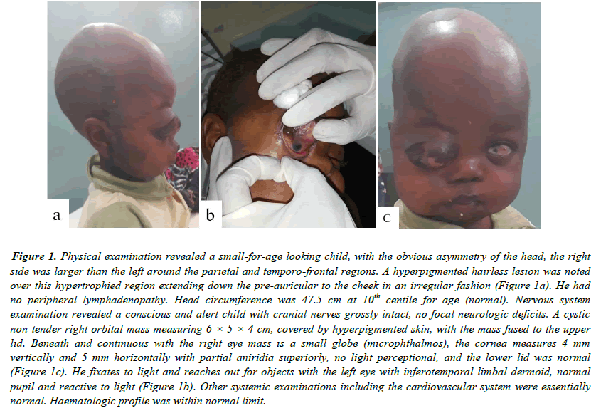
Figure 1. Physical examination revealed a small-for-age looking child, with the obvious asymmetry of the head, the right side was larger than the left around the parietal and temporo-frontal regions. A hyperpigmented hairless lesion was noted over this hypertrophied region extending down the pre-auricular to the cheek in an irregular fashion (Figure 1a). He had no peripheral lymphadenopathy. Head circumference was 47.5 cm at 10th centile for age (normal). Nervous system examination revealed a conscious and alert child with cranial nerves grossly intact, no focal neurologic deficits. A cystic non-tender right orbital mass measuring 6 × 5 × 4 cm, covered by hyperpigmented skin, with the mass fused to the upper lid. Beneath and continuous with the right eye mass is a small globe (microphthalmos), the cornea measures 4 mm vertically and 5 mm horizontally with partial aniridia superiorly, no light perceptional, and the lower lid was normal (Figure 1c). He fixates to light and reaches out for objects with the left eye with inferotemporal limbal dermoid, normal pupil and reactive to light (Figure 1b). Other systemic examinations including the cardiovascular system were essentially normal. Haematologic profile was within normal limit.
