- Biomedical Research (2011) Volume 22, Issue 3
Electrocardiogram (ECG) as a diagnostic tool for the assessment of Cardiovascular status in alcoholics
Venkatesh G*
Department of Physiology, Sri Siddhartha Medical College, Tumkur, India.
- *Corresponding Author:
- G. Venkatesh
“Vagdevi” nilaya, Near JP School,
Sharadadevinagara, Tumkur-572102
Karnataka India.
Accepted Date: December 11 2010
Abstract
The Electrocardiogram (ECG or EKG) is a graphic recording of electric potentials gener-ated by the heart. It is a simple and noninvasive, inexpensive test helps in assessing the car-diovascular status. Alcohol consumption causes ECG changes. Therefore this study is un-dertaken to detect the ECG changes in alcoholics and thereby assessing the cardio vascular status. The study protocol was explained to the participants and informed consent taken. 50 Alcoholic men of age group above 20years, confirming to DSM-IV Criteria were randomly selected from the general population. 50 age matched Controls were taken for the study. The groups were subdivided into three groups according to age (in years) as 20–39 (n=22), 40-49 (n=16) and > 50 (n=12). The ECG was recorded in resting state in lying down position. The ECG results were analyzed for Heart rate, P wave, PR interval, QRS duration, QTC in-terval, frontal axis, ST segment, T wave and TP interval. The results showed significantly increase in Heart rate and decrease in PR interval, QRS complex and TP intervals in alco-holic subjects. On comparison of subgroups, alcoholic subjects do not show statistically sig-nificant results. Our study results show cardiovascular risk in alcoholics. Therefore, earlier detection of ECG changes is useful in preventing the cardiovascular risk.
Keywords
Electrocardiogram, alcoholism, heart rate, age
Introduction
The Electrocardiogram (ECG or EKG) is a graphic re-cording of electric potentials generated by the heart. It is a simple and noninvasive, inexpensive, and highly versa-tile test helps in assessing the cardiovascular status. It is useful in detecting arrhythmias, conduction disturbances, myocardial ischemia and metabolic disturbances. The ECG waveforms are labeled alphabetically, beginning with the P wave, which represents atrial depolarization. The QRS complex represents ventricular depolarization, and the ST-T-U complex (ST segment, T wave, and U wave) represents ventricular repolarization [1].
Global alcohol consumption has increased in recent dec-ades with most of all of this increase occurring in devel-oping countries [1]. Alcohol consumption causes ECG changes which include cardiac conduction abnormalities, prolongation of the QT interval, prolongation of ventricu-lar repolarization and sympathetic stimulation [2]. Sinus tachycardia or a Supraventricular arrythmia, commonly atrial fibrillation and non-specific ST-T changes are also observed in alcoholics [3]. Alcohol alters the endocrinal function like increase in adrenocorticotrophic hormone, oxytocin and electrolytes, which may indirectly causes myocardial damage [4]. In a cross-sectional study, habit-ual alcohol intake was positively associated with Blood Pressure and Heart Rate compared with nondrinkers [5]. Studies have showed that alcohol abuse in men younger at 40 with a first myocardial infarction [6]. Moderate dose of alcohol intake is associated with an increase in maximum and the minimum P-wave duration which have been re-ported to represent an increased risk for atrial fibrillation in patients with no underlying disease [7]. Cardiomyopa-thy associated with ECG changes like sinus tachycardia or a supraventricular arrythmia, commonly atrial fibrilla-tion with a rapid ventricular response and non-specific ST-T changes [3]. Alcohol, through its effect on electro-lyte transport, could produce T-wave alterations [8].
Low voltage of the T waves is not unusual in old age, and is not necessarily abnormal. The aging myocardium is also liable to cause disorders of rhythm, ectopic Extrasys-toles are a very common incidental finding at all ages, they signify early myocardial degeneration or coronary sclerosis, so that some further assessment of their signifi-cance is important [9]. Therefore this study is undertaken to detect the ECG changes in alcoholics and thereby as-sessing the cardiovascular status.
Materials and Methods
This is a case control study undertaken in the department of Physiology JJMMC, Davangere. 50 Alcoholic men of age group above 20years, confirming to DSM-IV Criteria were randomly selected from the general population. 50 age matched Controls were taken for the study. The groups were subdivided into three groups according to age (in years) as 20–39 (n=22), 40-49 (n=16) and > 50 (n=12). The subject’s detailed history was taken. A de-tailed physical and systemic examination was done. Height, weight, pulse rate, blood pressure, respiratory rate were recorded. Body Mass Index (BMI) was calculated based on height and weight.
Following detailed assessment of the subjects, they were screened for the presence of inclusion and exclusion crite-ria and dropped if any of exclusion criteria were present.
Inclusion Criteria
Normal healthy individuals.
Men aged above 20 years consuming alcohol confirming to DSM-IV Criteria. [10].
Exclusion Criteria
Subjects aged below 20 years.
Subjects associated with Hypertension, Diabetes mellitus and cardiovascular diseases like arrhythmias, MI etc.
Subjects on drugs or other substances which affect con-ducting system of the heart e.g digitalis, beta blockers, cigarettes etc.
Female are excluded because drinking was more preva-lent in men than women cardiovascular diseases [11,12].
The subjects were made to answer CAGE questionnaire [13] that has been proven helpful in making the diagnosis of alcoholism. Though simple, the questionnaire has been shown to be extremely sensitive and specific. A positive answer to two or more questions should create high index of suspicion for alcoholism.
Recording of ECG
The subjects were made to rest for 5 minutes in supine position. All the electronic gadgets were taken away. A 12 lead electrocardiogram (Cardiant 108-T-MK-VI manu-factured by BPL Electronics Ltd.) was recorded at 25mm/sec and labeled with subjects name and age. It was later analyzed for Heart rate, P wave, PR interval, QRS duration, QTC interval, frontal axis, ST segment, T wave and TP interval.
Statistical Methods
Descriptive statistical analysis has been carried out in the present study. Results on continuous measurements are presented on Mean SD (Min-Max) and results on cate-gorical measurements are presented in Number (%). Sig-nificance is assessed at 5 % level of significance. Student t test ( two tailed, independent) has been used to find the significance of study parameters on continuous scale be-tween two groups (Inter group analysis) on metric pa-rameters, Chi-square/ Fisher Exact test has been used to find the significance of study parameters on categorical scale between two or more groups. Analysis of variance has been used to find the significance of parameters across the age groups in alcoholics.
Results
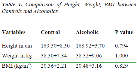
The results of this study are summarized in Tables 1–4. Mean age of controls and alcoholics were 35.8 10.1 years and 40.8 10.9 years respectively. Table 1 shows height, weight and Body Mass Index (BMI) measure-ments of controls and alcoholics subjects. BMI was calcu-lated based on height and weight. Values are within the normal range and there was no statistical significance
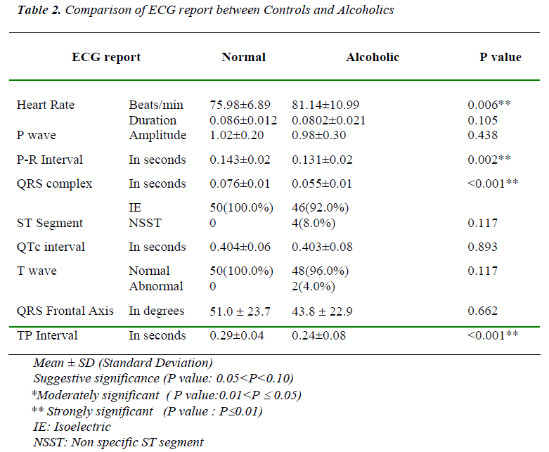
The electrocardiographic findings of controls and alcohol-ics subjects are listed in Table 2. Heart rate showed a sta-tistically significant increase in alcoholic groups com-pared to controls. Values of P-wave duration and ampli-tude do not show any statistically significance. P-R inter-val and QRS complex measurements showed statistically significant reduction in alcoholic group. Both Isoelectric (IE) and Nonspecific ST segment (NSST) values are within normal range and does not have any statistical sig-nificance. QTC interval, frontal axis and T-wave values are within normal limits. Although two subjects showed axis deviation of -30 and it was not statistically significant T-P interval values shows statistically significant decrease in alcoholic group compared to controls.
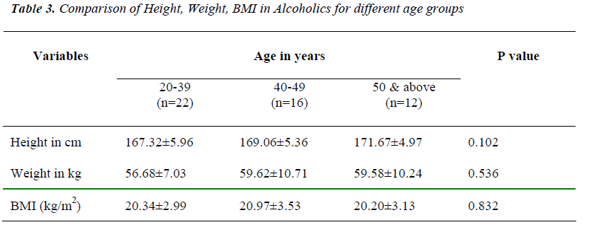
In Table 3 Height, weight and Body Mass Index (BMI) readings of alcoholics of different age groups, 20–39, 40-49 and > 50 years were shown. On comparison of all the three subgroups these dependent variables are within normal range and do not show any statistical significance.
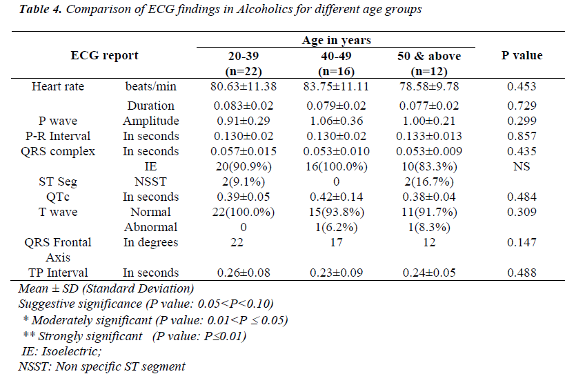
Table 4 shows ECG report of Alcoholics of 20–39, 40-49 and > 50 age groups. On comparison of these subgroups, electrocardiographic readings of Heart rate, P wave, PR interval, QRS duration, QTC interval, frontal axis, ST segment, T wave and TP interval values are within nor-mal limits and p value is >0.05 and thus the report does not have any statistical significance.
Discussion
Many workers did the work on effects of alcohol on elec-trocardiogram and showed positive results throughout the world over a long period of time. Excessive consumption of alcohol, in the absence of underlying organic heart dis-ease, may produce electrocardiographic abnormalities. These may at times imitate the changes produced by coronary artery disease, but the prognostic significance of the abnormal electrocardiogram would be quite different [8].
Ryan and Howes [14] in their study showed alcohol con-sumption is associated with reduced vagal activity, may be mainly due to a positive association between alcohol intake and heart rate in the age group of 33–68yrs.
Tetsuya Ohira et al [5] showed habitual alcohol intake was positively associated with Heart Rate compared with nondrinkers. This increase in heart rate could be due to decrease in peripheral resistance and increase in cardiac output. Our study results are also similar showing signifi-cant increase in heart rate in alcoholics.
The PR interval reflects the time needed to activate the atria, to conduct the impulse to the AV node and His bun-dle and start the ventricular depolarisation. QRS complex is because of ventricular depolarization. Lorsheyd A et al [15] showed that prolongation of the PR interval and QRS complex after acute ingestion of alcohol. In contrast our study there is statistically significant reduction P-R inter-val and QRS complex measurements.
QTC interval in the electrocardiogram includes both ven-tricular depolarization and repolarization times and varies inversely with the heart rate [1]. Lorsheyd A et al [15] studied acute effect of alcohol in healthy individuals and showed that QTC prolongation was seen in the subjects. In our study QTC interval does not show any statistical sig-nificance.
ST segment deviation from isoelectric line is a predictor of future coronary events in asymptomatic population. Klatsky [3] reported increasing intake of alcohol leads to cardiomyopathy and non specific ST-T changes in ECG. In our study three subjects showed non-specific ST changes in alcoholic group.
Robertson [9] in his study tells that as the age advances myocardium is also liable to cause disorders of rhythm, auricular fibrillation and low voltage of the T waves which may be due to early myocardial degeneration or coronary sclerosis. In our study on comparison of differ-ent age groups of alcoholic subjects does not show any statistically significant results indicating that electrocardi-graphic findings were similar in all 20–39, 40-49 and > 50 age groups.
In our study there is statistically significant decrease of TP interval in alcoholic group. TP interval change may be due to increased heart rate.
Conclusion
Excessive consumption of alcohol, in the absence of un-derlying organic heart disease, may produce electrocar-diographic abnormalities. In our study Heart rate was sig-nificantly increased indicating reduced vagal activity. There is reduction of P-R interval and QRS complex re-flecting the reduced spreading of depolarization from the sinus node to the atria and ventricles. TP interval reduc-tion may be due to increased heart rate. QTC interval does not show significant changes indicating no change in ven-tricular depolarization and repolarization. QRS frontal axis and ST segment does not show statistically signifi-cant results. In our study on comparison of different age subgroups of alcoholic subjects does not show any statis-tically significant results indicating that electrocardio-graphic findings were similar in all the age groups.
Our study results showed that alcoholics are prone for cardiovascular risk. Therefore, earlier detection of ECG changes is useful in preventing the cardiovascular risk.
References
- Goldberger AL. Electrocardiography. In: Braunwald E, Fauci AS, Kasper DL, Hauser SL, Longo DL, Jameson JL, Editors. Harrison’s Principles of Internal Medicine. Chapter 210. 16th ed. Newyork: Mc Graw Hill Co.; 2004: vol 1, pp. 1311-1319.
- Rossinen J, Sinisalo J, Partanen J, Nieminen MS, Viitasalo M. Effects of acute alcohol infusion on dura-tion and dispersion of QT interval in male patients with coronary artery disease and in healthy controls. Clin Cardiol.1999; 22: 591-594.
- Klatsky AL. Alcohol, coronary disease and hyperten-sion. Annu Rev Med.1996; 47: 149-160.
- Langer RD, Criqui MH and Reed DM. Lipoprotein and blood pressure as biological pathways for effect of moderate alcohol consumption on coronary heart dis-ease. Circulation.1992; 85: 910-915.
- Ohira T, Tanigawa T, Tabata M, Imano H, Kitamura A, Kiyama M et al. Effects of Habitual Alcohol Intake on Ambulatory Blood Pressure, Heart Rate, and Its Vari-ability among Japanese Men. Hypertension. 2009; 53:13
- William MJA, Restieaux NJ. Myocardial infarction in young people with normal coronary arteries. Heart.1998; 79: 191-194.
- Uyarel H, Ozdo¨l C, Karabulut A, Okmen E, Cam N. Acute alcohol intake and P-wave dispersion in healthy men – Original Investigation Anadolu Kardiyol Derg 2005; 5:289-93.
- Sereny G. Effects of Alcohol on the Electrocardiogram. Circulation 1971; 44; 558-564
- Robertson D. The Electrocardiogram in Senile Arterial Disease. Postgraduate Medical Journal; March 1955: 121-125
- Jorome HJ. Substance related disorders In: Sadock BJ, Sadock VA, editors Kaplan and Sadock’s Comprehen-sive Textbook of Psychiatry 7th ed. Philadelphia: Lip-pincott Williams and Wilkins Company; 1999, Vol. 2, pp. 928-932.
- Das SK, Balakrishnan V, Vasudevan DM. Alcohol: Its health and social impact in India. National Medical Journal of India. 2006; Vol.19, NO. 2
- D' Costa G, Nazareth I, Naik D, Vaidya R, Levy G, Patel V et al. Harmful Alcohol use in Goa, India, and its associations with violence: A Study in Primary Care. Alcohol and Alcoholism 2007; 42(2):131-137.
- Marc AS. Alcohol and alcoholism. In: Braunwald E, Fauci AS, Kasper DL, Hauser SL, Longo DL, Jameson JL, Editors. Horrison’s Principles of Internal Medicine. 15th ed. Newyork: Mc Graw Hill Co.; 2001: vol 2, pp. 2561-2566.
- Ryan JM and Howes LG. Relations between alcohol consumption, heart rate, and heart rate variability in men. Heart. 2002 December; 88(6): 641–642.
- Lorsheyd A, De Lange DW, Hijmering ML, Cramer MJM, Van de Wiel A. PR and QTC interval prolonga-tion on the electrocardiogram after binge drinking in healthy individuals. Neth J Med.2005; Feb; 63 (2): 59-63.


