Research Paper - Biomedical Research (2019) Volume 30, Issue 3
Effects of intervertebral disc degeneration on segmental rotation and intradisc pressure of c4-c6 cervical spine using finite element analysis
Varshini Karthik, Ashokkumar Devaraj*, Arun KuriakoseDepartment of Biomedical Engineering, SRM Institute of Science and Technology, Chennai, India
- Corresponding Author:
- Ashokkumar Devaraj
Department of Biomedical Engineering, SRM Institute of Science and Technology, Kattankulatur, Tamilnadu, India
Accepted date: March 6, 2019
DOI: 10.35841/biomedicalresearch.30-19-072
Visit for more related articles at Biomedical ResearchAbstract
The spinal segment C4-C6 of the cervical region is one of the most frequent site for injuries in spinal cord. Cervical degenerative disc disease develops due to loss of hydration in the intervertebral discs with increasing age. The spinal segment progresses to develop painful conditions due to high flexion and rotation movements in the upper extremity bones. The main aim of the study to analyse the effects of intervertebral disc degeneration on biomechanics of the cervical spine. The three dimensional computed tomography image was used and the finite element models of the cervical spine were developed with various levels of intervertebral disc degeneration at the C4-C6 functional spinal unit. The developed model is validated with the published data. The biomechanical effects of mild and moderate disc degeneration on the mechanical behaviour of cervical spine were studied using the developed Finite element models. The annulus fibrosis and nucleus pulposus material properties were varied to achieve moderate degeneration. The validated model is modified to various conditions with different mechanical properties and the effects intradisc pressure and progressive degeneration on the range of motion and on intradisc pressure. The analysis and the comparison is made between mild, moderate with normal human cervical spine in terms of progressive intervertebral disc degeneration. Based on this study the range of motion decreased with degeneration and the intra disc pressure increased with progressive degeneration.
Keywords
Disc degeneration, Finite element analysis, Intradisc pressure, Annulus fibrosus, Nucleus pulpous.
Introduction
The cervical vertebrae C1-C7 of spine has 7 vertebrae’s, which is referred to as neck region. The cervical vertebrae is flexible and strong which is subject to movements like flexion, extension and rotation and is responsible for the majority of the movements which our neck experiences. As compared to other vertebras of the spine, the cervical vertebra is much smaller and geometrically very complex.
In each of the seven vertebrae the intervertebral disc provides smooth elasticity to help in shock absorbance and also to prevent rubbing of the vertebrae with each other. C5-C6 is important because a particular spinal nerve that controls the arm and neck movements branches from the spinal nerve at this point. As a result cervical degenerative disc disease is responsible for the arm pain and neck pain thus limiting motion of the components.
More than 10 million cases per year are reported in India. It develops when the cushioning discs in the cervical spine starts to break down due to wear and tear. The material properties of the discs, the loading modes and the geometry of cervical spine are the key factors that help in modeling of the finite element model of C4-C6 cervical spine. Since cervical spine is a complex structure involving passive and active components, as a result we use finite element method which is cost effective and more reliable.
In early 1990, the 3D CT image was used assess the strength of the vertebral bone and also it used for finite element analysis (FEA) [1]. The FEA method is mainly applied to study the effect of human bone mechanics [2]. FEA method is used to determine the strength and mechanical stresses in various anatomical functions and also it can provide useful information [3,4]. The FE method offers the advantages in handling complex geometric configurations as well as material and geometric nonlinearity. The human cervical spine composed of hard tissue structures connected soft tissue like ligaments and intervertebral discs, as a result getting an accurate geometry is very complex.
In 1994, Natarajan RN et al. study exhibits the failure which starts in the end plate and not in the annulus, indicating that annulus is the weaker part in the spinal disc unit [5]. Yoganandan et al. provided information on modelling of cervical spine using finite element analysis [6]. Kumaresan et al. showed that the material properties of soft tissue structures have a major effect on the internal and external activities than the hard tissue structures [7]. Hong-Wan Ng, et.al provided the deeper view of various spinal components which provides stability and the results correlated with the experimental results and also with common clinical findings [8].
Later, Wang et al. found out through his study about the cervical spine response to the various properties of the ligament and he showed an influence on the intervertebral disc which is less with smaller range of motion (ROM) were the cervical ligaments are relaxed. He also suggested that neck posture control will have higher impact for the patients with a degenerated cervical spine [9] Kurowski et al. [10] in his study found out that the stress was more distributed toward the endplates and cortical wall in degenerated discs and highest effective stresses was concentrated near the spongy core.
The main aim was to conduct the study on von mises stress in the intervertebral disc with progressive degeneration and the level of degeneration was increased with various ROM for mild, moderately degenerated models under various movements.
Materials and Methods
Materials
The 3D CT image of a 35 year old male was taken from SRM Institute of medical sciences, Vadapalani, Chennai. The CT image is free from spine diseases and the image was in Digital Imaging and Communications in medicine (DICOM) format. The CT image is was imported to Materialise Interactive Medical Image Control System (MIMICS) (MIMICS 10.1, Materialise, Belgium) medical imaging software for extracting the surface model. This software used to design and model in 3D format. The imported image is extracted to create 3D surface model of C4-C6 of cervical vertebrae. The 3D surface model is imported to HYPERMESH software which will give the 3D meshed model and the meshed model was further imported to ANSYS software, where the material properties were assigned for analysis.
Methodology
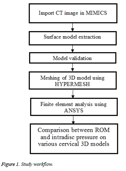
The detailed workflow of the study is given in Figure 1.
Image processing
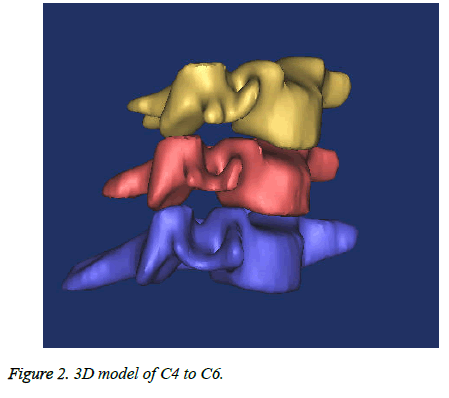
The CT images were obtained and it was imported in MIMICS for 3D segmentation of cervical spine model. The 3D image is pre-processed for segmentation. Segmentation process starts with thresholding. Thresholding is used to classify all pixels within a certain Hounsfield range as the same color or mask. By setting the lower threshold value, all pixels higher or equal to the set value will comprise the same mask. Region growing is useful for removing floating pixels. After segmenting all three axis (Sagittal coronal and axial), MIMICS can convert the surfaces in 3D geometry. The 3D model is smoothened and output of 3D segmental model is obtained for the cervical region (C4-C6). Figure 2 depicts the 3D surface model of C4- C6. The 3D model was next imported to HYPERMESH for the meshing procedure.
Meshing and analysis
Hypermesh is a pre-processing technique for finite element model which is used to analyze product design performance in highly interactive and visual environment. The advanced functions of hypermesh allows the user to efficiently manipulate geometry and mesh highly complex models. These functionalities include extensive meshing and model control, morphing technology to update existing meshes to new design proposals and the software focused on the process of 3D capture into usable 3D data so the model can be used in engineering and design.
The 3D segmented model obtained from mimics is imported to Hypermesh software for meshing where the model divided into smaller segments to get an accurate response from each segment. The smaller parts are the finite elements which will undergo meshing and the quality check is made. The interverteberal disc and endplates were developed using Hypermesh software without the actual geometry being lost. The material properties of the annulus fibrosus and nucleus pulposus were altered to achieve mild and moderate disc degeneration.
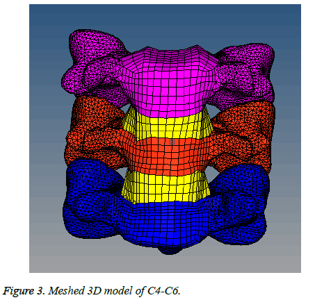
The meshed 3D model is validated with the published literature [8] where the element types of all models are solid 45. The material properties were assigned for every component in Hypermesh as specified in Table 1 [11,12] and once the validation has been done the model are ready for FEA using ANSYS. Figure 3 shows the 3D meshed model.
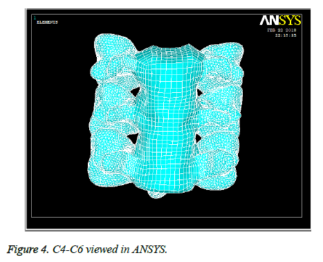
The 3D meshed model was given as input in ANSYS (ANSYS 14.0, Ansys, United States) and the analysis were done which is shown in Figure 4.
Results
Validation
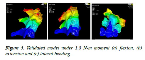
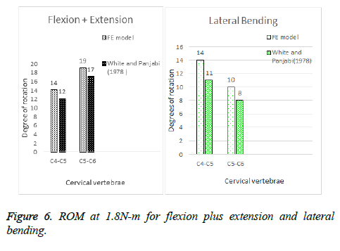
The 3D model was validated under different conditions such as flexion, extension and lateral bending, as it forms a link between the FE model developed using MIMICS and HYPERMESH. The detailed validated model is shown in Figure 5. The analysed values are compared with published papers with same load conditions. The spine components material properties are linear, homogeneous and isotropic. The boundary condition was such that the inferior-most vertebrae C6 was kept fixed (all degrees of freedom) and the superior cervical vertebrae was unconstrained from all degrees of freedom so that it could take the external load applied to achieve flexion, extension and lateral bending that is required for the validation. The initial and final position of the Finite element model is obtained by noting down the x, y and z coordinates. These coordinates are used to obtain to intersecting lines that will give the range of motion for the particular load in flexion, extension or lateral bending. The FE model ROM falls within the same range which is obtained by White and Panjabi [13] which is shown in Figure 6.
Influence of mild degeneration on ROM
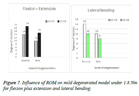
For mild degeneration, the nucleus pulposus material properties were altered i.e. Young’s modulus is taken as 0.3 MPa and Poisson Ratio is kept constant. When force moment 1.8 Nm was applied for flexion and extension to the mild degenerated model, the degrees of rotation slightly reduced from the values obtained from intact model when same load was applied as seen in Figure 7. For lateral bending, the degrees were slightly increased for C4-C5 level for both models i.e. mild and moderate degenerated FE model. From the study, we see that range of motion reduced as degeneration increased i.e. moderate degenerated model showing the least mobility. Further degeneration can lead to severe degeneration which can be later studied and is achieved by varying material properties and reduction of intervertebral disc height.
Influence of mild degeneration on intradisc pressure
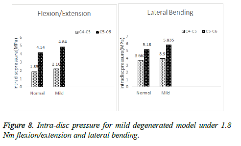
When 1.8 N-m load was applied to the mild degenerated model for flexion, extension and lateral bending the stress distribution in C4-C5 and C5-C6 showed an increase from the values obtained from intact model which can be seen in Figure 8.
Influence of ROM on moderate degeneration
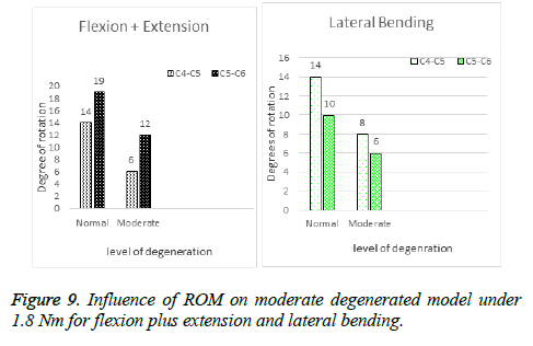
Moderate degeneration was achieved by altering the properties of annulus fibrous i.e. the Young’s modulus was kept as 3.0 MPa in addition to the dehydrated nucleus. ROM for C4-C5 and C5-C6 during flexion and extension in moderated degenerated model was obtained as 6 degree and 12 degree as shown in Figure 9 and for lateral bending C4-C5 and C5-C6 gave 8 degree and 6 degree which is shown in the same figure. When compared to mild, the moderate degenerated model showed less ROM during flexion/extension and lateral bending.
Influence on intradisc pressure for moderate degenerated model
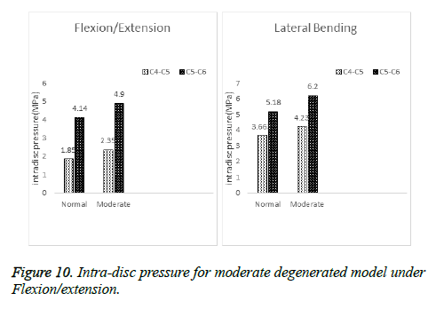
As we see in Figure 10, as severity of degeneration increases the von misses stress increases in moderate model as compared to normal and mild. The values showed a slight increase when compared to mild. This tells us that increase in degeneration can affect the intradisc pressure and biomechanics of the cervical spine. The pressure in the disc was calculated by selecting two elements on either ends of the intervertebral discs. Furthermore, a greater increase in degeneration can also cause bone spurs resulting from vertebrae rubbing against each other.
Discussion
The effects of degenerated intervertebral disc on ROM and various degenerated models with mild and moderate intradisc pressure were studied. The created 3D model of C4-C6 vertebrae with all material properties are used to achieve progressive degeneration. The segmented 3D model was carried out using MIMICS and similar kind of segmentation process was used in the cleft defect skull [14]. The 3D FE model was developed and analysed with a similar kind of process in a published literature [15].
The created 3D model was validated with published values of ROM. The degree of rotation of C4-C6 vertebrae are well within the range of published values [13]. The validated model was altered with a mild and moderate degeneration by assigning material properties for nucleus pulposus and annulus fibrosus. The material properties are taken as input with various degeneration levels which has obtained from the finite element modelling literature [16].
The results were obtained with changes in ROM of C4-C5 and C5-C6 vertebrae with an increment in disc degeneration. The applied load of 1.8 N-m force was fixed in C4-C6 by constraining all degrees of freedom. The intradisc pressure on C4-C6 was studied for different levels of degeneration.
Static, linear and steady state analysis was carried out. The ROM for moderate under flexion, extension and lateral bending was less as compared to the normal and mild condition. The intradisc pressure for moderate degenerated model showed a maximum stress as compared to mild and normal model when exercised under same load.
Artificial disc is a new technology which has a major impact on cervical vertebrae and it was common that patients with degenerative disc disease and other conditions have a cervical spinal fusion, which is an anterior cervical discectomy and fusion (ACDF). At present cervical disc replacement causes displacement, implant breakage and loosening, neurologic deficit as wells as adjacent disc degeneration. By studying the intradisc pressure and ROM on different degenerated models throws some light on developing disc implants with the appropriate materials like polyethylene, cobalt-chrome (CoCr) alloys, stainless steel, titanium (Ti) alloys, polyurethanes, Ti alloy-ceramic composites.
Conclusion
In summary, the effects of progressive degeneration on the intervertebral disc were studied using the FE model of the cervical vertebrae of spine (C4-C6) developed using the CT image and then it was validated against the existing literature to ensure that model behaves as a normal cervical spine under various loading conditions. The C5-C6 vertebrae disc showed an increase in stress than C4-C5 vertebrae disc. The range of motion for flexion/extension, lateral bending also reduced with increase in severity of degeneration. The results suggest that the cervical spine movement and behaviour is interconnected to the levels of degeneration. As degeneration increases, it becomes difficult for the cervical spine to attain that range of motion that is experienced by a healthy individual with a healthy disc. Since present methods do not offer much stability and restricts mobility, to an extent. This study shows the importance of developing disc implants. Further, intradisc pressure on intervertebral discs calculated with different loads throw light on the materials that can be used for the development of disc implants.
References
- Faulkner KG, Cann CE, Hasegawa BH. Effect of bone distribution on vertebral strength: assessment with patient-specific nonlinear finite element analysis. Radiology 1991; 179:669-674.
- Huiskes R, Chao EY. A survey of finite element analysis in orthopedic biomechanics: the first decade. J Biomech 1983; 16:385-409.
- Cattaneo PM, Dalstra M, Melsen B. The finite element method: a tool to study orthodontic tooth movement. J Dent Res 2005; 84:428-433.
- Su R, Campbell GM, Boyd SK. Establishment of an architecture-specific experimental validation approach for finite element modeling of bone by rapid prototyping and high resolution computed tomography. Med Eng Phys 2007; 29:480-490.
- Natarajan RN, Ke JH, Andersson GB. A model to study the disc degeneration process. Spine 1994; 19:259-265.
- Kumaresan S, Yoganandan N, Pintar FA. Finite element analysis of the cervical spine: a material property sensitivity study. Clin Biomech 1999; 14:41-53.
- Yoganandan N, Kumaresan S, Voo L, Pintar FA. Finite element applications in human cervical spine modeling. Spine 1996; 21:1824-1834.
- Ng HW, Teo EC. Nonlinear finite-element analysis of the lower cervical spine (C4–C6) under axial loading. J Spinal Disord 2001; 14:201-10.
- Wang K, Deng Z, Wang H, Li Z, Zhan H, Niu W. Influence of variations in stiffness of cervical ligaments on C5-C6 segment. J Mech Behav Biomed Mater 2017; 72:129-137.
- Kurowski P, Kubo A. The relationship of degeneration of the intervertebral disc to mechanical loading conditions on lumbar vertebrae. Spine 1986; 11:726-731.
- Kim YE, Goel VK, Weinstein JN, Lim TH. Effect of disc degeneration at one level on the adjacent level in axial mode. Spine 1991; 16:331-335.
- Yoganandan N, Kumaresan S, Voo L, Pintar FA. Finite element model of the human lower cervical spine: parametric analysis of the C4-C6 unit. J Biomech Eng 1997; 119:87-92.
- Panjabi MM. The basic kinematics of the human spine. A review of past and current knowledge. Spine 1978; 3:12-20.
- Balakumaran V, Harikrishnan P, Kumar DA, Magesh V. Three dimensional finite element model for a complicated craniofacial defect of human skull. InElectrical, Electronics, and Optimization Techniques (ICEEOT), International Conference IEEE 2016; 297-300.
- Kuriakose VA, Karthik V, Manickam PS, Kumar DA. A biomechanical study of cervical disc degeneration in C4-C6 using finite element analysis. InIOP Conference Series: Materials Science and Engineering 2018; 402:012007.
- Kumaresan S, Yoganandan N, Pintar FA, Maiman DJ, Goel VK. Contribution of disc degeneration to osteophyte formation in the cervical spine: a biomechanical investigation. J Orthop Res 2001; 19:977-984.
