Research Article - Current Pediatric Research (2022) Volume 26, Issue 3
Effects of core stabilization exercise and kinesio taping on pain, Cobb angle and endurance of trunk muscles in children and adolescents with idiopathic scoliosis.
Mohamed I Kamel1*, Ezzat El Sayed Elawh Moubarak2, Bassam A El-Nassag3, Ahmed Abd El-Moneim Abd El-Hakim4, Radwa S Abdulrahman5
1Department of Physical Therapy for Pediatrics, Cairo University, Cairo, Egypt
2Department of Physical Therapy, Cairo University Musculoskeletal Disorders and its Surgery, Zagazig University Hospital, Zagazig, Egypt
3Department of Physical Therapy for Neurology, Cairo University, Cairo, Egypt
4Department of Physical Therapy for Basic Sciences, Beni-Suef University, Cairo, Egypt
5Department of Physical therapy for Pediatrics, Cairo University, Cairo, Egypt
- Corresponding Author:
- Mohamed I Kamel
Department of Physical Therapy for Pediatrics, Cairo University, Cairo, Egypt
E-mail: mipt2002@hotmail.com
Received: 08 March, 2022, Manuscript No: AAJCP-22-53930; Editor assigned: 09 March, 2022, PreQC No: AAJCP-22-53930 (PQ); Reviewed: 14 March, 2022, QC No: AAJCP-22-53930; Revised: 21 March, 2022, Manuscript No: AAJCP-22-53930 (R); Published: 29 March, 2022. DOI:10.35841/0971-9032.26.3.1289-1296.
Abstract
Background/Objective of study: There are several types of therapeutic exercises in scoliosis rehabilitation. But there was a necessity to evaluate the effect of adding Kinesio Taping (KT) to Core Stabilization (CS) with traditional exercises on pain, Cobb angle and trunk extensor muscles’ endurance in children and Adolescents with Idiopathic Scoliosis (AIS). Materials and Methods: Sixty patients with AIS, cobb’s angle ranged 10°-25° and ages 9-16 years, were randomly allocated into two groups CS with traditional exercises and KT group A (n:30), and CS with traditional exercises only group B (n:30). The outcome measurements were, pain measured with Visual Analog Scale (VAS), Cobb angle measured with loaded poster anterior radiograph, trunk muscle endurance with Sorensen test, all were assessed at pretreatment and after intervention, using independent T-test for comparison between groups, and paired T-test for intergroup comparison. Subjects received 4 sessions every week, one hour every session, for both groups, for a total period of 12 weeks, and the KT was changed every week. Results: After three months of rehabilitation, both groups showed significant improvement in all measured variables, with high significant values in group (A) than group (B) patients at (p<0.05). Conclusion: Adding KT to CS exercises yields improvement on pain, reduction in Cobb’s angle and improvement in back muscle endurance more than CS alone in AIS.
Keywords
Core stabilization, AIS, Kinesio-taping, Pain, Cobb angle, Muscle endurance.
Introduction
Adolescent Idiopathic Scoliosis (AIS) is defined as a three-dimensional spine deformity with rotation of the spine in the horizontal plane and lateral bend in the frontal plane more than 10° when measured by x-ray, which produces a rib cage and flank muscles asymmetry [1], occurring in healthy pubertal children [2]. The prevalence of AIS with a curve angle of >10° is approximately 2.5% in the general population, so it is the most common deformity of the spine in the maturing population [3]. Adolescent idiopathic scoliosis is present in 2%–4% of children and occurs in ages 10 to 16 years old, girls being more at risk for severe progression by a ratio of 3.6 to 1 [4]. In AIS, improper mechanical forces acting on the spine led to physiological and biomechanical changes along the trunk segment. The trunk is the core segment of the body that controls the centre of gravity and maintains postural stability [5].
The causes of IS are not known, but it is considered to be multifactorial and includes imbalanced front and back vertebral growth. Genetic predisposition, abnormal muscle contraction mechanism, connective tissue and neurological abnormalities [6]. It mainly occurs in children and adolescents with a Cobb angle of 10 degrees or greater [6-8]. Scoliosis is a complex spinal deformity that can cause posture imbalance, decreased spinal movement, and muscle weakness in the spinal area, chronic pain, psychological problem, impaired exercise tolerance, muscle endurance and physical deconditioning which can also be early manifestations in patients with mild scoliosis [9]. Scoliosis also changes spinal mobility, flexibility, and recruitment of Para spinal muscles. It was considered that if the cobb angle exceeds the critical threshold (30° to 50°) before the completion of growth, and if the subjects untreated may experience one or more of following symptoms like hump on one side of spine, constipation, nerve compression, functional difficulties, and pain in many areas of the body as back, shoulders, and neck [10].
A subject with AIS has equal levels of self-reported physical activity as healthy subjects. Some authors have reported that staying in the sitting position for long periods of time and maintaining a static posture, without movement, and sometimes inadequate movement, can lead to postural alterations of the spine, such as scoliosis [11]. Treatment guidelines for IS are determined depending on age, residual growth, size and degree of the curvature, and underlying diseases. In particular, there is a high probability of incurvation of the spine being progressive in adolescents; therefore, aggressive, early treatment may be able prevent further spinal deformity to a certain degree [12].
Therapeutic approaches for IS include surgical and conservative treatments. Surgical treatment is considered when the Cobb angle is ≥ 40°, about 1 in 25 or 0.1% cases may need surgery. Gradually it is becoming obvious that if effective non-operative treatment is offered, more evidence in favour of conservative treatment for AIS is being gathered [13], whereas conservative treatment typically involves wearing braces, spinal adjustment therapy, and exercise therapy [14]. The goal of the treatment of scoliosis with a conservative approach is to prevent curve progression throughout pubertal growth. This will reduce or even avoid development of respiratory, cardiopulmonary problems and back pain. Treatment of scoliosis with exercises is a popular way to correct spine curvature. On the other hand, there are general physiotherapeutic exercises, like CS exercises, Pilates exercises, KT application, and yoga, which have recently been used in the conservative treatment of IS [15,16].
Core stabilization is a newly developed exercise approach aimed at increasing postural balance and avoiding compensatory movements by controlling the position of the trunk in static postures and functional activities. Core stabilization has been reported to improve muscle imbalance, particularly between Para spinal muscles, and multifidus thus enhancing local and global spinal stability. It has been found that CS to be more effective than general fitness exercises for improving spinal stabilization, decreasing Cobb’s angle and reducing pain scores in AIS [17-19]. Core stabilization exercises are more effective in reducing the angle of lumbar trunk rotation and pain than traditional exercises in moderate AIS [20]. Core stabilization improves the ability of the core muscles to correct and maintain the alignment of the spine. However, there are limited studies demonstrating the effectiveness of CS exercises and the most effective exercise method for the treatment of AIS remains controversial [21].
Kinesio Taping is linked to the tape that was developed by Dr. Kenzo Kase in 1979 and is still popular until now. It is an elastic tape designed to suit the properties of human skin surface. The tape flexibility is like the skin’s flexibility, with an elasticity ranged from 40 to 60% of its original length. It can also decrease the risk of skin irritation due to its latex adhesive. The correct application of this tape is designed to fulfil many goals: corrects muscle function by enhancing weak muscles, adjust movement of the joints through correcting misaligned joints by reducing muscle spasm and retrieving it back to the normal resting position, it can diminish pain through neurological suppression, stimulating receptors in the skin, and improving blood and lymph circulation beneath the skin [22].
Taping also has been shown to reduce sports injuries, osteoarthritis, and patellofemoral pain syndrome, reduces edema, myofascial pain syndrome, while also enhancing range of motion and improving endurance [23]. Kinesio Taping has also been shown to decrease discomfort. The effects of KT versus non elastic tape on children with IS were studied; in the KT group, the results showed a better spine angle and more muscular tone. The participating children’s everyday activities improved, and their pain decreased. Furthermore, KT has motivated the parents to encourage their children to exercise more [24].
Based on these information, CS exercise, traditional exercises and KT may be used effectively to increase back muscle endurance, relieve pain, improve muscle imbalance and decrease cobb’s angle, which are the causes of IS [25]. However, studies on the therapeutic effects of using CS exercises and KT in patients with AIS are still lacking. Accordingly, the objective of the present study was to identify the effects of CS with traditional exercises and KT methods on decreased the Cobb angle, relief pain and improves endurance of back muscles in patients with AIS.
Materials and Methods
Study design
Prospective randomized controlled clinical study conducted in outpatient clinic of Zagazig university hospital for 12 weeks. All parents and participants were informed that the collected data would be submitted for publication, and a consent form was signed before the study.
Ethical considerations
An ethical approval received from the Zagazig university institutional review board (ZU-IRB No. #6980, 2021) following the Declaration of Helsinki standards with a clinical trial registry in PACTR No, PACTR 202107516011646.
Sample size
All children and adolescents with AIS admitted to outpatient clinic during the period from 2021 to 2022 were enrolled in the study. The total numbers through this period (one year) were 61 subjects. One subject excluded from the study (not met the inclusion criteria) which made the sample as 60 subjects, divided randomly into two groups, 30 subjects in each group.
Patient enrolment
Sixty children and adolescents with IS were recruited to this study after evaluating by specialized orthopaedic surgeon, and confirmed by a radiologic examination. All of the evaluation and training procedures were explained before the beginning of the study, according to sample size, the participants’ demographic data, including age, sex and height are found in Table 1.
Inclusion criteria
Children and adolescents between the ages of 9-16 years old, having AIS confirmed by clinical and radiological examination with a Cobb’s angle of 10º-25º, patients’ history of complain of back pain for more than 3 months caused by scoliosis and able to participate and complete this study [26-29].
Exclusion criteria
Children and adolescents were excluded if they have any history of spinal surgery, history of allergies to KT, skin diseases such as eczema or psoriasis, congenital curve or due to (neuromuscular, rheumatologic, renal, cardiovascular, pulmonary or vestibular diseases), also patients with metabolic, infectious, traumatic conditions, psychological, psychiatric problems, and subjects with any other disorders which lead to changes in spinal curves or any back disorders, such as spondylolysis, spondylolisthesis and lumbosacral transitional anomalies that could be associated with back pain were excluded [30-33].
The sixty children and adolescents were randomly assigned into two groups by randomization through using graph pad software for subjects’ allocation to treatment groups. Group (A: n=30) received CS with traditional exercises and KT and group (B: n=30) received CS with traditional exercises only.
Procedure for assessment
Assessment of outcome was done before rehabilitation exercises for both groups and after 3 months of intervention program. Data were collected on the first and the last day, before and after rehabilitation exercise. The recorded data included: Pain severity (VAS) cm, Cobb’s angle, and back muscles endurance as the following:
Pain severity: Pain was measured using a subjective Visual Analogue Scale (VAS), which employs a 10 cm line with 0 (no pain) and 10 (extreme pain), where patient documents their level of pain on a straight line of 0 to 10 cm. Patient gave degree of their own sensation of pain without any input from other [30,31].
Cobb’s angle: The X-ray examination and Cobb angle measurement were performed by a single investigator who was a radiological technologist [32,33], the radiograph was taken with loaded poster anterior view, with the knees together, the chest and waist extended, the body weight equally distributed onto both lower limbs, and both arms elevated while holding one’s breath after exhaling a little. The angle obtained by tracing a line parallel to the superior endplate of one vertebra and tracing a line parallel to the inferior endplate of the vertebra was defined as the Cobb angle [34]. The intersection of these two lines detected the angle degree of deviation of the spine. The Cobb angle is used to know if the scoliosis degree improved or not, which is necessary to know the effectiveness of rehabilitation [35,36].
Endurance of back extensor muscle: Biering-Sorensen test was used to measure endurance of back extensor muscles. The subjects were positioned in a prone with a pillow under the lower abdomen, the upper edge of the iliac crests aligned with the bed edge and the upper trunk outside the bed. The lower limbs were strapped to the bed at pelvic level, knees, and ankles. Subjects were asked to maintain their original positions throughout the test as long as possible, with the arms folded across the chest. The test was terminated when the patient could not hold the test position any longer, and the time during which the subjects maintain the upper body horizontal and straight was measured. In patient who experienced no difficulty in maintaining the position, the test was stopped after 240 seconds [37,38].
Procedure for treatment include: Three months of core stabilization exercises with traditional exercises given to both groups (A and B), while KT was applied to group (A).
A program of core stabilization exercise: Both groups received, four sessions per week, one hour every session. The traditional exercises consisted of stretching, breathing and strengthening exercises. Stretching exercises aimed to correct the apex of the curve to the midline and passively over correct the curve. Strengthening exercises performed on the convex side of the curve, through positioning, and resistance graduation [39,40].
All exercises in the session included a 10 minute warm-up, a 40 minute CS exercise, and a 10 minute cool down. The twenty minutes of stretching, strengthening and breathing exercises (traditional exercises) made up the warm-up and cool down activities. The purpose of the CS exercises in scoliosis is to improve the ability of the core muscles to regain the dynamic control of internal and external forces over the spine. CS training gradually progressed from the stability of local core muscles (transversus muscles, multifidus, and diaphragm) in static positions to global muscle stability training (internal and external oblique abdominal muscles, psoas major, quadratus lumborum, and pelvic floor muscles), global muscle mobility, and strength training (rectus abdominis, back extensor muscles, and hamstring muscles) in dynamic body positions [41,42].
Core stabilization exercises as pelvic tilt, elbow toe, cat–camel posture (back rises), basic trunk curl (crunch), back bridge (with knee extension), side bridge, double-leg abdominal press, superman’s, arm/leg raises prone, quadruped arm leg raises, and hand walkout were among the core stability exercise, three sets of 12 repetitions were performed for each type of exercise or according patient tolerance [43]. Patients who could not manage to complete the program continued with the same exercises and performed a few simple exercises from the next level with fewer repetitions [44]. Kinesio Taping (KT) applications (group A): Taping was used for neurological facilitation of corrected positions. Facilitation on convex side of curve and mechanical correction on rib cage to help the individual for derotation and deflexion [45,46].
The KT was done with 25%-35% tension, ends with no tension, along the convex side of the spine. On the concave side, the tape was applied with zero tension. Taping was applied to the spinal is muscle for correction of the spine and was applied to the direction of the beginning and end of the curvature [47]. Taping was applied with trunk lateral flexion, rotation to the opposite side and inhalation. Each subject’s shoulder and back muscles connected with the spine were assessed for strength and tension [48-50]. The KT was then applied accordingly on the muscles that had been identified as being affected. If a muscle was very weak, KT was used to facilitate it, mounting it from the origin to the insertion of the muscle at 15 to 35 strains. Also, KT was used to achieve muscle inhibition its mounting should be from the insertion site to the origin of the muscle at 15%-25% tension. Every week the tape was changed and replaced with new tape (KT applied for five-days before removal and two days interval was maintained before the patient given another KT applications) [51-53].
Statistical analysis
All statistical measures were performed through the Statistical Package for Social Studies (SPSS) version 22 for windows. Before final analysis, data were screened for normality assumption and presence of extreme scores. To determine the homogeneity of the groups at baseline, subject age, height, and body weight were compared using independent t-tests. The current test involved two independent variables. The first one was the (tested group); between subjects’ factor which had two levels (group A receiving CS with KT and group B receiving same CS exercise). The within-subject factor which had two levels (pre and post). In addition, this test involved three tested dependent variables (VAS, Cobb’s angle and back muscle endurance).
Results
Demographic data and baseline characteristics
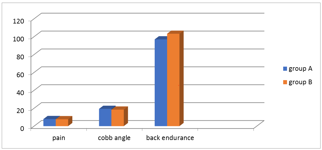
There were no significant differences between both groups in age, height, and weight (P>0.05). Also, there were no significant differences between the two groups pre-treatment in all variables, pain, cobb’s angle, and back muscle endurance (P>0.05), as shown in Table 1.
| Characteristics | Group A (n=30) Mean ± SD | Group B (n=30) Mean ± SD | p-value |
|---|---|---|---|
| Age (years) | 12.91 ± 1.401 | 13 ± 1.71 | 0.797 |
| Height (cm) | 153.01 ± 10.71 | 158.01 ± 10.51 | 0.483 |
| Weight (kg) | 44.11 ± 8.1 | 50.6 ± 10.01 | 0.228 |
| Sex (girls/boys) | 20/10 | 22/8 | |
| Pain (VAS) cm | 7.86 ± o.89 | 7.71 ± 0.79 | 0.513 |
| Cobb’s angle(o) | 19.29 ± 2.32 | 18.46 ± 1.98 | 0.144 |
| Back muscle endurance(second) | 96.06 ± 14.88 | 103.23 ± 14.71 | 0.106 |
Table 1. Demographic data and baseline characteristics of both groups: P>0.05 was considered no statistically significant differences, SD: Standard Deviation; VAS: Visual Analogue Scale.
Comparison of both groups’ post-treatment
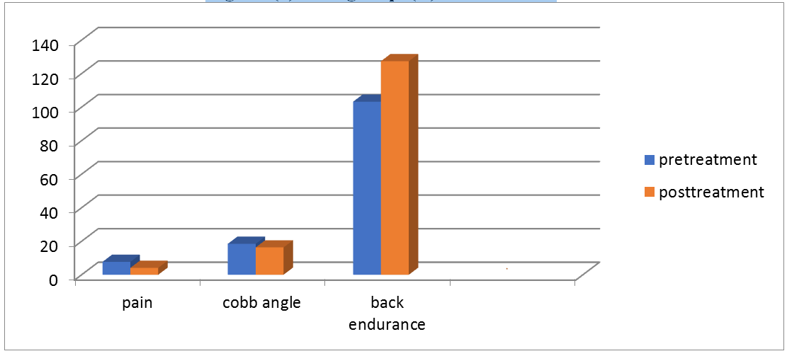
Post-treatment the group (A) showed significant differences in pain, Cobb’s angle, back muscles endurance between pretreatment and post-treatment (p<0.05). Also, group (B) showed significant differences in all variables after rehabilitation between pre-treatment and post-treatment (p<0.05) the detailed are presented in Table 2; Figure 1 and 2.
| Variables | Group A (n=30) Mean ± SD | Group B (n=30) Mean ± SD | ||||
|---|---|---|---|---|---|---|
| Pre-treatment | Post-treatment | P-value | Pre-treatment | Post-treatment | P-value | |
| Pain (VAS) cm | 7.86 ± 0.89 | 3.33 ± 0.71 | 0 | 7.71 ± 0.79 | 4.12 ± 0.41 | 0 |
| Cobb’s angle(o) | 19.29 ± 2.32 | 14.95 ± 2.38 | 0 | 18.46 ± 1.98 | 16.3 ± 2.11 | 0 |
| Back muscle endurance(second) | 96.96 ± 14.88 | 137.33 ± 12.89 | 0 | 103.23 ± 14.71 | 127.4 ± 11.31 | 0 |
Table 2. Intergroup comparison for each group pre and post rehabilitation. p<0.05 was considered statistically significant differences, SD: Standard Deviation, VAS: Visual Analogue Scale.
Comparison between groups after rehabilitation
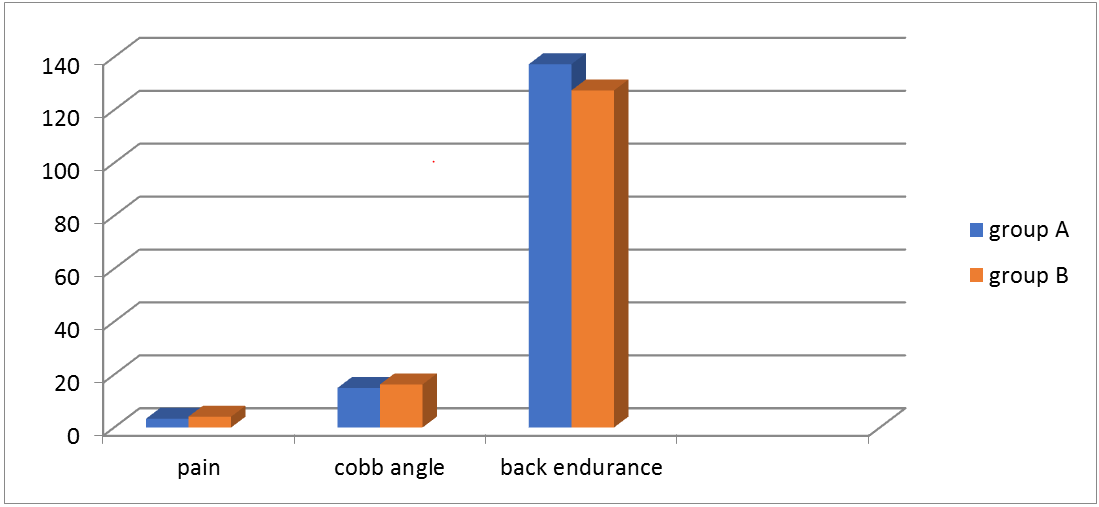
There were significant differences between groups regarding to pain, cobb’s angle, and back muscles endurance after rehabilitation with favor to group (A), (p<0.05), as shown in Table 3 Figure 4.
| Variables | Group | Pre-treatment | Post-treatment | ||
|---|---|---|---|---|---|
| Mean ± SD | P | Mean ± SD | P | ||
| Pain (VAS)cm | A | 7.86 ± 0.89 | 0.513 | 3.33 ± 0.71 | 0 |
| B | 7.71 ± 0.79 | 4.12 ± 0.41 | |||
| Cobb’s angle (o) | A | 19.29 ± 2.32 | 0.144 | 14.95 ± 2.38 | 0.024 |
| B | 18.46 ± 1.98 | 16.3 ± 2.11 | |||
| Back muscle endurance (second) | A | 96.96 ± 14.88 | 0.106 | 137.33 ± 12.89 | 0.002 |
| B | 103.23 ± 14.71 | 127.4 ± 11.31 | |||
Table 3. Comparison between groups (A and B). p<0.05 was considered statistically significant differences, SD: Standard Deviation, VAS: Visual Analogue Scale.
Discussion
The additional effect of Kinesio Taping (KT) to Core Stabilization training (CS) in the management of AIS was examined in this study. After 12 weeks of treatment, the study and control groups demonstrated a significant decrease in pain, a decrease in Cobb angle, and an increase in back muscle endurance, with favor to group A that had the KT in conjunction with the core stability exercise regimen.
The benefits of core stability exercise have been established for reducing pain in patients with persistent lower back pain, improving athletic performance, and preventing sports injuries in athletes conveyed from their study that, eight weeks of spinal stabilization exercises led to a signi?cantly reduced numeric pain rating scale score in subjects with IS and lower back pain. A study conducted by showed that six weeks of KT with exercises led to reduction of pain score on VAS scale in patient with scoliosis. Additionally, reported that pain severity was reduced in patients received either therapeutic exercise for scoliosis augmented by KT or therapeutic exercise only in female adolescents complained from scoliosis.
In their study, found that using KT with tension significantly relieves low back pain quickly after application and has a favorable effect on one's quality of life. Accordingly, KT could be an effective treatment for subject with AIS who suffer from back discomfort. Also studied the efficacy of physiotherapy on pain in subjects with adolescent and adult IS, and found significant reduction in VAS scale. Vercelli et al. proposed that, KT improved blood and lymphatic reflux by causing skin wrinkles, which increased the distance between the skin and the underlying connective tissue. It also improves proprioceptive input, pain suppression, stability, and range of motion. Furthermore, according to Paolini et al. the cutaneous stretch stimulation allowed by KT may interfere with the transmission of mechanical and painful stimuli; delivering afferent stimuli that facilitate pain inhibitory mechanisms and pain reduction. Hwang-Bo et al. demonstrated in a case report that, normalized muscle performance is believed to be the cause of pain relieve in acute back pain syndrome. Despite the fact that KT decrease pain severity, especially in the short term, all former reviews agreed that there is no high-quality evidence for its use in patients with musculoskeletal diseases.
Our study reported a statistically significant reduction in Cobb's angle in the post-treatment difference between studied groups with favor to the core stability with KT, besides the improvement within each group when comparing pre with post-treatment results. In the CS with KT and CS groups, the Cobb angle decreased by 4.34 and 2.16 degrees, respectively.
In consistence with the present study results of reduced cobb angle in all treated patients, Hirano et al. have found that CS exercise reduces thoracic kyphosis and lumbar lordosis in the sagittal plane of the spine and improves spinal alignment which could be attributed to the long duration of training for participated patients. It was reported that, a 10-week CS exercise lowered thoracic and lumbar Cobb angles in primary school children with scoliosis, according to Gür et al. and Park et al. also conveyed that, college students with scoliosis had decreased their Cobb angle after 10-week of CS program. Ko KJ et al. stated that, 12 weeks of CS training for adolescents with IS reduced the thoracic and lumber cobb angle but the differences between the exercise and the control groups were significant only in the lumber angles. Also Balne et al. found that there is less curve correction may be up to 2 degrees, and rate of progression was reduced after application of CS exercises and KT application of physiotherapy in subjects with adolescent and adult idiopathic structural scoliosis.
In contrast to the results of current study, Weinstein et al. claimed that, no definite evidence has found that physiotherapy or bracing decrease the risk of scoliotic angle progression, normalize the existing deformity, or reduces the need for operation, also Helmy et al. reported no improvements in the Cobb's angle between pre and post treatment evaluations in patients treated with KT and exercises or the control groups. Brooks et al. found that Scoliosis-specific exercises, in combination with other types of physical therapy, have been recommended as a way to slow or stop curve advancement. Furthermore, a case report by Negrini et al. stated an 18.5 Cobb angle reduction after 12 months of scoliosis specific exercises workouts. In previous research that focused on the effect of intervention on Cobb's angle; the authors found that pilates-based exercises reduced Cobb's angle and that the core muscle release technique corrected Cobb's angle in scoliotic patients better than general exercise and electrotherapy Lee et al.
Studies reported a “substantial” change in the cure angle following rehabilitation, were in fact; of modest magnitude and did not take into consideration the claimed inter or intra-observer error rate. These studies had insufficient analysis and failed to mention if the slight improvements observed were sustained over time Simon et al. revealed that, in both AIS and adult scoliosis patients, the KT is used to help subjects to 'maintain' their corrected posture as applied in (schroth method, scientific exercises approach to scoliosis); decrease of pain in long-standing scoliosis subjects with postural collapse; and assistance with pulmonary function in subjects with neuromuscular or IS scoliosis. This was supported by research investigated the effects of applying KT combined with strengthening exercises on the curve angle, the stiffness and tone of upper trapezius muscle in subject with AIS, in which all variables were significantly reduced after the rehabilitations.
Eva et al. studied the short-term (3-day) influence of KT applied to the muscular groups implicated in the establishment of functional scoliosis, on muscle tonus and elasticity parameters for the assessed muscles. The post-intervention results showed a significant increase in flexibility in the tested muscles, indicating that kinesiological tape can alter the muscle's "structure" as well as the postural behavior of non-structural scoliosis, providing the necessary support to rectify a posture deficiency.
As showed in the findings of this study; endurance of back muscles was raised in both studied groups following the intervention program. It came in line with Roongtip et al. who researched the effects of eight weeks of three-dimensional schroth exercises and KT on trunk muscle strength and endurance in children with idiopathic scoliosis. The study revealed an increase in the back extensor muscles’ endurance. Schreiber et al. findings; who discovered that scoliosis patients who received conventional therapy and underwent schroth exercises had greater back muscle strength and endurance than those who simply received standard care.
In people with low back pain, athletes, and healthy people; CS exercise that focused on trunk coordination and control was shown to be effective for increasing the endurance and strength of back stabilizer muscles, improving intersegmental trunk coordination, and maintaining a neutral spine [54].
In individuals with lower back discomfort, CS exercise increases lumbar and truncal muscular strength, according to Moon et al. added that, CS exercise is helpful for increasing muscle strength and range of motion for lumbar spine in subjects with IS. This might be due to the fact that CS exercise aids in the enhancement of muscular functions through deep muscle co-contraction and surface muscle cooperation. Consequently, in individuals with AIS, CS exercise can be deemed to have a favorable influence on the normal physiological curvature of the spine [55].
Alvarez et al. demonstrated that KT had a good effect on low back muscle exhaustion by displaying an increase in back extensor muscle resistance, which is an essential factor in back pain therapy. These findings corroborate those of Castro et al. who found that persons with back pain who had KT had a considerably lower level of impairment and improved trunk muscular functional endurance. Our findings have significant implications for schools and school-based health professionals involved in the prevention and early identification of spinal postural abnormalities [56].
There are a few limitations in this study; first, because there was no defined recorded, dose-specific home-exercise maintained during rehabilitation, the effect of the rehabilitation program on patients' psychological effects such as quality of life was not evaluated, and because the number of participants was small, generalizing the findings of this study would be difficult. Furthermore, because this study used a short-term intervention, it would be important to do a follow-up to assess the exercise's long-term benefits. Strengths of this study were the application of same inclusion and exclusion criteria to both groups for a homogeneous comparison [57].
Recommendation
Future studies should use the randomized controlled trial study design to better assess, and compare the effects of combining many different types of exercises or physical therapy modalities, in schools with different levels and hospital outpatient clinics.
Conclusion
The current study’s results showed that in the CS with traditional exercises and KT group, the pain severity reduced, the Cobb angle decreased and trunk muscle endurance increased, more than CS with traditional exercises alone. Therefore, adding KT to CS with traditional exercises is an effective method for decreasing pain, improving curve angle and increasing back extensor muscle endurance in patients with AIS.
References
- Ghandehari H, Mahabadi MA, Mahdavi SM, et al. Evaluation of patient outcome and satisfaction after surgical treatment of adolescent idiopathic scoliosis using scoliosis research society-30. Arch Bone Jt Surg 2015; 3(2): 109-13. [Crossref][Google Scholar][Indexed]
- Negrini S, Donzelli S, Aulisa AG, et al. 2016 SOSORT guidelines: Orthopaedic and rehabilitation treatment of idiopathic scoliosis during growth. Scoliosis Spinal Disord 2018; 13: 3. [Crossref][Google Scholar][Indexed]
- Weinstein SL, Dolan LA, Cheng JC, et al. Adolescent idiopathic scoliosis. Lancet 2008; 371(9623): 1527-37. [Crossref][Google Scholar]
- Konieczny MR, Senyurt H, Krauspe R. Epidemiology of adolescent idiopathic scoliosis. J Child Orthop 2013; 7 (1): 3-9. [Crossref][Google Scholar][Indexed]
- Monticone M, Ambrosini E, Cazzaniga D, et al. Active self-correction and task-oriented exercises reduce spinal deformity and improve quality of life in subjects with mild adolescent idiopathic scoliosis. Results of a randomised controlled trial. Eur Spine J 2014; 23: 1204–1214. [Crossref][Google Scholar][Indexed]
- Ayhan C, Unal E, Yakut Y. Core stabilisation reduces compensatory movement patterns in patients with injury to the arm: A randomized controlled trial. Clin Rehabil 2014; 28(1): 36–47. [Crossref][Google Scholar][Indexed]
- Jada A, Mackel CE, Hwang SW, et al. Evaluation and management of adolescent idiopathic scoliosis: A review. Neurosurg Focus 2017; 43(4): E2. [Crossref][Google Scholar][Indexed]
- Weinstein SL, Dolan LA. The evidence bases for the prognosis and treatment of adolescent idiopathic scoliosis: The 2015 orthopaedic research and education foundation 'clinical research award. J Bone Joint Surg Am 2015; 97 (22): 1899-903. [Crossref][Google Scholar][Indexed]
- Balne NK, Afshan SJ, Mathukumall N: Efficacy of physiotherapy on spinal mobility parameters and pain in persons with adolescent and adult idiopathic structural scoliosis. Indian J Physiother Occup Ther 2021; 17: 179-187. [Crossref][Google Scholar]
- Diarbakerli E, Grauers A, Moller H. Adolescents with and without idiopathic scoliosis have similar self-reported level of physical activity: A cross-sectional study. Scoliosis Spinal Disord 2016; 11: 17. [Crossref][Google Scholar][Indexed]
- Ko KJ, Kang SJ. Effects of 12-week core stabilization exercise on the Cobb angle and lumbar muscle strength of adolescents with idiopathic scoliosis. J Exerc Rehabil 2017; 13(2): 244-249. [Crossref][Google Scholar][Indexed]
- Hagit B, Victoria AL, Bettany-Saltikov J. Physiotherapy scoliosis specific exercises–a comprehensive review of seven major schools. Scoliosis Spinal Disord 2016; 11. [Crossref][Google Scholar]
- Sungyoung Y, Min-Hyung R. Effect of physical therapy scoliosis specific exercises using breathing pattern on adolescent idiopathic scoliosis. J Phys Ther Sci 2016; 28(11): 3261–3263. [Crossref][Google Scholar][Indexed]
- Alves de Araujo ME, Bezerrada SE, Bragade MD, et al. The effectiveness of the pilates method: Reducing the degree of non-structural scoliosis, and improving flexibility and pain in female college students. J Bodyw Mov Ther 2012; 16(2): 191–198. [Crossref][Google Scholar][Indexed]
- Gür G, Ayhan C, Yakut Y. The effectiveness of core stabilization exercise in adolescent idiopathic scoliosis: A randomized controlled trial. Prosthet Orthot Int 2017; 41(3): 303–310. [Crossref][Google Scholar][Indexed]
- Hides J, Wilson S, Stanton W, et al. An MRI investigation into the function of the transversus abdominis muscle during "drawing-in" of the abdominal wall. Spine 2006; 31(6): E175–E178. [Crossref][Google Scholar][Indexed]
- Shin SS, Lee YW, Song CH. Effects of lumbar stabilization exercise on postural sway of patients with adolescent idiopathic scoliosis during quiet sitting. J Phys Ther Sci 2012; 24(2): 211–215. [Crossref][Google Scholar]
- Emery K, De Serres SJ, McMillan A, et al. The effects of a pilates training program on arm–trunk posture and movement. Clin Biomech 2010; 25(2): 124–130. [Crossref][Google Scholar][Indexed]
- Bassett KT, Stacey LA, Ellis RF. The use and treatment efficacy of kinaesthetic taping for musculoskeletal conditions: A systematic review. New Zealand J Physiother 2010; 38, 56-62.
- Williams S, Whatman C, Hume PA, et al. Kinesio taping in treatment and prevention of sports injuries, A meta-analysis of the evidence for its effectiveness. Sports Med Arthrosc Rev 2012; 42, 153-64. [Crossref][Google Scholar][Indexed]
- Álvarez-Álvarez S, José FG, Rodríguez-Fernández AL, et al. Effects of Kinesio Tape in low back muscle fatigue: Randomized, controlled, doubled-blinded clinical trial on healthy subjects. J Back Musculoskelet Rehabil 2014; 27: 203-12. [Crossref][Google Scholar][Indexed]
- Zakaria AA, Hafez AR, Buragadda S, et al. Stretching versus mechanical traction of the spine in treatment of idiopathic scoliosis. J Phys Ther Sci 2012; 24: 1127-31. [Crossref][Google Scholar]
- Barr KP, Griggs M, Cadby T. Lumbar stabilization: Core concepts and current literature, Part 1. Am J Phys Med Rehabil 2005; 84(6):473-80. [Crossref][Google Scholar][Indexed]
- Roongtip D, Teerapat L, Numchai R, et al. Effects of Three-Dimension Schroth Exercises and Kinesio Taping on General Mobility of Vertebrae, Angle of Trunk Rotation, Muscle Strength and Endurance of Trunk, and Inspiratory and Expiratory Muscle Strength in Children with Idiopathic Scoliosis. Walailak J Sci Tech 2019; 16(12): 965-973. [Crossref][Google Scholar]
- Yagci G, Yakut Y. Core stabilization exercises versus scoliosis-specific exercises in moderate idiopathic scoliosis treatment. Prosthet Orthot Intl 2019; 43(3): 301–308. [Crossref][Google Scholar][Indexed]
- Haefeli MEA. Pain assessment. Eur Spine J 2005; 15(suppl 1): S17-24. [Crossref][Google Scholar][Indexed]
- Harrison DE, Cailliet R, Harrison DD, et al. Reliability of centroid, Cobb, and harrison posterior tangent methods. Spine (Phila Pa 1976) 2001; 26: E227–E234. [Crossref][Google Scholar][Indexed]
- Malfair D, Flemming AK, Dvorak MF, et al. Radiographic evaluation of scoliosis: Review. AJR Am J Roentgenol 2010; 194: S8–S22. [Crossref][Google Scholar][Indexed]
- Deacon P, Flood BM, Dickson RA. Idiopathic scoliosis in three dimensions. A radiographic and morphometric analysis. J Bone Joint Surg Br 1984; 66: 509-512. [Crossref][Google Scholar][Indexed]
- Modi HN, Chen T, Suh SW, et al. Observer reliability between juvenile and adolescent idiopathic scoliosis in measurement of stable Cobb's angle. Eur Spine J 2009; 18(1): 52-58. [Crossref][Google Scholar][Indexed]
- Biering-Sorensen F. Physical measurements as risk indicators for low-back trouble over a one-year period. Spine 1984; 9(2): 106-19. [Crossref][Google Scholar][Indexed]
- Ebnezar J. Essentials of orthopedics for physiotherapists. (2nd edn) 2011; 4: 288-93.
- Ru Ed. The possibilities of using elastic therapeutic (Kinesio) tape in patients with scoliosis. 2014, (suppl 1) P12. [Crossref][Google Scholar]
- Yunus A, Canan GA, Aysegul A, et al. The effect of Kinesio taping on back pain in patients with Lenke Type 1 adolescent idiopathic scoliosis: A randomized controlled trial. Acta Orthop Traumatol Turc 2017; 51: 191-196. [Crossref][Google Scholar][Indexed]
- Gözde Y, Elif T, Yavuz Y. Effect of elastic scapular taping on shoulder and spine kinematics in adolescents with idiopathic scoliosis. Acta Orthop Traumatol Turc 2020; 54(3): 276-286. [Crossref][Google Scholar][Indexed]
- Choi T-S, Choi W-S, Choi J-H. Effects of Kinesio-taping and Strengthening Exercise on Cobb Angle and Muscle Tone in Patients with Idiopathic Scoliosis. Research J Pharm Tech 2019; 12(9): 4438-4442. [Crossref][Google Scholar]
- Akuthota V, Ferreiro A, Moore T, et al. Core stability exercise principles. Curr Sports Med Rep 2008; 7: 39-44. [Crossref][Google Scholar][Indexed]
- Huxel Bliven KC, Anderson BE. Core stability training for injury prevention. Sports Health 2013;5:514-522. [Crossref][Google Scholar][Indexed]
- Zapata K, Parent EC, Sucato D. Immediate effects of scoliosis-specific corrective exercises on the Cobb angle after one week and after one year of practice. Scoliosis Spinal Disord 2016; 11(Suppl. 2): 36. [Crossref][Google Scholar][Indexed]
- Vercelli S, Colombo C, Tolosa F, et al. The effects of kinesio taping on the color intensity of superficial skin hematomas: A pilot study. Phys Ther Sport 2017; 23: 156–161. [Crossref][Google Scholar][Indexed]
- Bischoff L, Babisch C, Babisch J, et al. Effects on proprioception by Kinesio taping of the knee after anterior cruciate ligament rupture. Eur J Orthop Surg Traumatol 2018; 28(6): 1157–1164. [Crossref][Google Scholar][Indexed]
- Kasawara KT, Mapa JMR, Ferreira V, et al. Effects of Kinesio Tapi on breast cancer-related lymphedema: A meta-analysis in clinical trials. Physiother Theory Pract 2018; 34: 337–345. [Crossref][Google Scholar][Indexed]
- Gulpinar D, Tekeli Ozer S, Yesilyaprak SS. Effects of rigid and kinesio taping on shoulder rotation motions, posterior shoulder tightness, and posture in overhead athletes: A randomized controlled trial. J Sport Rehabil 2019; 28: 256–265. [Crossref][Google Scholar][Indexed]
- Hwang-Bo G, Lee JH. Effects of Kinesio taping in a physical therapist with acute low back pain due to patient handling: A case report. Int J Occup Med Environ Health 2011; 24: 320–323. [Crossref][Google Scholar][Indexed]
- Castro-Sanchez A, Inmaculada C, Guillermo A, et al. Kinesio taping reduces disability and pain slightly in chronic non-specific low back pain: A randomised trial. J Physio 2012; (58): 89-95. [Crossref] [Google Scholar][Indexed]
- Added MA, Costa LO, Fukuda TY. Efficacy of adding the Kinesio taping method to guideline-endorsed conventional physiotherapy in patients with chronic nonspecific low back pain: A randomised controlled trial. BMC Musculoskelet Disord 2013; 14: 301. [Crossref][Google Scholar][Indexed]
- Hirano K, Imagama S, Hasegawa Y, et al. Effect of back muscle strength and sagittal spinal imbalance on locomotive syndrome in Japanese men. Orthopedics 2012; 35: e1073-1078. [Crossref][Google Scholar][Indexed]
- Park YH, Park YS, Lee YT, et al. The effect of a core exercise program on Cobb angle and back muscle activity in male students with functional scoliosis: A prospective, randomized, parallel-group, comparative study. J Int Med Res 2016; 44: 728-734. [Crossref][Google Scholar][Indexed]
- Brooks WJ, Krupinski EA, Hawes MC. Reversal of childhood idiopathic scoliosis in an adult, without surgery: a case report and literature review. Scoliosis 2009; 4: 27. [Crossref] -->


