Research Article - Biomedical Research (2021) Volume 32, Issue 1
Effect on malondialdehyde, superoxide dismutase and reduced glutathione and its metabolizing enzymes in vitamin D deficient newly diagnosed Type- 2 diabetic patients.
Kuldip Singh*
Department of Biochemistry, Government Medical College, Patiala, Punjab, India
- Corresponding Author:
- Kuldip Singh
Department of Biochemistry
Government Medical College
Patiala Punjab
India
Accepted on December 07, 2020
Abstract
Background: India, with 32 million diabetic individuals, currently has the highest incidence of diabetes worldwide. Recently, World Health Organization (WHO) reported that these numbers are predicted to increase to 80 million by the year 2030. Vitamin D regulates glucose metabolism and oxidative stress is a well-established factor of this multi-factorial disease. Aim: Present, study was designed to evaluate certain oxidative stress markers like malondialdehyde, superoxide dismutase, reduced glutathione, glutathione reductase, Glutathione Peroxidase (GPx) along with total antioxidant activity in vitamin D deficient newly diagnosed Type 2 diabetics. Methods: 150 vitamin D deficient newly diagnosed type 2 diabetics’ and equal number of healthy subjects of both genders were recruited. Fasting blood was collected for evaluation of glucose, 25(OH) D and oxidative stress markers. Results: Significant (P<0.01) increase in malondialdehyde by 51.03% while a significant decrease in oxidative stress markers like SOD, GSH, GR, GPx and total antioxidant activity by 27.91 (P<0.05), 26.14% (P<0.05), 32.04% (P<0.01), 25.41% (P<0.05) and 35.85% (P<0.01) respectively was recorded in vitamin D deficient newly diagnosed type 2 diabetics with respect to healthy controls. Conclusions: A fore mentioned observations suggested that vitamin-D deficient newly diagnosed Type-2 diabetics of North-West Indian’s are associated with oxidative stress, a hallmark of various diseases like Diabetes Mellitus, cardiovascular diseases, osteoporosis etc. Therapeutic interventions in combinations of lifestyle and dietary modification might be beneficial to prevent further risk of development of Diabetes Mellitus and cardiovascular diseases like atherosclerosis in North-West Indians.
Keywords
Diabetes Mellitus, Vitamin-D, Malondialdehyde, Oxidative stress markers, Total anti-oxidant, Cardiovascular diseases.
Introduction
With the Vitamin D deficiency is a major public health problem worldwide. The incidences of Diabetes Mellitus (DM) is increasing prevalence of DM all over the world, it is expected that this disorder will remain as one of the main causes of morbidity and mortality [1-3]. The World Health Organization (WHO), predicts that the global prevalence of Type 2 diabetes increase to 366 million people in 2030 [4]. According to a recent World Health Organization (WHO) report, India with 32 million diabetic individuals, currently has the highest incidence of diabetes worldwide; these numbers are predicted to increase to 80 million by the year 2030 [4,5].
The literature reports revealed [6,7] that Vitamin D deficiency to alter insulin synthesis and secretion and type 2 Diabetes Mellitus. Vitamin D replenishment improves glycaemia and insulin secretion in patients with Type 2 diabetes with established hypovitaminosis D, thereby suggesting the role of vitamin D in the pathogenesis of Type 2 Diabetes Mellitus. The presence of vitamin D receptors and vitamin D binding proteins in pancreatic tissue and the relationship between certain allelic variations in the of vitamin D receptors and vitamin D binding proteins genes with glucose tolerance and insulin secretion have further supported this hypothesis. It is well documented that vitamin D status is important to regulate some pathways related to Type 2 diabetes developments. Because the activation of inflammatory pathways interferes with normal metabolism and disrupts proper insulin signalling, it is hypothesized that vitamin D could influence glucose homeostasis by modulating inflammatory response [8].
Hyperglycemia generates Reactive Oxygen Species (ROS), which in turn cause damage to the cells in many ways. Damage to the cells ultimately results in secondary complications in Diabetes Mellitus [5,9]. Increased oxidative stress is a widely accepted, participant in the development and progression of diabetes and its complications [10]. Many etiological factors including genetic, environmental, lifestyle and nutritional habits have been implicated in the causation of Diabetes Mellitus. There is ample evidence [11-14] that oxidative stress is important risk factors for increased incidence of diabetes associated complications. Therefore, we designed this study to assess the certain oxidative stress markers like by measuring malondialdehyde, superoxide dismutase, reduced glutathione, glutathione reductase, Glutathione Peroxidase (GPx) along with total antioxidant activity in vitamin D deficient newly diagnosed Type 2 diabetics patients of North-West Indian population.
Materials and Methods
The present cross sectional study was carried out in the Department of Biochemistry, Govt. Medical College Patiala in collaboration with medicine department; Rajindra hospital Patiala on 150 vitamin- D deficient newly diagnosed Type- 2 diabetics (Group-1) and 150 normal healthy subjects (Group-1). The subjects of both groups were taken from rural and urban area of general community of both genders (male and female) in the age ranging of 18-35 years.
Inclusion criteria
The participants of both genders (male and female) in the age range of 18-35 years newly diagnosed type 2 diabetics with fasting blood glucose ≥ 140 mg/dl and Vitamin D deficiency with serum 25(OH)D less than 20 ng/ml with symptoms of Diabetes Mellitus polyuria, polydipsia, fatigue, weight loss and equal numbers of normal healthy subjects (male and female), in same age group ranging from 18-35 years were recruited as control group.
Exclusion criteria
A known Type 2 diabetics, hypertension, hypothyroidism, renal failure, hepatic diseases, acute illnesses, recurrent myocardial infarction, unstable angina and those not on any weight loss treatment, subjects taking vitamin D supplementation and pregnant and nursing mothers were excluded from the study.
All subjects recruited for the study were vegetarian, non-smokers and non-alcoholic, with no positive family history of diabetes, Cardiovascular Diseases (CVD). Data was obtained from subjects using an interviewer-based questionnaire where the detailed information on their lifestyle, medical history, diet etc. and after obtaining the written consent these were considered for the study. BP was measured 3 times to the nearest of 2 mm Hg in the sitting position, using a mercurial sphygmomanometer and appropriately sized cuffs.
Study design
This study was a case control cross-sectional prospective study. This study protocol was approved by the Institution Ethical Committee. A detailed history, physical, and systemic examination, including measurement of height, weight, heart rate, blood pressure in the sitting position, and Body Mass Index (BMI), complete lipid profile, fasting glucose levels was carried out in every subject who entered the study as per a pre-designed proforma for assessing the signs of chronic heart failure, diabetes and also the presence of any exclusion criteria.
Data collection
Measurements of Anthropometric Parameters: The examination of body weight was done by taking weight in kilogram (kg) and height was measured in centimeters. The BMI was calculated from the formula: BMI = weight in kg/ (height in meters)2.
Blood sampling: A volume of 5 ml of peripheral venous blood was collected by vein puncture using a dry, disposable syringe between 8 and 9 AM after an overnight fast from both groups (Control and newly diagnosed Type 2 diabetic subjects) in potassium oxalate: sodium fluoride vial heparin containing vial. Blood samples were immediately centrifuged at 4000 rpm at 4 °C and plasma was stored at -20 °C until analysis. The 100 μl of RBC were taken and lysed with 2.0 ml ice cold water and the clear lysate obtained after spinning down the cell debris at 8500 g for 10 min at 4 °C was used for various biochemical assays.
Biochemical Assays
1. Estimation of glucose levels: Fasting glucose levels were estimated spectrophotometrically using an enzymatic test kit based on GOD-POD method supplied by Transasia Biomedical Private Limited, Mumbai (India).
2. Estimation of hemoglobin (%): Hemoglobin (Hb) from whole blood was estimated spectrophotometrically by using a kit supplied by Transasia Biomedical Private Limited, Mumbai (India).
3. Estimation of Vitamin D: Serum 25 (OH) D levels was estimated by using a kit supplied by Calbiotech, Inc (USA) based on ELISA method.
4. Estimation of Malondialdehyde (MDA) levels: MDA level, a product of lipid peroxidation of erythrocytes was assayed using a diagnostic kit supplied by Transasia Biomedical Private Limited, Mumbai (India).
5. Estimation of superoxide dismutase (EC 1.15.1.1): SOD levels were measured in erythrocyte by using Jaiswal et al. [15] method in RBC hemolysate.
6. Estimation of reduced Glutathione (GSH): The level of GSH from whole blood was estimated by applying the method of Giustarini et al. [16].
7. Estimation of glutathione peroxidase (EC 1.11.1.9): The activity of GPx in erythrocytes was estimated spectrophotometrically by applying the method of Paglia et al. [17].
8. Estimation of glutathione reductase (1.6.4.2): Erythrocyte GR activity was determined by using the method described by Worthington et al. [18].
9. Estimation of Total Antioxidant Activity (TAA): Plasma total antioxidant capacity was estimated in plasma by the FRAP (Ferric Reducing Ability of Plasma) assay by applying the method of Benzie and Strain, 1996 [19].
Statistical analysis
The data was expressed as Mean ± SD and analyzed with the SPSS 16.0.7 statistical software package. Differences between the obese and control subjects were evaluated using the Student’s independent samples t test. Differences were considered statistically significant at P<0.05.
Results
Anthropometric parameters
The Anthropometric measurements of both obese and normal healthy control subjects are summarized in the Table 1. The body weight, height, BMI, BP systolic and BP-diastolic was 74.34 ± 4.95 kg, 169.92 ± 6.83 cm, 25.78 ± 7.30 Kg/m2, 123.71 ± 3.24 mmHg and 80.81 ± 3.91 mmHg respectively in type 2 Diabetic patients w. r. t. 67.68 ± 5.17 kg, 168.23 ± 6.12cm, 23.32 ± 5.11 Kg/m2 83.72 ± 4.32 mmHg and 79.11 ± 3.89 mmHg respectively of healthy control subjects.
| Anthropometric Assays | Vitamin-D Deficient Newly Diagnosed Type 2 Diabetic Patients (Group-2) [n=150] |
Normal Healthy Control Subjects (Group-1) [n=150] |
|---|---|---|
| Subject Number | 150 | 150 |
| Gender (Male/Female) |
88/62 | 86/64 |
| Height (cm) | 169.92 ± 6.83 | 168.23 ± 6.12 |
| Weight (kg) | 74.34 ± 4.95 | 67.68 ± 5.17 |
| Age (years) | 29.21 ± 7.50 | 30.21± 6.25 |
| Body mass index (Kg/m2) | 25.72 ± 4.45 | 23.91 ± 4.71 |
| Blood pressure systolic (mmHg) | 123.71 ± 3.24 | 83.72 ± 4.32 |
| Blood pressure diastolic (mmHg) | 80.81 ± 3.91 | 79.11 ± 3.89 |
Table 1. General characteristics of Vitamin-D deficient newly diagnosed type 2 diabetic are patients and healthy control subjects.
Glucose, serum 25(OH) D and hemoglobin
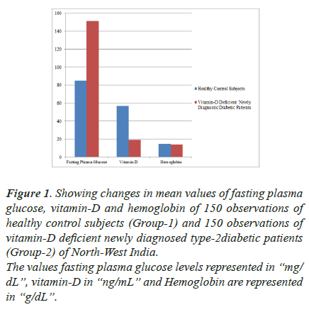
The levels of fasting plasma glucose, serum 25(OH) D and hemoglobin levels are summarized in (Figure 1. A significant (P<0.001) increase in plasma glucose by 77.93% from 84.96 ± 3.67 mg/dL to 151.17 ± 8.18 mg/ dL was found in newly diagnosed type 2 diabetic patients with respect healthy control subjects while a significant (P<0.001) decreased serum 25(OH) D by 66.37% from 56.86 ± 9.01 ng/ml to 19.12 ± 4.18 ng/ml was recorded in newly diagnosed type 2 diabetic patients with respect to normal healthy subjects. A statistical non -significant change from 13.89 ± 2.91 g/dL to 14.81 ± 1.16 g/dL was seen in hemoglobin levels in study group (Figure 1).
Figure 1. Showing changes in mean values of fasting plasma
glucose, vitamin-D and hemoglobin of 150 observations of
healthy control subjects (Group-1) and 150 observations of
vitamin-D deficient newly diagnosed type-2diabetic patients
(Group-2) of North-West India.
The values fasting plasma glucose levels represented in “mg/
dL”, vitamin-D in “ng/mL” and Hemoglobin are represented
in “g/dL”.
Lipid peroxidation
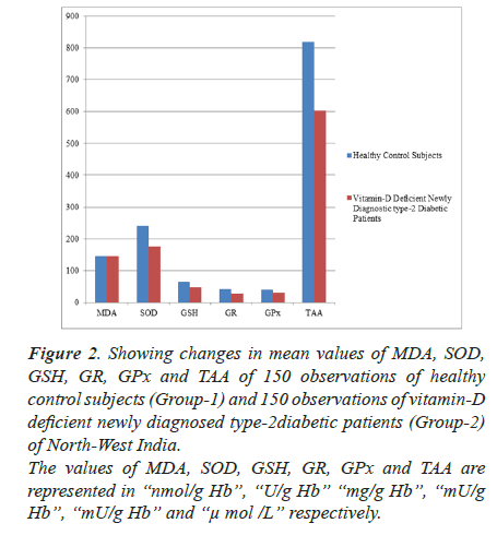
A significant increase was recorded in the MDA levels (from 145.23 ± 5.11 nmol MDA/gHb to 219.34 ± 22.11nmol MDA/gHb) in newly diagnosed Type 2 diabetic patients by 51.03% (P<0.001) with respect to normal healthy subjects (Figure 2).
Figure 2. Showing changes in mean values of MDA, SOD, GSH, GR, GPx and TAA of 150 observations of healthy
control subjects (Group-1) and 150 observations of vitamin-D
deficient newly diagnosed type-2diabetic patients (Group-2)
of North-West India.
The values of MDA, SOD, GSH, GR, GPx and TAA are
represented in “nmol/g Hb”, “U/g Hb” “mg/g Hb”, “mU/g
Hb”, “mU/g Hb” and “μ mol /L” respectively.
Superoxide dismutase and reduced glutathione and its metabolizing enzymes (GR and GPx):
A significant fall in the levels of SOD (from 241.21 ± 6.01 U/gHb to 175.38 ± 5.16 U/g Hb), GSH (48.22 ± 5.36 mg/g Hb to 48.22 ± 5.36 mg/g Hb), GR (from 42.26 ± 3.12 mU/g Hb to 28.72 ± 4.29 mU/g Hb) and GPx (from 39.78 ± 3.29 mU/g Hb to 29.67 ± 4.51 mg/gHb) by 27.91 (P<0.05), 26.14% (P<0.05), 32.04% (P<0.01), 25.41% (P<0.05) respectively was observed in erythrocyte of newly diagnosed type 2 diabetic patients in comparison to normal healthy control subjects (Figure 2).
Total antioxidant activity
The levels of Total Antioxidant Activity were found 603 ± 8.07μ mol/L in plasma of newly diagnosed Type 2 diabetic patients and 819.23 ± 11.19 μ mol/L in normal healthy control individuals. This fall in plasma total antioxidant activity by 35.85% was found to be statistically significant (P<0.01) in newly diagnosed type 2 diabetic patients with respect to control healthy subjects (Figure 2).
Discussion
A significant increase (P<0.001) by 61.07%) in MDA (representing the lipid peroxidation) levels in newly diagnosed type 2 diabetic patients was seen in comparison to healthy control individuals (Figure 2). Lipid peroxidation causes polymerization of membrane components and induces changes in the membrane permeability resulting in hemolysis, would relate to the degree of intravascular Red Blood Cell (RBC) destruction. Extravascular mechanisms of RBC destruction may involve changes in cell deformability and antigenicity. Previously, from our own laboratory, we reported that newly diagnosed Type-2 diabetic patients of North-West India are suffering with dyslipidemia [20]. Our observation of significantly increased malondialdehyde levels and hyperlipidemia is agreement with the literature reports and to decrease the membrane fluidity, deformability, visco-elasticity and life span of erythrocytes in the study group [21-24], which might be responsible for the initiation of complications such as diabetes, hypertension etc. in newly diagnosed type 2 diabetic patients later on in the north western Indian population.
Superoxide dismutase, a superoxide radical scavenging enzyme is considered the first line of defense against the deleterious effect of oxygen radicals in the cells and it scavenges reactive oxygen radical species by catalyzing the dismutation of O2- radical to H2O2 and O2 [25-28]. A significant decrease by 26.14% in SOD activity in newly diagnosed type-2 diabetics (Figure 2) may results in an increased flux of O2.- radical and hence reflects the tissue damage/injury.
Glutathione (GSH), a tripeptide is maintained in reduced state by an efficient glutathione peroxidase/glutathione reductase system. Glutathione is a potent endogenous antioxidant that helps to protect cells from a number of noxious stimuli including oxygen derived free radicals [27,29,30]. In the present work, the level of glutathione significantly decreased by 26.14% (P<0.05) in newly diagnosed Type-2 diabetics patients with compared to healthy control subjects (Figure 2). A significant fall in GSH levels, confirm an increased susceptibility to oxidative damage. The activity of GPx, a seleniumcontaining enzyme was found to be decreased by 25.41% (P<0.01) in newly diagnosed type-2 diabetics patients in comparison to healthy control subjects (Figure 2). Glutathione peroxidase catalyzes the reduction of variety of hydrogen peroxide (ROOH and H2O2) using glutathione as a substrate, thereby protecting mammalian cells against oxidative stress [31]. The significant inhibition (P<0.001, 32.04%) in the activity of GR in newly diagnosed Type- 2 diabetics patients (Figure 2) attributed to increased oxidation or decreased synthesis of GSH. The less availability of NADPH may also cause a decrease in GR activity [32]. Our observations of significant reduction in GSH levels are is an agreement with the literature reports that inverse relationship exists between MDA and GSH status, can impair the cell defense against the toxic action of xenobiotic and may lead to cell injury/death [30]. A significant decrease in GPx activity may render the tissue more susceptible to LPO damage. Accordingly, in the present work, we observed a significant decrease in GPx activity upon increase in MDA level. This observation is in accordance with the hypothesis that MDA and GPx might play a role in tissue damage [33-36] that oxidative stress is induced in the newly diagnosed Type-2 diabetics patients. Our observations of significant decrease (P<0.001) in total antioxidant activity by 35.85% in vitamin-D deficient newly diagnosed type 2 diabetic patients w. r. t. healthy control subjects further suggested that oxidative stress induced in the newly diagnosed Type-2 diabetics patients (Figure 2).
Conclusion
Aforementioned observations suggested that vitamin-D deficient newly diagnosed Type-2 diabetic patients of North Indian Punjabi population are associated oxidative stress, hallmark of various diseases such as Diabetes Mellitus, cardiovascular diseases, osteoporosis etc. Therapeutic interventions in combinations, lifestyle and dietary modification might be beneficial to increase the vitamin D levels and hence to reduce oxidative stress should be included as a part of treatment to prevent further risk of development of Diabetes Mellitus and other various cardiovascular diseases like atherosclerosis in North- West Indians.
Acknowledgement
Authors would like to acknowledge Department of Medicine, Govt. Medical College and Rajindra Hospital – Patiala, India for recruitment of vitamin D defiant newly diagnostic patients for this study.
Ethics approval and consent to participate
The author received no financial support for the research, and/or publication of this article.
Declaration of conflicting interests
Author declared no potential conflicts of interest with respect to the research, authorship, and/or publication of this article.
References
- Mohammad AB, Rogheyeh A, Bahar B, Fayyaz S. Status of Vitamin-D in diabetic patients. Caspian J Intern Med 2014; 5: 40-42.
- Aparna P, Muthathal S, Nongkynrih B, Gupta SK. Vitamin D deficiency in India. J Family Med Prim Care 2018; 7: 324-330
- Daga RA, Laway BA, Shah ZA. High prevalence of vitamin D deficiency among newly diagnosed youth-onset diabetes mellitus in north India. Arq Bras Endocrinol Metabol 2012; 56: 423-428.
- Wild S, Roglic G, Green A, Sicree R, King H. Global prevalence of diabetes: Estimates for the year 2000 and projections for 2030. Diabetes Care 2004; 27: 1047-1053.
- Leung GM, Lam KS. Diabetic complications and their implications on health care in Asia. Hong Kong Med J 2000; 6: 61-68.
- Palomer X, Gonz

