Review Article - Journal of Parasitic Diseases: Diagnosis and Therapy (2022) Volume 7, Issue 4
Echinococcosis in south Asia; the present status.
Zulfiqar Ahmad*
Department of Biochemistry and Biophysics, University of Rochester Medical Center, Rochester, United States
- Corresponding Author:
- Zulfiqar Ahmad
Department of Biochemistry and Biophysics
University of Rochester Medical Center
Rochester, United States
E-mail: vet.zulfiqar@gmail.com
Received: 29-Jun-2022, Manuscript No. AAPDDT-22-44354; Editor assigned: 01-Jul-2022, PreQC No. AAPDDT-22-44354 (PQ); Reviewed: 15-Jul-2022, QC No. AAPDDT-22-44354; Revised: 20-Jul-2022, Manuscript No. AAPDDT -22-44354 (R); Published: 27-Jul-2022, DOI:10.35841/aapddt-7.4.119
Citation: Ahmad Z. Echinococcosis in south Asia; the present status. J Parasit Dis Diagn Ther. 2022;7(4):119
Abstract
Cystic Echinococcosis and Alveolar Echinococcosis, caused by Echinococcus granulosus senso strict and Echinococcus multilocularis respectively, are endemic in South Asia. Various species of this zoonotic parasite are maintained in domesticated and wild mammals through predator-prey interaction. Different approaches for diagnosis were followed for this zoonotic parasite such as CT scan, MRI, Ultrasound, X-ray and surgical operations for hydatid cyst. A limited data is available of molecular characterization such as genotyping, subtyping, population genetic, western blotting and immune-proteomics for E.granulosus senso strict. Molecular analysis is used to help differentiate infecting parasite species and genotype with a goal of better understanding the life cycle of the parasite in order to help in the planning and implementation of the control program. This review reports the present status of Echinococcosis in different countries of South Asia.
Keywords
Cystic echinococcosis, diagnosis, X-ray and surgical operations, Metacestode.
Introduction
Cystic Echinococcosis (CE), a global zoonosis caused by larval stage of Echinococcus granulosus sensulato. Adult tapeworms caused infection in carnivores (definitive hosts), while the metacestode caused the cystic echinococcosis in herbivores (intermediate hosts) [1]. The definitive hosts for Echinococcus granulosus are dogs and wild canids while sheep, goats, cattle, camels and pigs are the major intermediate hosts agudelo Human are infected by the accidently ingestion of contaminated eggs present in food, soil and water and also by direct contact with the definitive hosts.
The intermediate hosts become infected by the ingestion of eggs and the metacestode develop as hydatid cysts in the internal organs of the hosts. Transmission of this zoonotic parasite occur in those area where human are directly contacted with stray dogs and other wildlife [2]. Control and prevention of this parasite should consider the life cycle of the parasite (species/genotype). Different studies have been summarized the status of the echinococcosis in Asia and worldwide Studies have been revealed that Echinococcus granulosus senso lato include five species. The human cystic echinococcoses around the world mainly caused by the Echinococcus granulosus senso stricto belong to G1 family.
Echinococcous has vast variety of species and there is a lot of diversity in species, advancement in differentiation is due to different molecular techniques [3]. Four different molecular approaches are reported in different studies. The first molecular technique is single genes using mitochondrial DNA. The second approach is the analysis of microsatellite markers showing polymorphic DNA loci consist of repeated nucleotide sequen Some studies have reported the full genomic analysis of this zoonotic parasite. The fourth approach is the comparison of mitochondrial DNA and nuclear DNA for species hybridization which was conducted for E. Canadensis G6/7, G8 and G10 [4].
Cystic Echinococcosis
Status of CE senso lato is endemic in those areas where the animal husbandry practices are in abundant throughout the world. The life cycle of the parasite is maintained by the farmers who feed the definitive host (dogs) with offal of the slaughtered sheep and cattle in Asia and other regions. Hyenas and lions are considered definitive host for E. felidis in Africa. Some studies have hypothesized about Asia that this region may has E. felidis and also thought that lions are also originated from this region [5].
Organotropism and genotype
Usually liver is considered the most predilection site for CE but CE is also reported from unusual organ such as brain, kidney, heart muscle, spleen and lungs in Pakistan similarly brain CE in children reported in hospital based case study in Mongolia caused by E. Canadensis. Iran has also similar reports of CE in children but these children don’t have liver and lung involvement. These reports have confirmed that E.canadensis has tendency to brain CE [6].
Literature Review
Confirmation of CE in human and animals in South Asia
Pakistan: The most endemic province for CE in Pakistan is the Punjab where case studies of CE are reported. Studies by 2010 provide the molecular status of CE in the province of Punjab. Molecular confirmation of E.granulosus G1 genotype was reported by 2014. Alveolar Echinococcois was reported in the Punjab from different hospital based studies the prevalence of CE was reported in sheep and buffalo and cattle in the Punjab [7].
In Pakistan, the second most CE prevalent province is Sindh, where molecular characterization of targeting cytochrome oxidase 1 Study by revealed the prevalence of CE in sheep and goat in abattoir of Larkana. Different predilection and unsual site of hydatid cyst in different hospitals of the province were studied and reported by [8].
Khyber Pukhtonkhwa is considered less endemic than Punjab and Sindh, where case studies were reported of CE in two different hospitals of the capital city. A study by 2018 stated that 1297 animals were examined for CE from September 2015 to September 2017. The high prevalence was recorded in buffaloes. The current status of the hydatid cyst is summarized in the Table 1 [9].
| Definitive host | Infection rate | Country | Author | Reporting time |
| Dogs | 19/289 | India | Ingole R.S and his team | Ignore R.S et al. 2018 |
| Dogs | 10/166 | Bhutan | Thapa N.K and his team | Thapa N.K et al. 2017 |
| Dogs | 144/3670 | Bangladesh | Tarafdar M and Samad M.A | Tarafdar M and Samad M.A (2010) |
Table 1. The details of definitive host are reported in the below.
India: The whole country of India is considered endemic for CE, while northern hilly areas are also considered for this zoonotic parasite. Different cases of different predilection sites of hydatid cyst were reported from different regions of the India and different diagnostic and surgical procedures were also reported amplified ATP6 and nadII genes (Nuclear and Mitochondrial) through Single-stranded conformation polymorphism in cattle, buffaloes, sheep and goats [10]. A dog study using conventional floatation and copro-PCR revealed the highest prevalence of CE (6.57%) through amplification mitochondrial cytochrome oxidase 1 gene as compared to conventional floatation technique (4.84%). The tehsil wise prevalence was also reported and tehsil akola (9.84%) was found highest prevalent tehsil. There are various prominent works on CE from various parts of India [11]. The more recent studies have shown the presence of E.granulosus genotypes (G3, G1 and G2) through nucleotide polymorphism in sheep and presence of 4 genotypes of E.granulosus, sheep strain (G1), Buffalo strain (G3), cattle strain (G5) and camel strain (G6) in human samples targeting Cox1 gene. Molecular characterization of different samples of different species (sheep, goats, cattle, buffalo and pigs) amplifying cox1 gene CE in human and buffaloes were reported and confirmed by western blotting and latex agglutination test [12].
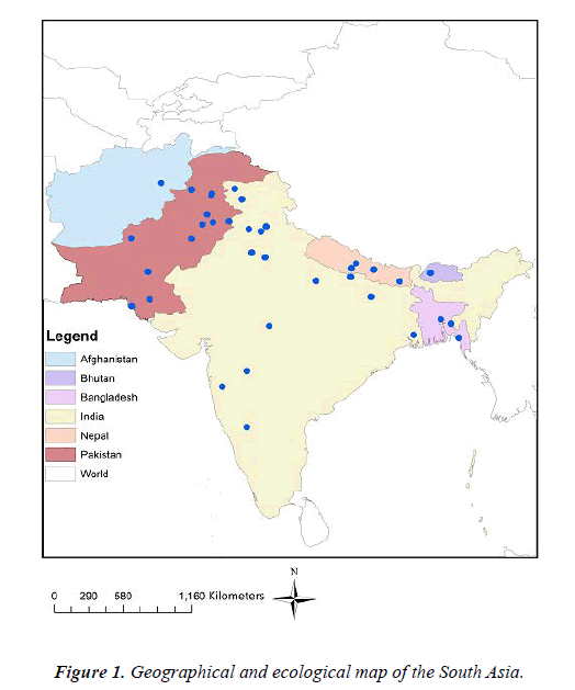
Nepal: Cystic Echinococcosis and Echinococcol cyst in buffaloes, goats, sheep and pig from abattoir of Kathmandu, Nepal and these finding were reviewed by. Human CE cases were reported from different areas of the country Figure 1. Different approaches of diagnosis (CT scan, MRI, and X-RAY) and surgical procedures (Laprotomy, thoractomy, splenctomy) were revealed the present situation of human CE in Nepal [13]. A study has revealed the presence of E.granulosus s.s (G1) in livestock, human and dogs and presence of E.ortleppi (G5) in cattle and E. Canadensis (G6) in human To date there is no study of alveolar echinococcosis in human and cattle reported from Nepal while CE is endemic in near countries including India, Pakistan and China. It is immense obligation for carrying out different studies and to be assured to update the present status of CE in the country [14].
Bangladesh: CE is endemic in northern part of the Bangladesh. There are several studies reported on CE in dogs, cattle and human confirmed through serological and histopathology examination. Reported 130 human cases of CE, female were found more infected with hydatid cyst. The only study on molecular confirmation of hydatid cyst in goats of Chittagong was reported by. In the study, 12S rRNA gene was amplified through semi nested PCR and phylogenetic analysis was also carried out. The CE considered endemic in Bangladesh so it is dire need to present the most advance genotyping and subtyping of the zoonotic parasite [15].
Bhutan: CE was reported in only one study in Bhutan. In this study, total 183 dog samples were examined. 20 out of 183 dog fecal samples were found positive for CE through classic technique (Microscopally). 16 samples were found PCR positive. Bhutan is located near with India, China, Sri Lanka and Nepal are considered endemic for AE and CE, so these two zoonotic parasite are also considered to be present in Bhutan [16].
Sri Lanka: There is no case reported on CE in the country but the hydatid cyst is endemic in all neighbor countries, so it is believed to be endemic in Sri Lanka.
Maldives: Maldives is an Island and less populated country in South Asia but being in South Asia, CE is considered to be prevalent in the country but no single case is reported in the country till today. The neighbor countries have reported the CE, so it is believed that CE should be studied and presents the situation for the betterment of the livestock industry [17].
Afghanistan: There are two cases report of a 20 year man having sheep and cows and 28 year old (Shepherd) with CE. However there is lack of studies on CE in the country [18].
Perspectives
Among the countries in the South Asia, where Echinococcus granulosus are highly prevalent and presence of the one or more species of the zoonotic parasite in the region. Pakistan and India are appeared to be the most impacted in terms of case number and species diversity [19]. These eight countries generally and specifically India and Pakistan where several species are sympatrically distributed and different diagnostic techniques such as CT SCAN, X-RAY, feacal floatation method, ultrasound and histopathology examination were studied in these countries but there is lack of advanced molecular techniques such as genotyping [20]. Sub typing and protein estimation and cloning of this zoonotic parasite. Molecular identification is very important for better understanding the distribution and variation within the species in this region [21]. The control strategies include deworming of definitive hosts, however this plan can be difficult which require too much funds and this can be too laborious.
Discussion
Definitive host and its infection
The prevalence of definitive host was presented after performing the copro-PCR in district Akola, India. Total of 289 samples were processed by floatation method, only fourteen samples were found positive through classical diagnostic method [22]. The Polymerase chain reaction was also performed for these 289 samples. The results revealed that only 19 samples were amplified at base pair of 225 of mitochondrial cytochrome oxidase-1 gene. The eleven fecal positive samples were found negative. The highest prevalence (6.5%) was recorded in PCR samples as compared to floatation method [23]. The pilot study was carried in the Thimpu city and samples were found positive for taeniid eggs. These samples were processed through floatation and sieve method. Out of 166 samples, 20 samples were processed through PCR. Ten samples were found positive taeniid eggs [24].
Conclusion
Better prevention is possible when proper diagnosis of the hydatid disease is carried out and effectively. In south Asia, different diagnostic techniques (classical diagnostic and molecular methods) are followed by different researchers, techniques include MRI, CT scan, X-ray, fecal samples, serological, ultrasonography and PCR are followed for the better diagnosis and prevention. The control strategies are totally depend upon meat inspection, upgrading the farm practices, public awareness, dog population control, proper detection of the parasite in intermediate host as well as in final host and complete elimination of the parasite from the final host. But the failure is considered to be present in different areas is decline in vulture population, improper disposal of meat, huge population of stray dog, unhygienic slaughtering, roaming of dogs in the premises of abbatoir, contamination of pastures with the feces, and the most important inadequate use of anthelmintic and drug resistance.
Control Strategies: After discussing the epidemiological factor, current status of echinococcosis in the region, control and preventive measure, mal practices of drugs and social awareness or public awareness of the zoonotic importance of the parasite and unhygienic slaughtering are the main causes of the failure for the eradication of the hydatid diseases. In south Asia, there is no study carried out for the development of vaccine against the echinococcosis, so this study will solve the issue with the most advances style and will develop the vaccine against this very important disease to control the economic and social losses in control strategies.
References
- Acharya S, Ghimire B, Khanal N. Spontaneous rupture of isolated splenic hydatid cyst without acute abdomen: A case report. Clin Case Rep. 2019;7(11):2064-7.
- Addy F, Wassermann M, Banda F, et al. Genetic polymorphism and population structure of Echinococcus ortleppi. Parasitol. 2017;144(4):450-8.
- Ahmad Z, Uddin N, Memon W, et al. Intrahepatic biliary cystadenoma mimicking hydatid cyst of liver: a clinicopathologic study of six cases. J Med Case Rep. 2017;11(1):1-7.
- Amin MU, Mahmood R, Manzoor S, et al. Hydatid cysts in abdominal wall and ovary in a case of diffuse abdominal hydatidosis: Imaging and pathological correlation. J Radiol Case Rep. 2009;3(5):25.
- Bajpai J, Jain A, Kar A, et al. Necklace in the lung: Multilocularis hydatid cyst mimicking left-sided massive pleural effusion. Lung India: Official Organ of Indian Chest Society. 2019;36(6):550.
- Beigh AB, Darzi MM, Bashir S, et al. Gross and histopathological alterations associated with cystic echinococcosis in small ruminants. J Parasit Dis. 2017;41(4):1028-33.
- Budke CM, Deplazes P, Torgerson PR. Global socioeconomic impact of cystic echinococcosis. Emerg Infect Dis. 2006;12(2):296.
- Casulli A, Bart JM, Knapp J, et al. Multi-locus microsatellite analysis supports the hypothesis of an autochthonous focus of Echinococcus multilocularis in northern Italy. Int. J. Parasitol. 2009;39(7):837-42.
- Craig PS, Budke CM, Schantz PM, et al. Human echinococcosis: a neglected disease? Trop Med Health. 2007;35(4):283-92.
- Dasbaksi K, Haldar S, Mukherjee K, et al. A rare combination of hepatic and pericardial hydatid cyst and review of literature. Int J Surg Case Rep. 2015;10:52-5.
- Devleesschauwer B, Ale A, Torgerson P, et al. The burden of parasitic zoonoses in Nepal: A systematic review. PLoS Negl Trop Dis. 2014;8(1):e2634.
- Faruk MO, Siddiki AM, Masuduzzaman M, et al. Identification and molecular characterization of Echinococcus granulosus from domestic goat in Chittagong, Bangladesh. Trop Biomed. 2017;34(4).
- Fatimi SH, Naureen S, Moizuddin SS, et al. Pulmonary hydatidosis: clinical profile and follow up from an endemic region. ANZ J Surg. 2007;77(9):749-51.
- Fomda BA, Khan A, Thokar MA, et al. Sero-Epidemiological survey of human cystic echinococcosis in Kashmir, North India. PLoS One. 201;10(4):e0124813.
- Griffin DO, Donaghy HJ, Edwards B. Management of serology negative human hepatic hydatidosis (caused by Echinococcus granulosus) in a young woman from Bangladesh in a resource-rich setting: A case report. ID Cases. 2014;1(2):17-21.
- Gudewar J, Pan D, Bera AK, et al. Molecular characterization of Echinococcus granulosus of Indian animal isolates on the basis of nuclear and mitochondrial genotype. Mol Biol Rep. 2009;36(6):1381-5.
- Hamalainen S, Kantele A, Arvonen M, et al. An autochthonous case of cystic echinococcosis in Finland, 2015. Euro Surveill. 2015;20(42):30043.
- Hazra NK, Batajoo H, GHimire S, et al. Open conservative surgical management of cystic echinococcosis in a tertiary care hospital, Nepal. Clin Diagnostic Res. 2015;9(7):PC01.
- Ingole RS, Khakse HD, Jadhao MG, et al. Prevalence of Echinococcus infection in dogs in Akola district of Maharashtra (India) by Copro-PCR. Vet Parasitol: Reg Stud Rep. 2018;13:60-3.
- Islam MS, Das S, Islam MA, et al. Pathological affections of lungs in slaughtered cattle and buffaloes at Chittagong Metropolitan Area, Bangladesh. Adv Anim Vet Sci. 2015;3(1):27-33.
- Jan Z, Zeb S, Shoaib A, et al. Hydatid cyst involving right pectoralis major muscle: A case report. Int J Surg Case Rep. 2019;58:54-6.
- Johnson WE, Eizirik E, Pecon-Slattery J, et al. The late Miocene radiation of modern Felidae: A genetic assessment. Sci. 2006;11(5757):73-7.
- Kapoor SK, Kataria H, Patra SR, et al. Multi-organ hydatidosis with extensive involvement of the hemi-pelvis and ipsilateral femur. Parasitol Int. 2013;62(1):82-5.
- Karim MF, Brunetti E, Rahman S, et al. Abdominal cystic echinococcosis in Bangladesh: a hospital-based study. J Infection Develop Countries. 2015;9(01):070-5.
Indexed at, Google Scholar, Cross Ref
Indexed at, Google Scholar, Cross Ref
Indexed at, Google Scholar, Cross Ref
Indexed at, Google Scholar, Cross Ref
Indexed at, Google Scholar, Cross Ref
Indexed at, Google Scholar, Cross Ref
Indexed at, Google Scholar, Cross Ref
Indexed at, Google Scholar, Cross Ref
Indexed at, Google Scholar, Cross Ref
Indexed at, Google Scholar, Cross Ref
Indexed at, Google Scholar, Cross Ref
Indexed at, Google Scholar, Cross Ref
Indexed at, Google Scholar, Cross Ref
Indexed at, Google Scholar, Cross Ref
Indexed at, Google Scholar, Cross Ref
Indexed at, Google Scholar, Cross Ref
Indexed at, Google Scholar, Cross Ref
Indexed at, Google Scholar, Cross Ref
Indexed at, Google Scholar, Cross Ref
Indexed at, Google Scholar, Cross Ref
