Research Article - Biomedical Research (2017) Artificial Intelligent Techniques for Bio Medical Signal Processing: Edition-I
Dysfunction of elbow joint treated by minimally invasive elbow arthroscopy
Hao Chen, Lin Guo, Pengfei Yang, Dejie Fu, Lingchuan Gu, Yang Peng and Liu Yang*Centre for Joint Surgery, Southwest Hospital, Third Military Medical University, Chongqing, PR China
- *Corresponding Author:
- Liu Yang
Centre for Joint Surgery, Southwest Hospital
Third Military Medical University, PR China
Accepted date: January 31, 2017
Abstract
Clinically, elbow joint dysfunction is more common. It is mainly caused by many traumatic factors, such as elbow joint injury and elbow surgery. Anatomic structure of elbow joint is complex. In the past, there were limitations and deficiencies in the diagnosis and treatment of elbow joint disease. Current minimally invasive arthroscopic treatment of elbow joint disease has opened up an important way. It has broad application prospects and it also has been the focus of foreign and domestic research institutions. Based on this, in view of structure and function of the elbow joint, the difficulty and operation process of elbow arthroscopic mini-invasive surgery in the treatment of elbow dysfunction were studied. 74 patients who had difficulty in elbow flexion and extension received arthroscopic treatment of elbow joint in West China Hospital of Sichuan Province. After the operation, the rehabilitation function training was carried out according to the unified standardization program. Mayo elbow function score was used to compare the degree of flexion and extension of the elbow joint before and after the operation. The results of the study were analyzed, and the advantages, considerations and application prospect of arthroscopic elbow surgery were discussed. Practice has proved that the elbow arthroscopy miniinvasive surgery has the advantages of small trauma and fast recovery, which is an effective way to treat elbow flexion and extension dysfunction.
Keywords
Elbow joint, Arthroscopy, Mini-invasive, Dysfunction
Introduction
With the continuous improvement of the quality of modern social life, people’s demand for sports has become more and more intense. People lack scientific exercise guidance and some of them have inevitable special occupation (such as miners, bricklayer, cooks, heavy physical labor, professional athlete); besides, they often believe that exercise is beneficial to the body; thus, one-sided emphasis on the intensity and activity amount of exercise makes the joint experience longterm high strength and stress. In the end, the joints are in a state of disorder. If we do not prevent this unreasonable amount of exercise, the joints will continue to be destroyed due to vicious spiral. Joints will be in total loss of compensation, leading to joint swelling, pain, joint instability as well as joint flexion and extension dysfunction. This even causes joint stiffness in the worst situation.
According to statistics organized by Hai-Meng et al. in the clinical application of elbow arthroscopy, the common indication is the removal of the loose body. Elbow joint loose body can occur in a variety of diseases, such as synovial cartilage, osteoarthritis and bone cartilage lesions. The patient complained of pain, compression, locking and other discomforts [1]. Because of the extensive use of arthroscopy, the understanding of the disease has increased. Moon et al. reported 7 cases of synovial cartilage disease using elbow arthroscopy to remove the loose body and clean up the synovial. All the patients were followed up for 31 months, and their operation incision healed. According to Mayo elbow score, the results were good and excellent, and there are no related complications or postoperative recurrence [2]. Peng et al. adopted arthroscopy to clean up and take the loose body out of 16 patients with traumatic loose body of elbow joint. Patients returned to their daily activities about 2 weeks after operation and they were followed up for 5 to 29 months. Most of them had obvious pain relief, and their motor function was improved significantly [3].
Olecranon bursitis is a common disease of elbow joint. As to this disease, surgical treatment is rarely required and nonoperative treatment is the first choice. However, when the outer wall of the slide sac is obviously thickened, especially when the loose body occurs, the surgical treatment should be considered. In this circumstance, the surgical incision will be larger, residual paralysis scar tissue is likely to lead to local incision, causing "provoke" phenomenon [4]. Jiang et al. found that up to 20% of patients had post-operative complications of skin problems [5]. Ramsey et al. used arthroscopy for the treatment of 17 cases of olecranon bursitis. All the incisions healed well after the operation. These patients were followed up for 9 to 24 months. There were no scar left and no joint swelling, and joint range of motion returned to normal [6]. Singh et al. defines the normal flexion motion of the elbow joint as flexion 0 -146° [7]. Boileau et al. determined the range of motion of the elbow joint is flexion 30-130°. This 100° range is the range of activities that most of the daily life needs. You cannot complete the basic daily activities if less than this range of activities [8]. The elbow stiffness can be mostly found in the post traumatic complications. It also occurs in a variety of arthritis, congenital malformations, burns and various types of brain injury [9]. Keener et al. treated 243 cases of traumatic and degenerative contracture through arthroscopic surgery. After the operation, 212 cases were followed up for an average of 58 months. There was a remarkable improvement in pain, activity, stability and function [10]. In this paper, we studied the elbow arthroscopy in the treatment of elbow joint disorder. By following the experimental analysis, the effect of arthroscopic treatment and its application prospect have been researched.
Elbow Joint Dysfunction
Elbow joint
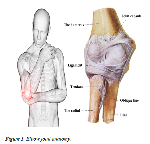
The elbow is the link of the arm (also called the upper arm) and small arm (also called the forearm) [11]. In anatomy, the elbow is a composite joint, which is composed of the joint capsule of humerus, radius and ulna and elbow as well as the surrounding ligament. When it comes to the elbow that we usually mentioned, seemingly it is a specific joint. In actuality, the elbow joint consists of the three joints of the humero-ulnar joint, humeroradial joint and the upper ulnar joint. The Figure 1 shows the elbow joint anatomy.
At the front and back of the elbow, joint capsule with ligament can be found. On both sides of the elbow joint, the brachiocubital ligament and the brachioradial ligament are used to strengthen in order to prevent excessive adduction and abduction of elbow [12]. At the same time, the radial head neck is wrapped with annular ligament tissue, which is very important for maintaining the stability of the radial head.
Function of elbow joint
The elbow is a joint with two axes of movement and two degrees of freedom. In other words, elbow flexion and extension can be both carried out to bend arms and straight arms; meanwhile, elbow can perform the forward and backward rotation of small arms. Turn the small arms to let the palm of your hand up (forward) and the palm of your hand down (backward). In all kinds of movements of our daily life, usually two kinds of movements coordinate; at the same time, shoulder joint rotation function also involves. Therefore, flexion and extension are easy to be noticed and the rotation function is often neglected. The normal angle of elbow flexion and extension is approximately 0°~150°. A number of people (especially women) have the problem of over-extension of elbow, which is about 10~15°.The degrees are 80°~90° both before and after the rotation. There is also a physiological angle called the carrying angle, which is about 15° [13].
Joint dysfunction
In the treatment, fixation for a certain time to a certain degree is required after the injury of elbow joint or a fracture around it, such as humeral fractures and forearm fractures. Common treatment has the suspension protection of scarf bandage, plaster fixation, splint and protection of fixator. Either way the soft tissue around the elbow occur inflammation and braking. This leads to changes in the muscles, ligaments and joint capsules around elbow joints in terms of shape, structure, bio-mechanics and so on, causing the joint adhesion and the atrophy of the muscles around the elbow [14].
Elbow joint adhesion is mainly manifested in the limitation of elbow flexion and extension function. But in reality, if only the elbow extension is limited, the impact on the function is not very significant. It is not good-looking because the arm is not straight. However, if the forward and backward rotations of forearms have been restricted at the same time, the daily life would be seriously affected. For example, when you wash your face, the forearm must be rotated if you intend to make the palm of your hand reach the face. The palm also must be up if you need to hold the bowl when you are eating; besides, take a pen to write and hold chopsticks to dish must be completed through forearm rotation. If the forearm does not flex, definitely, the problem can be more serious. Even if you have food to eat, you cannot reach it if you do not bend your arms. Every time you have to stretch out the neck to find food, which is miserable and awkward. In view of these dysfunctions, the rehabilitation treatment and functional exercises are absolutely necessary if elbow joint movement is limited. Through this way, functions will be improved and restored.
Elbow Joint Dysfunction Treated by Mini- Invasive Surgery
Difficulties in open surgery and mini-invasive surgery
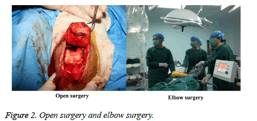
The Figure 2 shows the open surgery and elbow surgery. Elbow arthroscopic surgery has plenty of advantages. Open surgery is often adopted when the elbow joint movement has been restricted. The main steps are to cut the joint capsule, ligament and soft tissue, and then to clean up the loose body, synovial and cartilage damage in the joint cavity, and eventually to remove the osteophyma. However, the surgery usually causes big trauma and much bleeding, and functional exercise might be affected due to incision pain after the operation; besides, it is prone to complications such as heterotopic ossification. Finally we realized that it is difficult to achieve satisfying surgical effect. In this group of patients, arthroscopic surgery is most practical when we detect loose body, synovitis, osteophytes and cartilage injury or degeneration.
Mini- invasive elbow arthroscopy in the treatment of elbow joint dysfunction
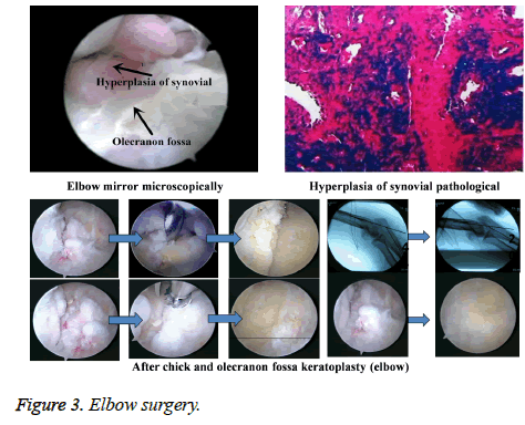
Elbow arthroscopic surgery is performed only through 2 to 3 incision less than 5 mm in length around the elbow. Based on the surgery, we are able to make clear diagnosis of elbow joint disease. Osteophytes which hinder joint flexion and extension can be removed by arthroscopy. After that, repair and reconstruction operation of tendon and ligament will be carried out. This operation has the advantages of small incision, beautiful appearance, low infection rate and fast recovery, representing the development trend of elbow joint disease treatment. The Figure 3 shows the elbow surgery.
Operative procedure of elbow arthroscopy
Removal of heterotopic ossification and loose bodies (total joints): small incision surgery is suitable for cases with large loose bodies assisted by arthroscopy. It is also suitable for posterior incision. All arthroscopic operation is applicable in the cases with small loose bodies. We can remove the loose bodies to relieve joint internal pressure. Through this process, we need to avoid loose bodies to be left in the channel when we take them out. If necessary, we must expand the incision to remove or use the C-arm fluoroscope to confirm. Anterior capsular release/amputation (anterior cubital region): through the arthroscopic mini-invasive surgery, elbow joint adhesion can be released and cut off to make joint flexible. It should be noted that the space can be formed from the front blunt dissection of the anterior capsule if there is no anterior joint capsule gap. Then the joint capsule was cut off. Anterior elbow impact is released through fossae coronoidea formation (anterior cubital region). After that, posterior elbow impact is also relieved through olecranon and anconal fossa formation (outer margin of elbow). Finally, by removing capitulum radii and releasing the triceps tendon adhesion, the triceps tendon was cutting off proximally to enhance elbow flexion [15].
Clinical Data
Data of laboratory personnel
There were 74 patients with different degrees of elbow dysfunction in the course of disease received by West China Hospital of Sichuan Province. Among them male patients were 44 and female patients were 30. They aged from 20 to 65 years old and their average age was 42.5 years old. The average time of onset was 16 years. Left hand patients were 20 cases and right hand patient were 54 cases. All patients had no obvious abnormal bony structure and malformation.
Preoperative assessment
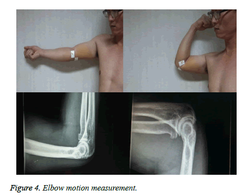
In order to evaluate the patients’ improvement of preoperative and postoperative elbow functions, the motion degree of the elbow joints before and after operation must be accurately measured through elbow flexion and extension angle measurement devices. Besides, it is necessary to photograph the maximum extension position and flexion position of the elbow before and after the operation as well. In the operation, the loose bodies in the joint cavity should be removed as much as possible; therefore, we must be very clear about the number of loose bodies and the situation of osteophyma before the whole surgery. Only X-ray film is not enough, CT plain scanning and three-dimensional reconstruction of elbow joints are needed to further positioning and judge the joint cavity. The Figure 4 shows the elbow motion measurement [16].
For the pre-operative need to further clarify the cartilage damage and the surrounding soft tissue damage, it is necessary to photograph MRI. Before the actual operation, the elbow joint lateral stress test should be carried out to judge the injury of medial and lateral collateral ligament, which can further guide the surgical procedures. Before operation, it is also crucial to assess whether the patient is associated with vascular injury. We need to use the Moyo Rating Scales to make functional evaluation of patient’s elbow [17]. General examinations are blood routine, coagulation routine, ECG and other projects. X-ray examination of the elbow aims to observe whether there is a significant change in the structure of the bone. For patients suffering from certain special cases, they must have spiral CT examination or MRI examination.
Surgical instruments and methods
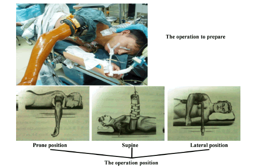
Surgical instruments are No.18 lumbar puncture needle, marker pen, 50 ml syringe, No.11 blade, hemostatic forceps, probe hook, belt gear grasping forceps, penetration device, Smith and Nephew arthroscopic system, electric planning machine, plasma radio frequency vaporization instrument and electric drill. Among them the arthroscope is 30 degree arthroscopy with diameter of 2.7 mm. Surgery adopts brachial plexus anesthesia. The patient is in the supine position, and his proximal arm is tied with pueumatic tourniquet; meanwhile, he needs to maintain the 90° flexion and 90° extension of the shoulder joints. The preparation for operation is shown in Figure 5. Common approaches of elbow joint are the direct lateral approach (that is, the soft point), proximal lateral approach, anterior lateral approach, anterior medial approach and posterior lateral approach. The direct lateral approach can be used to observe and deal with the oboro torsion joint. Proximal lateral approach is selected for observation of the anterior compartment, including radial head and the front and lateral humerus head. Anterior lateral approach is adopted to observe the lower part of the lateral approach, medial joint capsule, medial synovial folds, coronary process, trochlea and fossae coronoidea. Anterior medial approach can be exchanged with the proximal lateral approach as a way of observing and working. Posterior lateral approach enables us to observe the olecranon tip, olecranon fossa and the posterior trochlea. Anterior transposition of the ulnar nerve must be applied to patients who have ulnar neuritis.
Exploration and treatment of elbow joint: Congested and hyper-plastic synovial tissue can be mostly found under arthroscopy. Flexion and extension activities of elbow joints are usually accompanied by joint clearance synovial squeeze, scar adhesion, thickening and contracture of joint capsule, loose body and osteophyte formation. In the operation, planning machine will be used to clean up the hyper-plastic synovium and thickened joint capsule. Forceps or nucleus forceps are used to remove the loose body and bite the hyper-plastic osteophyte. In addition, we clear the synovium and loose the joint capsule by using plasma radio frequency vaporization apparatus, which is good for wound hemostasis. Drainage tube is placed at elbow joints after the operation.
Treatment and rehabilitation after the operation
Elastic bandage for elbow joint and local ice compression are necessary after the operation. Besides, routine prophylactic use of antibiotics to prevent infection is also crucial. Besides, patients need to take non-steroidal anti-inflammatory and analgesic drugs for 3 weeks to prevent the formation of heterotopic ossification. 2 days after the operation patients mainly exercise their muscle strength, including flexion and extension of wrist finger joint and some exercise of fingers, in order to avoid premature action to aggravate the injury of the elbow joint, causing joint adhesion and stiffness. 2 days later, active and passive flexion and extension exercises of elbow joint should be performed. Then the range of activities gradually increases. Patients who have difficulty in flexion and extension should perform passive activity assisted by CPM. Through this way the elbow joint can reach the normal range of motion in 3-4 weeks after the operation. All functional exercises should be practiced regularly, but not in pain. It is noticed that follow-up review cannot be neglected after the surgery. According to the recovery of joint function, further guidance for treatment and rehabilitation exercise should be carried out.
Statistical methods
We compare the elbow flexion and extension before and after the operation by adopting paired t test (a=0.05); at the same time, Mayo elbow function score for elbow is necessary. ≥ 90 is excellent, 75-89 is good, 60-74 is acceptable and <60 is poor. In addition, we need to observe the wound healing and complications [18].
Experimental Results and Discussion
Experimental result analysis
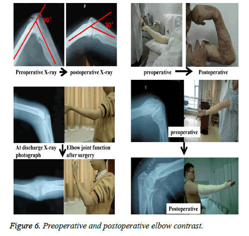
All 74 patients were followed up after operation for an average period of 18 months (8-28 months). After 8-28 months followup, as many as 70 patients with preoperative elbow dysfunction were well removed, and there was an obvious improvement of flexion and extension activities. Recovery of swelling and pain symptoms, postoperative elbow stability and special function were satisfactory. Daily life and exercise can be well performed without obvious discomfort. There were two cases in 3 months after operation occurring obvious stiffness of elbow motion and apparent lack of joint mobility. Through follow-up survey we detected that patients missed early regular exercise due to the elbow pain. 3 months after the operation, the elbow joint function failed to achieve very satisfactory results. Then the patients took the second arthroscopic release and had been told to carry out the early regular rehabilitation exercise. This time the joint mobility and function were significantly improved 6 months after operation without affecting the patient's daily life and activities. Due to leakage during operation, the elbow recurrent ulnar neuritis occurred in 1 case. However, through positive symptomatic treatment after the operation, their ulnar nerve symptoms were recovered after follow-up for 6 months. There was also a case of poor prognosis. After actively changing dressing, the wound healing was good. During the follow-up period, 74 patients underwent X-ray examination. There were no loose bodies and new osteophytosis occurred. Preoperative and postoperative elbow contrast is shows in Figure 6.
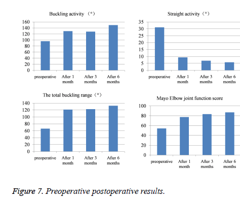
In order to analyze whether differences in activity of elbow joint (flexion activity, extension activity and total flexion and extension range), Mayo elbow function score and preoperative evaluation results exist or not 1 month, 3months and 6 months after the operation, we are suggested to take the repeated measures of the general linear model to obtain the significant probability through analysis. If the standard is a=0.05, based on F test, the corresponding P values can be obtained by F, and rating results are shown in Table 1.Through the calculation we know that the corresponding P values are less than 0.05, and the differences are statistically significant. Then we can believe that there are obvious statistic differences in the activity of elbow joint (flexion activity, extension activity and total flexion and extension range), Mayo elbow function score and preoperative evaluation results. Therefore, it can be determined that the activity and function of elbow are greatly improved after having arthroscopic treatment [19].
| Buckling activity (°) | Straight activity (°) | The total buckling range (°) | Mayo Elbow joint function score | |
|---|---|---|---|---|
| Preoperative | 96.1 ± 13.7 | 31.1 ± 16.7 | 66.1 ± 33.7 | 54.1 ± 22.7 |
| After 1 month | 129.3 ± 10.6 | 9.3 ± 2.6 | 121.3 ± 6.9 | 77.3 ± 15.6 |
| After 3 months | 127.8 ± 4.2 | 6.9 ± 5.6 | 122.6 ± 8.6 | 83.3 ± 15.2 |
| After 6 months | 149.3 ± 8.4 | 5.8 ± 6.6 | 132.3 ± 14.6 | 86.8 ± 14.1 |
Table 1: Elbow joint mobility and Mayo scores before and after operation.
The Figure 7 shows the preoperative postoperative results. In this group, 74 cases were followed up for 8-16 months, and the average period is 11.3 months. Among them, elbow flexion and extension activities of 21 patients were remarkably enhanced. Pre-operative average active extension activity was (31.1 ± 16.7°) and flexion activity was (96.1 ± 13.7°). 6 months after operation, the average active extension activity was (5.8 ± 6.6°) and flexion activity was (149.3 ± 8.4°). There were statistically significant differences in the activity of flexion and extension before and after the operation (P<0.05°). The total pre-operative flexion and extension range was (61.8 ± 30.9°). There were statistically significant differences in the activity of flexion and extension before and after the operation (P<0.05°). Mayo elbow function score was used to compare and we found good in 4 cases, acceptable in 8 cases and poor in 52 cases. Fine rate was 5.45%. 6 months after operation, we found excellent in 10 cases, good in 57 cases and acceptable in 7 cases. Fine rate was 70.9%. 1 patient had transient ulnar nerve palsy because the elbow swelling compressed the cubital nerve. The other 1 patient had elbow heterotopic ossification without any complications.
The cause and pathological changes of elbow joint activity limitation
Elbow joint movement restriction is a common complication of elbow injury and some certain diseases. The etiology of the disease is divided into intra-articular type and extra-articular type. In this group, the intra-articular factors are damage, fracture, loose body, synovitis etc., and extra-articular factors are contracture and scar of skin, joint capsule and lateral collateral ligament. The main pathological features include scar tissue and adhesion in intra-articular cavity, contracture and thickening of joint capsule, cartilage degeneration in some areas and osteophyma. Besides, bone hyperplasia and intra-articular loose bodies which seriously affect the daily-life are also included. A small number of patients have severe cartilage damage as well.
Considerations in elbow arthroscopy
Pre-operative routine X-ray examination of the elbow aims to confirm whether it is bone block or elastic barrier. As to the operation position, this group took supine position. Patients maintained 90° of flexion and extension of shoulder joint and continued to pull their upper arms. The force should be appropriate to prevent nerve damage [20]. Surgical incision must avoid blood vessels and nerves. Person who performs this operation is required to grasp the knowledge of elbow joint anatomy. He needs to acutely cut the skin and bluntly separate the subcutaneous soft tissue. The person who does the operation must be skilled in the operation of knee arthroscopy. Early cold compress after the operation helps to relieve pain and reduce bleeding. Meanwhile, analgesic drugs should be used to establish the confidence of patients in rehabilitation. Only when patients actively cooperate with rehabilitation exercise can they achieve good results.
The application prospect of arthroscopic technology in arthroscopic disease
Arthroscopic technology is mostly used for the evaluation and treatment of knee and shoulder joint diseases, but rarely used for elbow joints. Obvious disability can be caused by the loss of motion of elbow joints. If the elbow flexion and extension activities are slightly limited or injured for a short time, most can return to the range of functional activities after regular physical therapy and rehabilitation exercise. Because there are nerves and vascular bundles closely related to elbow joints around the elbow, in arthroscopic surgery, orthopedic surgeons are required to be familiar with the anatomy of the joint. Because elbow arthroscopy has characteristics of small trauma, less bleeding, rapid recovery and clear visual field; furthermore, scholars have a gradual deeper understanding of elbow joint disease, elbow arthroscopy has developed rapidly in recent years. By making use of arthroscope, arthroscopic debridement is carried out if elbow joint movement is restricted, and the effect is satisfactory.
Conclusions
With the popularization and development of the arthroscopic technology, elbow arthroscopic surgery is increasing year by year and the scope of application is also more and more extensive. At present, it has become a safe and effective method of surgery. People's demand for medical technology is getting higher and higher, and they do hope to obtain the most effective treatment for complex diseases with the least cost and minimal trauma. In view of elbow joint dysfunction, this paper analyzed the difficulties and treatment process. Based on the comparative analysis of 74 patients with elbow diseases before and after operation, this paper also studied the preoperative and postoperative elbow joint activities as well as Mayo elbow function score changes. It is concluded that open surgery is more difficult; however, the use of arthroscopic technology can make complex elbow surgery simple and minimally invasive, reducing the complications and shortening the recovery period. Through this way, the curative effect and the level of diagnosis and treatment of elbow disorders will be enhanced. Therefore, the elbow arthroscopy technology has broad application prospects. The experimental results showed that the adoption of mini-invasive elbow arthroscopy to treat elbow joint dysfunction can greatly reduce the suffering of patients and postoperative complications.
References
- Hai-Meng MA, Zhao-xun P, Xiao-ming Y. Arthroscopic treatment of elbow joint motion limitation. Chinese J Bone Jotnt Injury 2013.
- Moon JG, Bilaris S, Jeong WK. Clinical results after arthroscopic treatment for septic arthritis of the elbow joint. Arthroscopy J Arthroscopic Related Surgery 2014; 30: 673-678.
- Peng J, Zhu Y, Lu H. Arthroscopic Management for Juvenile Elbow Loose Bodies: A Retrospective Review of Fifteen Cases. J Biomaterials Tissue Eng 2015; 5: 539-543.
- Rezende CMF, Melo EG, Malm C. Arthroscopical treatment of elbow joint disease. Arquivo Brasileiro De Medicina Veterinária E Zootecnia 2012; 64: 9-14.
- Jiang LL, Liu YQ. Curative effect observation of arthroscopic treatment for 12 cases of elbow joint osteoarthritis. J Med Sci Yanbian University 2013.
- Ramsey ML, Getz CL, Parsons BO. What's new in shoulder and elbow surgery. Am J Bone Joint Surgery 2008; 90: 1719-1727.
- Singh H, Nam KY, Moon YL. Arthroscopic management of stiff elbow. Orthopedics 2011; 34: 167.
- Boileau P, Gonzalez JF, Chuinard C. Reverse total shoulder arthroplasty after failed rotator cuff surgery. J Shoulder Elbow Surgery 2009; 18: 600-606.
- Meluzinová P, Kopp L, Edelmann K. Elbow arthroscopy in the surgical treatment of post-traumatic changes of the elbow joint. Acta Chirurgiae Orthopaedicae Et Traumatologiae Cechoslovaca 2014; 81: 399-406.
- Keener JD, Galatz LM. Arthroscopic management of the stiff elbow. J Am Acad Orthop Surg 2011; 19: 265-274.
- Millett PJ, Horan MP, Pennock AT. Comprehensive Arthroscopic Management (CAM) Procedure: Clinical Results of a Joint-Preserving Arthroscopic Treatment for Young, Active Patients With Advanced Shoulder Osteoarthritis. Arthroscopy J Arthroscopic Related Surgery 2013; 29: 440-448.
- Maclean SB, Oni T, Crawford LA. Medium-term results of arthroscopic debridement andcapsulectomy for the treatment of elbow osteoarthritis. J Shoulder Elbow Surgery 2013; 22: 653-657.
- Lattermann C, Romeo AA, Anbari A. Arthroscopic debridement of the extensor carpi radialis brevis for recalcitrant lateral epicondylitis. J Shoulder Elbow Surgery 2010; 19: 651-656.
- Kozin SH, Boardman MJ, Chafetz RS. Arthroscopic treatment of internal rotation contracture and glenohumeral dysplasia in children with brachial plexus birth palsy. J Shoulder Elbow Surgery 2010; 19: 102-110.
- Jesalpura JP, Chung HW, Patnaik S, Choi HW, Kim JI, Nha KW. Arthroscopic treatment of localized synovial chondromatosis of the posterior knee joint. Orthopedics 2010; 33: 49.
- Inagaki K. Current concepts of elbow-joint disorders and their treatment. J Orthop Sci 2013; 18: 1-7.
- Urney MAG, Ysnik MR, Omerford EJC. Intra-articular morphine, bupivacaine or no treatment for postoperative analgesia following unilateral elbow joint arthroscopy. J Small Animal Practice 2012; 53: 387-392.
- Blonna D, O'Driscoll SW. Delayed-onset ulnar neuritis after release of elbow contracture: preventive strategies derived from a study of 563 cases. Arthroscopy J Arthroscopic Related Surgery 2014; 30: 947-956.
- Samoy YC, De BE, Van VD. Arthroscopic treatment of fragmented coronoid process with severe elbow incongruity. Long-term follow-up, in eight Bernese Mountain Dogs. Vet Comparative Ortho Traumatol 2013; 26: 27-33.
- Merolla G, Buononato C, Chillemi C. Arthroscopic joint debridement and capsular release in primary and post-traumatic elbow osteoarthritis: a retrospective blinded cohort study with minimum 24-month follow-up. Musculoskel Surgery 2015; 99: 83-90.
