- Biomedical Research (2013) Volume 24, Issue 2
Coherence in muscle activity of the biceps brachii at middle, proximal and distal tendon region among the arm wrestling contestants
Nizam Uddin Ahamed1*, Kenneth Sundaraj1, R. Badlisha Ahmad1, Matiur Rahman2, Md. Anamul Islam1 and Md. Asraf Ali11AI-Rehab Research Group, Universiti Malaysia Perlis (UniMAP), Kampus Pauh Putra, 02600 Arau, Perlis, Malaysia.
2College of Computer Science and Information System, Najran University, Kingdom of Saudi Arabia.
- *Corresponding Author:
- Nizam Uddin Ahamed
AI-Rehab Research Group(School of Mechatronic Engineering)
Universiti Malaysia Perlis (UniMAP)
Kampus Pauh Putra, 02600Arau
Perlis, Malaysia.
Accepted Date: February 06 2013
Abstract
The aim of this study was to analyze the electromyographic (EMG) activity of biceps brachii (BB) muscle under the same muscle contraction in three different locations. For this reason, arm wrestling contest was conducted to record the EMG signal from ten male subjects. Electrodes were placed on the three locations of upper arm BB; i.e. middle (belly) of BB (M), lower part (L) and upper part (U) of the BB belly. Average EMG (EMGAVG), root mean square (EMGRMS) and highest peak of the signal [EMGHigh(pk)] were calculated from the sum of EMG activity. The analysis of the effect of electrode placement location using ANOVA (analysis of variance) tests yielded a number of statistically significant differences. The results indicated, 1) majority of the EMG results confirmed the muscle activity was higher in the order of L, M and U, 2) among the 16 comparisons among the muscles (from winners and losers), there was main interaction found between the entire BB of winners and losers, also another 7 results displayed same interaction (p<0.05), but remaining 8 locations did not significant (p>0.05), 3) in the loser, the BB was forced to perform eccentric contraction as the forearm is being pronated and elbow was gradually being extended, on the other hand, contraction of the BB was concentric in the winner, and 4) winners (during concentric contraction) did not always produce highest EMG peak and some of the results (muscle activity) of loser’s (during eccentric contraction) revealed higher than the winners. The findings of the study contain a precious contribution to rehabilitation, biomedical and sports medicine by describing an experimental set up to measure muscle electrophysiology during physical activity in the case of arm activities.
Keywords
Electromyography, biceps brachii, electrode, arm wrestling, contraction, winner, loser.
Introduction
Electromyography (EMG) signals are generated in the human skeleton muscle from muscle fiber contraction and these signals are always random [1]. Surface EMG is extensively used to detect and record the function of skeleton muscle. Moreover, this technique provides effortless access to the physiological processes that cause muscles to generate force, produce movement, accomplish the essential functions of everyday life and can show the functional status of muscles [2,3]. Upper arm BB muscles are one of the vital locations to produce and detect the EMG signal. The BB muscle is characteristically described as a two-headed muscle, which consists of a long head and a short head that originates proximally [4]. Usually, therapists preferred location for electrode placement is the middle of the BB muscle (or muscle belly). The next two choices are the muscle below the proximal tendon (upper part of muscle belly) and the upper muscle of the distal tendon (lower part of muscle belly) [5,6]. The multiplicity of the sEMG detection and processing methods on the BB results in a large number of physiological applications, including signal processing, the study of muscle fatigue, torque relationship, kinesiology, sports science, the study of ergonomics, exercise assessment, and laboratory examination [6,7].
Arm wrestling is a prominent research area where muscles are contracted and EMG signals are generated. It is a simple game during which the results (winner/loser) can be decided within a very short period [8]. It is a sport in which two competitors use a single arm to produce maximum force to win the game. The competitors each place their elbow on a solid surface and clasp each other’s palms. During the game, each competitor tries to push the other’s arm until it hits the surface [9]. The strength and activity of the biceps muscle can therefore be measured during this game[10]. A few studies have reported the assessment of muscle strength using electromyography sensor during arm wrestling. For example, Hong et al. analyzed the activities of the six upper limb muscles including BB of two competitors [11]. Silva et al. evaluated the muscle activity of the pectorials major, biceps brachii, pronator teres and flexor carpi ulnas from simulated arm wrestling [12], but did not measured during the winning or losing condition. A method to predict the winner of an arm wrestling match using the activity of the BB and TB muscles was determined by Gang et al[8]. It noted that, different types of contraction are produced during the arm wrestling game, like it generates isometric contraction when elbow are fixed, then in the loser, the BB is forced to perform eccentric contraction as the forearm is being pronated and elbow is gradually being extended, on the other hand, contraction of the BB is concentric in the winner. In this experiment, we have analyzed EMG signal during all these three conditions.
Researchers have investigated that results from electromyography signals are varied and influenced by the placement of the electrodes [3,13,14]. Some studies have examined the effects the EMG signal from the BB muscles during voluntary muscle contraction and the results of these vary depending on the inter-electrode distance. For example, Beck et al. described the relationship between isokinetic and isometric torque and found that an electrode distance between 20 mm and 60 mm does not affect the EMG amplitude [15]. Gerdle et al. examined the effects of an electrode distance of 10, 20, or 30 mm on the EMG signal from the BB during elbow flexion and different maximal voluntary contractions [16]. Mesin et al. surveyed the issue of electrode location within different muscles of the body and identified the three most effective BB locations, which are mentioned earlier[17]. A survey study was published by Hermen et al. which analyzed different BB electrode placements, sensor locations and skin preparations[5]. In their literature, researchers recommended 21 sensor placement descriptions in the BB, of which the three aforementioned locations were commonly mentioned. SENIAM (Surface Electromyography for the Noninvasive Assessment of Muscles) is part of a larger project that studies sEMG sensors and sensor placement locations in the human skeleton and body muscles, including the BB.
Previous researches have examined the physiological measurements of the BB during different muscle contractions with a variety of protocols, subjects, activities and sensor locations. However, the EMG activity of the BB using different electrode placement locations during arm wrestling among the competitors has not been studied previously. We have conducted arm wrestling course to create the muscle force for long time. Then measured the strength and compared the results between the muscles of the participants (winner and loser). We hope these results provided a solid foundation for further research studies on rehabilitation, sports science and other physiological measurement concerns that involve the upper extremities.
Materials and Methods
Subjects
Ten healthy right-handed male subjects were participated in this experiment. The mean and standard deviation of the age, height, weight, and arm dimension during relaxation and during extension of the participants were 24.5±3.5 years, 168±6.7 cm, 70.5±8.3 kg, 11.82±1.5 inch, and 13.3±0.8 inch, respectively. The study was approved by the university research and development review board for human subjects. The ten subjects were divided into five pairs. The demographics characteristics of the two players in each group were almost identical. All participants gave written consent to take part in the study.
Experimental Procedures
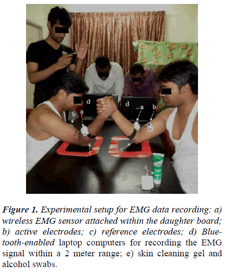
Two players were requested to sit on chairs with a small table between them. Each player kept his right elbow within the circle that was drawn on the table in front of him. Their palms were tied to each other and their left arms were folded along the back of their bodies. All the rules and constitutions delineated by the world arm wrestling federation (WAF), which is located in Canada, were followed; these can be viewed in their entirety at http://www.worldarmwrestlingfederation.com. Figure 1 depicts the full experimental setup during the arm wrestling match. A referee was responsible for conducting the game. Although no time limit was imposed, the referee kept track of the elapsed time as the two participants attempted to push their arms towards the left and cause their opponent’s arm to fall on the table. The winner was therefore the participant that successfully pushed his opponent’s arm to the table, i.e., the winner’s palm is on top of the loser’s. Each single pair faced each other three times with each electrode placement; therefore, there were a total of 3×3=9 matches between each pair of competitors, all of which were conducted in the same day with a 20-min rest between each. The data from the two matches, out of the three with the same electrode position, in which the same participant won were recorded. If the same participant won three times, then the two shortest matches were considered.
Electromyography recording
A wireless, touch proof and Bluetooth-enabled three channel EMG signal storage device, called SHIMMERTM Model SH-SHIM-KIT-004 (Real-time Technologies Ltd., Ireland), was used to record the EMG data. The raw EMG signal was recorded at a sampling frequency of 1 KHz and were preamplified with band-pass filter (10–500 Hz). EMG Meditrace noninvasive electrodes were used in the experiments. These are single, pre-gelled Ag/AgCl electrodes with biopotential sensors, which are capable of identifying the flow of ions through a nerve fiber in a human body. The skin was prepared using a skin cleaning gel (sigma gel) and an alcohol swab to obtain better EMG signals and avoid artifacts. Also, skin was prepared and the electrodes were placed in accordance with SENIAM [18].
Electrode placement location
At first, the pair of electrode was placed as parallel over the muscle belly (M). The second placement involves positioning the electrodes on the lower part of the muscle belly (L), which is between the biceps muscle endplate region and the distal tendon insertion. The last placement that was studied was the placement of two electrodes over the medial belly of each head (long and short head) (U), parallel to the muscle fibers and below the proximal bicep tendon [5,19]. The distance between the center of electrodes located at U and M and between M and L was 4 cm, whereas the distance between the center of the electrodes at U and L was 8 cm. The reference electrode was set on the bony part that is located underneath the elbow and slightly above the joint (i.e. on the back of the dominating arm). The resulting inter electrode distance was 2 cm (center to center). However, the electrodes were not placed on the three locations at the same time. Instead, because the device used is capable of being connected to only two active channels, two electrodes were placed on the same place in each of the two players; for example, if two electrodes were located on the muscle belly of the BB of one player, then an electrode pair was placed at the same location on the opposite player’s arm.
Statistical analysis
EMGAVG, SD, EMGRMS and EMGHigh (pk) during the muscle contraction were calculated for each participant. These values were then comparatively evaluated using analysis of variance (ANOVA) test. All statistical tests were performed using the Minitab statistical software (MINITAB® Release 14.12.0). Statistical significance was set at p<0.05 (95%).
Results
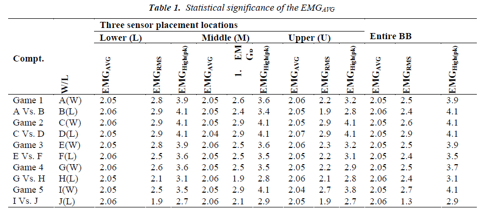
All the statistical output data is arranged by the EMG sensor placement location on the BB muscle of each competitor. Table 1 presents the individual results for the each pair of competitors, Table 2 shows the overall results and Table 3 gives a summary of the statistically significant differences (p-value).
Sensor on lower part of the BB muscle (L)
EMGAVG: If we reflect on individual pairs of participants, the EMGAVG value of the loser is higher than the winner’s in three instances; in the other two pairs, the winner is higher than the loser. In addition, when the total average EMGAVG of the five competitor pairs was calculated; the average EMGAVG of the losers (2.05mV) is slightly lower than the winners (2.06mV) (Table 2).
EMGRMS
The average EMGRMS of the winning wrestlers is significantly higher than the losers; this trend is maintained for all players and matches with two exceptions: in one case, the loser is greater than that of the winner, and in another case, the values of both players are equal. Likewise, the overall value of all the winners (2.72mV) is higher than the losers (2.47mV) (Table 2).
EMGHigh (pk)
The calculation of the highest peak of the EMG signal shows some differences among the competitors. When the five pairs of players are analyzed, three of the champions show a higher peak value than the respective losers, whereas only one loser has a higher peak value than the respective winner; the remaining pair exhibit equivalent values. In addition, the average highest peak EMGHigh (pk) for the winners is higher (3.84mV) than the losers (3.49mV).
Sensor on middle of the BB muscle (M)
EMGAVG
In this location, the EMG mean values of two winners are higher than their opponent’s and two losers are higher than respective winners. The remaining pair of competitors has the same value. Overall, the average of all the winners (2.06mV) is higher than all the losers (2.05mV).
EMGRMS
The average EMGRMS is higher in the winners of three of the pairs and the same in the remaining two pairs. Therefore, the value of all the winners (2.67mV) is greater than the corresponding value of the losers (2.37mV).
EMGHigh (pk)
Four winners obtain higher a peak value than their opponent’s; the remaining pair of competitors attains the same peak value. In addition, the average peak values of the winners and the losers are 3.77mV and 3.34mV, respectively.
Sensor on upper part of the BB muscle (U)
EMGAVG
The average activity of three of the losers is higher than their opponent’s; two winners, however, show more strength than their respective losers. On the whole, the average value of the losers (2.07mV) is higher than the winners (2.05mV).
EMGRMS
The average EMGRMS value of all the winners (2.42mV) is larger than the losers (2.21mV). In addition, if we consider the individual pairs (Table 1), in four cases, the winners have higher values than their respective losers and, in the remaining case, the pair of competitor’s exhibit equal result.
EMGHigh (pk)
Similar to the RMS value, the average peak value of the winners (3.43mV) is higher than the losers (3.11mV). In four of the individual cases, the amplitude of winners is greater than their respective losers; in the remaining pair, both competitors exhibit equal peak values.
P-value
Among the sensor placement locations. As shown, the lower part of the BB on both participants (winner and loser) have significant difference (P=0.04). Significant difference also found between the winners and losers (P=0.03) when compared between lower and middle parts. The results show significant between the winner (measured on the lower part) and the loser (measured on the upper part) with a P value of 0.01. A significant difference was also found when the muscle activity of the loser was measured on the upper part and the muscle activity of the winner was measured either on the middle (P=0.01) or upper portion (P=0.01). However, there was no difference found between the winner’s middle or upper portion with the loser’s lower (P=0.41 or P=0.18 respectively) or middle portion (P=0.34 or P=0.15, respectively). In addition, for winners there was no difference (P>0.05) between the different sensor placement locations within the same participant. For example, when the results is compared with the lower and middle part, the P value is 0.12; the same is true when the lower part le is compared to the upper portion (P=0.53), and when the middle part is compared to the upper portion (P=0.46). However, for losers two results show the significant differences (P<0.05) within the same participant. At this point, there were significant differences found between the lower and upper part (P=0.01); and between the middle and upper area (P=0.01). But, no interaction between the measurements obtained from the lower and middle locations of the losers (P=0.86). Lastly, there was a significant difference (P<0.05) between the entire right arm BB of the winner and the loser (P=0.0).
Discussion
This article discussed the processes for quantifying muscle activity during arm wrestling and summarized the key research findings. It focused on the comparison of the muscle activity during contraction, which was measured in one of three locations on the BB, between the winner and the loser of an arm wrestling match. Some EMG analyses have been previously performed on the BB; these have studied sensor placement location, age variations, muscle contractions with different ranges of motion, etc. in the fields of sports science, biomedical, clinical activities, and ergonomics[6,20,21]. Researchers have also found that the EMG results are varied on the interelectrode distance [22-24].
However, we did not find any previous study that investigated the muscle activity with the following conditions: 1) only the BB muscle of the dominating arm of each player was studied, 2) the data was collected during arm wrestling course where muscle was contracted as maximum level, and 3) three specific sensor placement locations of the muscle were compared. In this study, we clarified the electromyographic activity on three locations of the upper arm biceps brachii muscle of winning and losing arm wrestlers. In the statistical analysis of this experiment, we treated each pair of competitors as a unique experiment and discovered the following results:
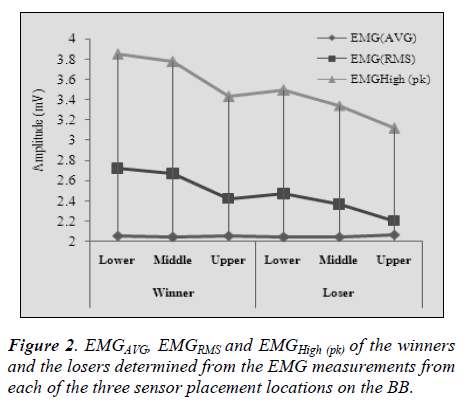
• The most important finding of the present investigation was that the EMG signal couldn’t determine the winner and loser in an arm wrestling match because, in some cases, the muscle activity of the loser was higher than that of the winner (Table 1 and Fig 2).
• A total of 90 results (ten participants, three electrode placement locations and three statistical measurements (Table 1, individual results) were recorded, which results in a total of 45 comparisons (pair). Among these, 27 comparisons (60%) found that the winners exhibited higher activity than the losers, 11 comparisons (24.44%) found that the losers had higher muscle activity than the winner and the remaining 07(15.56%) comparisons obtained equal results for both competitors.
• The consolidated statistical results for the entire biceps show (Table 2) the average EMGRMS (2.61±0.2) and average EMGHigh (pk) (3.69±0.3) values of the winners are higher than the respective values of the losers. In contrast, the average EMGAVG (2.07±0.4) value of the losers is greater than those of the winners (2.06±0.3).
• The placement of the electrode on the lower portion of the muscle measures higher muscle activity, in both the winners and the losers, than placement in the middle or upper part of the muscle. As a result, the muscle activity of the BB during arm wrestling is highest in the lower portion of the muscle, is decreased in the middle of the muscle and is even lower in the upper portion of the muscle (Fig 2).
• In Table 3, among the 16 statistical analyses (pvalues) shown, 8 comparisons show significant differences (p<0.05) and remaining 8 comparisons show not significant differences (p>0.05).
• This study highlights that the specific placement of EMG sensors that will be used to record muscle strength should be decided with the utmost concern. Thus, researchers need to use anatomical knowledge and a suitable understanding of the effect of electrode placement to ensure accurate results; these types of expert information are described by a number of well-known references.
Our experiment and findings are limited by some factors. First, we used only the right arm biceps muscle in our study although there are other muscles that are affected during arm wrestling, such as the shoulder, wrist, triceps, latissmus dorsi, and pectoralis major. In addition, the EMG data acquisition system used was only able to measure three channels. Furthermore, there was a 20- minute rest between each match and we did not confirm the occurrence of muscle fatigue. However, we highlight that our results should not be used to justify improper EMG sensor placement on the biceps muscle and the results are effective for physiological measurements of the bicep in sports science studies that are concerned with the upper arm.
Conclusion
Although during the arm wrestling competition other muscles of upper arm are moving, however our experiment was only focused on the BB muscle activity among the arm wrestling participators. We analyzed the influence of electrode location on the measurement of the muscle activity under the same contraction. It recommends that, electromyography signal cannot distinguish the winner or loser, because signals are varied due to force and muscle activeness. The investigation also revealed that, the signals were higher in the order of lower, middle and upper part on the BB muscle. Our findings assist further researcher to develop rehabilitation program, systems or specific exercise protocols for arm wrestling or related upper limb activities. Lastly, these findings might bring new knowledge for strength and arm associated coaches to improve resistance training protocols in a performance and prophylactic point of view. Further investigation is needed to achieve a better understanding of the biomechanics of arm wrestling, the mechanisms of resulting injuries and the function of various risk factors in injury causation.
References
- Komi PV, Viitasalo JHT. Signal Characteristics of EMG at Different Levels of Muscle Tension. Acta Physiologica Scandinavica. 1976; 96(2): 267-276.
- Ebenbichler G. Dynamic EMG: a clinician's perspective. IEEE Eng Med Biol Mag.2001; 20(6):34-5.
- Luca CJD. The use of surface electromyography in biomechanics. J Appl Biomech. 1997; 13(2): 135-163.
- Rai R, Ranade AV, Prabhu LV, Pai MM, Prakash. Third head of biceps brachii in an Indian population. Singapore. Med J. 2007; 48(10): 929-931.
- Hermens HJ, Freriks B, Disselhorst-Klug C, Rau G. Development of recommendations for SEMG sensors and sensor placement procedures. J Electromyogr Kinesiol. 2000; 10(5): 361-374.
- Ollivier K, Portero P, Maïsetti O, Hogrel J-Y. Repeatability of surface EMG parameters at various isometric contraction levels and during fatigue using bipolar and Laplacian electrode configurations. J Electromyogr Kinesiol. 2005; 15(5): 466-473.
- Stegeman DF, Blok JH, Hermens HJ, Roeleveld K. Surface EMG models: properties and applications. J Electromyogr Kinesiol. 2000;10(5): 313-326.
- Gang L, Haifeng C, Jungtae L. A Prediction Method of Muscle Force Using sEMG. Int. Association of Computer Science and Information Technology- IEEE Spring Conference, 17-20 April 2009, pp.501-505.
- ArmWrestling. Canada. [Update 2004; cited 2012 March 15]. 2004; Available from: http://www.armwrestling.com.
- Quanjun S, Bingyu S, Jianhe L, Zhen G, Yong Y, Ming L, et al., Prediction of Human Elbow Torque from EMG Using SVM Based on AWR Information Acquisition Platform, IEEE Int. Conf. on Information Acquisition; 20-23 Aug. 2006, pp.1274-1278.
- Hong M-K, Lin C-Y, Liao Y-S, Hong C-K, Wang L-H. Kinematic and Electromyographic Analysis of Upper Extremity in Arm Wrestling. Portuguese Journal of Sport Sciences. 2011; 11(2): 267-270.
- Silva DCdO, Silva Z, Sousa GdC, Silva LFGe, Marques KdV, Soares AB, et al. Electromyographic evaluation of upper limb muscles involved in armwrestling sport simulation during dynamic and static conditions. J Electromyogr Kinesiol. 2009; 19(6): 448-457.
- Mercer JA, Bezodis N, DeLion D, Zachry T, Rubley MD. EMG sensor location: Does it influence the ability to detect differences in muscle contraction conditions?. J Electromyogr Kinesiol. 2006; 16(2): 198-204.
- Basmajian JV. Muscles Alive. Their Functions Revealed by Electromyography. Academic Medicine. 1962; 37(8): 802.
- Beck TW, Housh TJ, Johnson GO, Weir JP, Cramer JT, Coburn JW, et al. The effects of interelectrode distance on electromyographic amplitude and mean power frequency during isokinetic and isometric muscle actions of the biceps brachii. J Electromyogr Kinesiol. 2005; 15(5): 482-495.
- Gerdle B, Eriksson NE, Brundin L. The behaviour of the mean power frequency of the surface electromyogram in biceps brachii with increasing force and during fatigue. With special regard to the electrode distance. Electromyogr Clin Neurophysiol. 1990; 30(8): 483- 489.
- Mesin L, Merletti R, Rainoldi A. Surface EMG: The issue of electrode location. J Electromyogr Kinesiol. 2009; 19(5): 719-726.
- Hermens HJ, Freriks B, Merletti R, Stegerman D, Block J, al. GRe. SENIAM: European recommendations for surface electromyography Roessingh Research and Development, Enschede. http://www.seniamorg/ 1999.
- Sargon MF, Tuncali D, Çelik H. An unusual origin for the accessory head of biceps brachii muscle. Clinical Anatomy. 1996; 9(3): 160-162.
- Elfving B, Liljequist D, Mattsson E, Németh G. Influence of interelectrode distance and force level on the spectral parameters of surface electromyographic recordings from the lumbar muscles. J Electromyogr Kinesiol. 2002; 12(4): 295-304.
- Cram JR, Kasman GS, Holtz J. Introduction to Surface Electromyography. Aspen Publications, Gaithersburg, MD. 1998: 408.
- Beck TW, Housh TJ, Cramer JT, Weir JP. The effects of electrode placement and innervation zone location on the electromyographic amplitude and mean power frequency versus isometric torque relationships for the vastus lateralis muscle. J Electromyogr Kinesiol. 2008; 18(2): 317-328.
- Beck TW, Housh TJ, Mielke M, Cramer JT, Weir JP, Malek MH, et al. The influence of electrode placement over the innervation zone on electromyographic amplitude and mean power frequency versus isokinetic torque relationships. J. Neurosci. Methods. 2007; 162 (1–2): 72-83.
- Campanini I, Merlo A, Degola P, Merletti R, Vezzosi G, Farina D. Effect of electrode location on EMG signal envelope in leg muscles during gait. J Electromyogr Kinesiol. 2007; 17(4): 515-526.


