- Biomedical Research (2016) Volume 27, Issue 3
Classification of multi-channel EEG signals for migraine detection.
S. Batuhan Akben1*, Deniz Tuncel2, Ahmet Alkan31Bahce Vocational School, Osmaniye Korkut Ata University, 80500, Osmaniye, Turkey
2Department of Neurology, Faculty of Medicine, KSU, 46500 Kahramanmaras, Turkey
3Department of Electrical and Electronics Engineering, KSU, 46100, Kahramanmaras, Turkey
Accepted date: February 25, 2016
Abstract
Migraine can only be detected by expert medical doctors. But recent studies showed that the migraine analysis can be done also by using EEG. These analyses are concerned with migraine diagnostic methods done by using EEG. T5-T3 channel of EEG was generally used in these proposed methods. However, the suitability of other channels in the diagnosis of migraine has not discussed. It is very important to find out which EEG channels and brain lobes are more important to learn the characteristics of migraine. The aim of this study is to analyze the each EEG channel separately for migraine patients. Analysis of this study is based on method in the literature that related to magnitude increase amount under flash stimulation. For this aim, beta band of each EEG channel’s data were pre-processed by using Burg-AR method. Then these features were applied to a support vector machine (SVM) classifier to observe which channel is the more definitive. As a result of this study, it is proposed that T3, F7, O1 and O2 channels are the most decisive for diagnosis of migraine, based on PSD magnitude increase under flash stimulation. Also, which brain lobes are more affected from triggering factors of migraine attack is proposed. Furthermore, asymmetry feature of migraine is approved by EEG and alternative migraine diagnosis methods is proposed for future researches according to reaction type of physiological structure of scalp to flash stimulation.
Keywords
EEG, Migraine, PSD, SVM.
Introduction
Migraine is one of the important brain disorders and its causes have not known definitely yet [1,2]. Since, pain description of patients is known as unique diagnosis method of migraine; its diagnosis is a difficult task for a neurologist. Studies are continuing for automatic diagnosis and determination of the causes of it [3,4]. Electrical activity changes of brain during the migraine attack were a critical point to determine the cause and automatic diagnosis of migraine [5]. For this aim, the electrical activity changes of brain obtained by triggering factors have been usually used as a method. But practical migraine diagnosis method approved by authority, has not been defined yet. Although behavioural, environmental, infectious, dietary, chemical, or hormonal factors are known as triggering factors of migraine, cause of it hasn’t been known definitely [6]. Recent studies have proposed that EEG is the commonly used tool for determining characteristics of migraine. By using EEG, brain electrical activity changes obtained by triggering factors for migraine patients, can be measured and studied for migraine diagnosis easily. Flash stimulation was used as a triggering method for activating migraine without aura in these studies. Phase synchronization changes of alpha rhythm in migraine patients under flash stimulation and revealing the existence of magnitude increasing in migraine patients under flash stimulation are important examples of these diagnosis methods by using EEG [7-9]. Since these former studies are focused on T5-T3 channels of EEG to detect the migraine by using computerized EEG diagnosis software, which EEG channels (brain lobes) are more valuable has not been determined completely yet. Therefore the electrical activity change amount of each EEG channel under flash stimulation is very important subject to define the cause of migraine and for automatic diagnosis system.
It is reported that flash stimulated EEG data of migraine patients at the beta band (13Hz-30Hz) of T5-T3 channels have a PSD magnitude increase while healthy subjects haven’t any magnitude changes in literature [7-11]. In this study, EEG data is obtained from both migraine patients and healthy subjects through different EEG channels under flash stimulation.
The flash stimulated and non-stimulated EEG signals of each channel are filtered to obtain the beta band and pre-processed by using Ar-burg method to obtain PSDs. PSD differences of the pre-processed data as two classes (migraineurs and healthy subjects) are used as the features that will be applied to a support vector machine (SVM) classifier. As a result of this study it is determined that which brain lobes are more affected from triggering factors (flash stimulation) of migraine and which EEG channels have more importance on the automatic diagnosis of migraine. Comparing the findings of the study, an alternative migraine diagnosis method depending on the physiological basis of face and scalp is proposed. Furthermore, asymmetry feature of migraine is approved by EEG.
Data Acquisition
Subjects
Migraineurs and healthy subject’s EEG data is obtained from Neurology Department of Kahramanmaras Sutcu Imam University. Migraineurs (without aura) group consisted of thirty patients (nine males, twenty one females), and were diagnosed according to the diagnostic criteria proposed by the International Headache Society (IHS). Control group (healthy subjects) consisted of thirty healthy subjects (eleven males, nineteen females) which has not any neurological or psychiatric disease. Both healthy and migraineur groups’ age ranges are between 20-40 years. Subjects (migraineurs and healthy subjects) had not taken any drug before the recordings and all were in the interictal (pain-free) state. They were tested in a dimly lit room, while in a couchant position. All the subjects were instructed to relax during the experiment, keeping their eyes closed.
Data recording
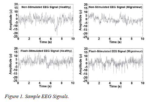
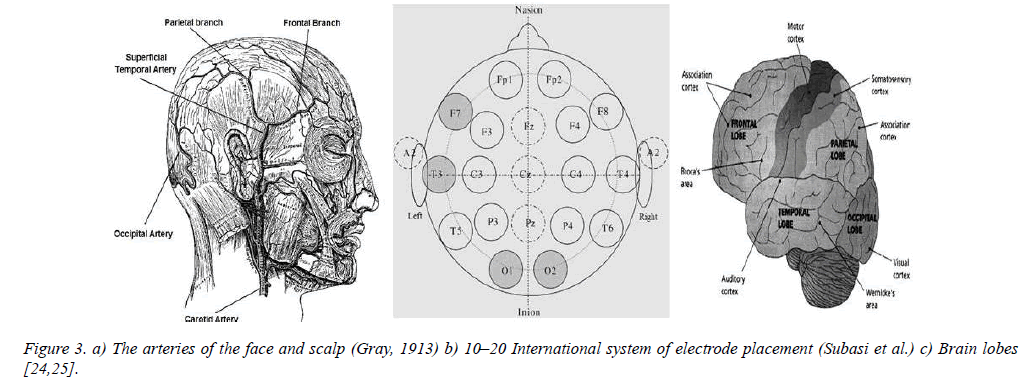
EEG recordings were obtained with an 18-Channel Nicolet One Machine. Electrodes were positioned according to the international 10–20 system (Figure 4b), at Fp1, Fp2, F7, F3, Fz, F4, F8, T3, C3, C4, T4, T5, P3, Pz, P4, T6, O1 and O2. The reference electrode was positioned at the linked earlobes (A1-A2) and the EEG signals were sampled at a rate of 256 Hz. Each recording process was taken minimum 20 minutes that contain 30s hyperventilation and 30s flash stimulation periods. In these 30 seconds flash stimulation time periods; stimulation frequency was 2, 4 and 6 Hz. Since the best definitive stimulation frequency is reported as 4 Hz in previous study [7], this stimulation frequency of 4 Hz is used. Sample EEG signals are given in figure 1.
Methods
Pre-processing of EEG signals
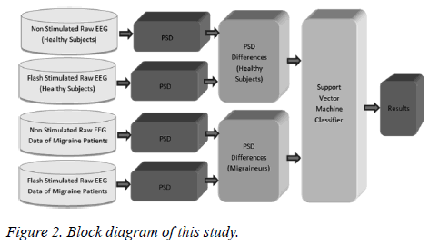
Feature extraction or sample reduction of EEG signals is a crucial step that influences the performance of the classifier. In this study, the beta band of EEG data is preprocessed to get the low-dimensional feature vectors that are frequency-magnitude relations. Power spectral density (PSD) of EEG data is obtained by using Burg-Ar method (Figure 2). This method is a model-based (parametric) method which models the data sequencex(n) as the output of a linear system characterized by a rational structure. In the model-based methods, for estimating the spectrum firstly parameters are estimated from a given data sequencex(n), 0 ≤ n ≤ N-1. Afterwards, by using these estimates, the power spectral density estimate can be computed. Details can be found in literature [11-13].
One of the better known criteria for selecting the model order has been proposed by Akaike called the Akaike information criterion (AIC) [13]. In this study, our selected model order of the AR method was 10 by using AIC. The differences between flash-stimulated and non-stimulated EEG PSDs are calculated as the last step of the pre-processing. These PSD differences are used as the feature vectors to be applied to the classification step as seen in figure 2.
In this study the noise and artifact removal algorithms were not used since the EEG recordings made and used by experts are the mostly raw data. Also, a lot of pre-processing and classification methods have tried for this study but could not find better than the proposed method.
Support vector machines
Support vector machines (SVMs) are well-known supervised learning methods that were developed by C. Cortes and V. Vapnik for binary classification and regression [14-16]. SVMs classify data in two steps: first, given a set of training examples, each marked as belonging to one of two categories by hyperplane. The hyperplane is a classifier which leaving the largest possible fraction of points of the same class on the same side, while maximizing the distance of either class from the hyperplane. It is determined by a subset of the points of the two classes, named as support vectors. Then SVM training algorithm builds a model by assistance of these two categories separated by hyperplane that predicts whether a new example falls into one category or the other according to positions of hyperplane [16-18].
In this study, linear support vector machines are used to classify the EEG data for migraine analysis. Differences between PSDs of flash-stimulated and non-stimulated data were used as inputs for SVM classifier. 90% of overall data were used for training and the rest of the data were used for testing. To obtain an objective classification accuracy rate 10- fold cross validation was used.
Performance measures
The aim of the SVM classification is to assign the input patterns to one of the two classes, usually represented by outputs restricted to lie in the range from 0 to 1 (0:Normal, 1:Migrainuer), so that they represent the probability of class membership. After classification step, the classification performance of data is evaluated by using sensitivity, specificity and accuracy measures. Terms of sensitivity, specificity and accuracy values are formulated as below [17]:

TP = True Positive, FN = False Negative,
TN = True Negative, FP = False Positive


Also Cohen's kappa coefficients are calculated to analyze the EEG data. The Cohen's kappa coefficient is a statistical method that measures the reliability of the agreement between two raters. This method determines if there is an agreement between two raters by chance with a percentage [19]. Implementation of the method is as follows: If the predictions of raters are determined as PA and PB.
The value of Kappa is defined as:

Where,

And

Finally;

Kappa values are easily interpreted for the following:
K=1: Raters fully complies with each other
K=0: Compliance for raters determined only by chance (There is no agreement).
The other values can be interpreted with the following table (Landis et al. [20])
Results
The SVM classification achievements of each channel are given in table 3. According to these results, one can see that accuracy rates change between 55.7% and 88.4% depending on the EEG channels. By examining the table it can also be seen that the best classifiable data were from T3, F7, O1 and O2 channels which have comparatively higher accuracy rates between 81.8% and 88.4% respectively. As a consequence, we can say that the data from T3, F7, O1 and O2 channels exhibits considerably better performance than other channels.
| Second Rater Predictions | |||
|---|---|---|---|
| First Rater Predictions | Prediction of A Value (percentage) |
Prediction of B Value (percentage) |
|
| Prediction of A Value (percentage) |
PAA | PAB | |
| Prediction of B Value (percentage) |
PBA | PBB | |
Table 1: Calculation of Kappa values.
| Interpretation | Poor agreement | Fair agreement | Moderate agreement | Good agreement | Very good agreement |
|---|---|---|---|---|---|
| K Value | <0.2 | 0.2-0.4 | 0.4-0.6 | 0.6-0.8 | 0.8-1 |
Table 2: Interpretation of Kappa values.
| Left Lobe | Accuracy % | Sensitivity % | Specificity % |
|---|---|---|---|
| Fp1 | 68.7 | 70.7 | 66.7 |
| F7 | 85 | 90 | 80 |
| T3 | 88.4 | 90 | 86.7 |
| T5 | 72.2 | 64.3 | 80 |
| O1 | 81.8 | 83.6 | 80 |
| P3 | 65.7 | 51.4 | 80 |
| C3 | 69.1 | 51.4 | 86.7 |
| F3 | 68.8 | 64.3 | 73.3 |
| Fz | 68.8 | 64.3 | 73.3 |
| Right Lobe | Accuracy % | Sensitivity % | Specificity % |
| Fp2 | 55.7 | 51.4 | 60 |
| F8 | 62.4 | 51.4 | 73.3 |
| T4 | 65.6 | 57.9 | 73.3 |
| T6 | 65.6 | 57.9 | 73.3 |
| O2 | 81.8 | 83.6 | 80 |
| P4 | 65.4 | 70.7 | 60 |
| C4 | 59.1 | 51.4 | 66.7 |
| F4 | 68.8 | 64.3 | 73.3 |
| Pz | 68.8 | 64.3 | 73.3 |
Table 3: SVM accuracy rates of EEG channels for migraine diagnosis.
To see whether these results are true or not by chance, we applied the classification results to kappa test. Kappa results are given in table 4 that satisfies the classification results with the higher kappa values. T3, F7, O1 and O2 channels have comparatively higher kappa values between 0.64 and 0.77 respectively. Regarding the accuracy rates and kappa values, it can be concluded that T3, F7, O1 and O2 channels are more helpful or dominant for the migraine analysis.
| Fp1 | F7 | T3 | T5 | O1 | P3 | C3 | F3 | Fz | |
| Kappa | 0.45 | 0.70 | 0.77 | 0.44 | 0.64 | 0.31 | 0.38 | 0.37 | 0.37 |
| Fp2 | F8 | T4 | T6 | O2 | P4 | C4 | F4 | Pz | |
| Kappa | 0.11 | 0.24 | 0.31 | 0.31 | 0.64 | 0.31 | 0.18 | 0.37 | 0.37 |
Table 4: Kappa values of EEG channels for migraine diagnosis.
Discussion
Migraine is a common neurological disorder that is not known its causes definitely yet. Since, the diagnosis of migraine is a difficult task for a neurologist, automatic diagnosis and determination of it has a great importance. In this study, flash-stimulated and non-stimulated EEG signals are used to diagnose or analyse migraine without aura. It often begins with a dull ache and then develops into a constant, throbbing and pulsating pain that you may feel especially at the superficial temporal artery, as well as the occipital artery of the head [21,22]. It is also reported that during the migraine attack facial arteries of head, shrink or dilate [23].
In human anatomy, auditory cortex is placed in the temporal lobe and visual cortex is placed in the occipital lobe of brain (Figure 3c). The superficial temporal artery is the major artery of the head. It begins in the substance of the parotid gland, behind the neck of the mandible, and passes superficially over the posterior root of the zygomatic (cheekbone) process of the temporal bone; about 5 cm. Above this process it is subdivided into two branches (frontal and parietal) [26]. The mean diameter of the temporal artery at the zygomatic arch was determined as 2.73 ± 0.51 mm. The diameters of the frontal branch were bigger than those of the parietal branch [27]. The second major artery of head is occipital artery. The occipital artery arises from the external carotid artery opposite the facial artery; its path is below the posterior belly of digastric (Small muscle located under the jaw) to the occipital region. Diameter of occipital artery is smaller than temporal artery [28]. Arteries of face, scalp and brain lobes are shown in figure 3.
If these physiological realities are examined together with the results of this study (Table 3 and Table 4) one can see a good consistency between them. The highest classification accuracy rate (88.7%) and kappa value (0.77%) were obtained with the T3 channel of the EEG data which is positioned over the temporal artery. F7 electrode is positioned over the frontal branch of temporal artery has the second best accuracy rate (0.85) and kappa value (0.70) according to SVM results. O1 and O2 channels recorded over occipital artery have the third (slightly less) accuracy rate (81.8%) and kappa value (0.64). According to analysis results, other EEG channels have less accuracy rates (≤ 69.1%) and kappa values (≤ 0.45) than that of T3, F7, O1 and O2 channels. These channels are placed on the auditory cortex and visual cortex. Also they are positioned over thinner diameter of vessels proportion to temporal and occipital artery except F8 and T4 channels.
Another critical point that was shown from SVM results is that; although temporal arteries are existed on both sides of head, T3 and F7 channels which have the highest accuracies are positioned left side of head over the temporal artery and frontal branch. If so, it can be said that the asymmetry feature of migraine is approved by using EEG. According to these results it can be proposed that EEG data recorded from T3, F7, O1 and O2 channels are more definitive for diagnosis of migraine without aura. In addition, it is observed that; for migraines left side of head is more affected from triggering factor (flash stimulation) than right side of head.
After the pre-processing and classification steps, it can be concluded that; measuring the T3, F7, O1 and O2 channels of EEG are sufficient for diagnosis of migraine. If the obtained results are examined, one can see that these channels are positioned over the visual and auditory cortex of brain. Crackling sound and repetitive light of flash stimulation process may stimulate these cortexes. It is also known that sound and light are triggering factors of migraine; obtained results are relevant with this physiological structure of the brain. Since, flash stimulation affects these cortexes; they may need more blood during the flash stimulation that can be studied in future by measuring blood flow of arteries.
Acknowledgement
This study was supported by the individual research project “Applications of Classification and clustering Techniques in Diagnosis of Migraine (Siniflandirma ve Kümeleme Tekniklerinin Migren Teshisinde Uygulamaları)”, BAP project (2011/2-11M) of Kahramanmaras Sutcu Imam University.
References
- Welch KMA. Migraine: A bio-behavioral disorder. Arch Neurol 1987; 44: 323-327.
- Pietrobon D, Striessnig, J. Neurobiology of migraine. Nature Reviews Neuroscience 2003; 4: 386-398.
- Ulrich V, Gervil M, Kyvik KO, Olesen J, Russell MB. Evidence of a genetic factor in migraine with aura: A population-based Danish twin study. Ann Neurol 2001; 45: 242-246.
- Cao ZH, Ko LW, Lai KL, Huang SB, Wang SJ, Lin CT. Classification of migraine stages based on resting-state EEG power. In Neural Networks (IJCNN), 2015 International Joint Conference on pp. 1-5.
- Lai KL, Hsiao FJ, Liao KK, Fuh JL, Wang SJ. Normalization of EEG power spectrum in patients with migraine during peri-ictal periods. Clinical Neurophysiology 2014; 125: 287.
- Martin V, Behbehani M. Toward a rational understanding of migraine triggers factors. Medical Clinics of North America 2001; 85: 911-941.
- Akben SB, Subasi A, Tuncel D. Analysis of EEG Signals under flash stimulation for migraine and epileptic patients. Journal of medical systems 2010; 35: 437-443.
- De Tommaso, M Marinazzo, D Guido, M Libro, G Stramaglia, S Nitti, L Lattanzi, G Angelini, L Pellicoro M. Visually evoked phase synchronization changes of alpha rhythm in migraine: correlations with clinical features. Int J Psychophysiol 2005; 57: 203-210.
- Trotta G, Stramaglia S, Pellicoro M, Bellotti R, Marinazzo D, De Tommaso M. Effective connectivity and cortical information flow under visual stimulation in migraine with aura. In Advances in Sensors and Interfaces (IWASI), 2013 5th IEEE International Workshop on pp. 228-232.
- De Tommaso, Marina. Functional and effective connectivity in EEG alpha and beta bands during intermittent flash stimulation in migraine with and without aura. Cephalalgia 2013; 33: 938-947.
- Proakis JG, Manolakis DG. Digital signal processing principles, algorithms, and applications. New Jersey: Prentice-Hall 1996.
- Stoica P, Moses R. Introduction to spectral analysis, New Jersey: Prentice-Hall 1997.
- Akaike H. A new look at the statistical model identification. IEEE Trans. Autom. Control AC 1974; 19: 716-723.
- Cortes C, Vapnik V. Support vector networks. Machine Learning 1995; 20: 273-297.
- Alkan A, Koklukaya E, Subasi A. Automatic seizure detection in EEG using logistic regression and artificial neural network. Journal of Neuroscience Methods 2005; 148: 167-176.
- Cristianini N, Shawe-Taylor J. An Introduction to Support Vector Machines and other kernel-based learning methods. Cambridge University Press 2000.
- Alkan A, Günay M, Identification of EMG signals using discriminant analysis and SVM classifier. Expert Systems with Applications 2012; 39: 44-47.
- Vapnik VN. Statistical Learning Theory. Wiley-Interscience 1998.
- Cohen J. A coefficient of agreement for nominal scales. Educational and Psychological Measurement 1960; 20: 37-46.
- Landis JR, Koch GG. The measurement of observer agreement for categorical data. Biometrics 1977; 33: 159-174.
- Lance JW, Anthony M. Some clinical aspects of migraine, A prospective survey of 500 patients. Arch Neurol 1966; 15: 356-361.
- Friedman AP. The migraine syndrome. Bull N Y Acad Med 1968; 44: 45-62.
- Kruuse C, Thomsen LL, Birk S, Olesen J. Migraine can be induced by sildenafil without changes in middle cerebral artery diameter. Brain 2003; 126: 241-247.
- Gray H. Anatomy Descriptive and Applied. New York: Lea & Febiger 1913
- Subasi A, Erçelebi E, Alkan A, Koklukaya E. Comparison of subspace-based methods with AR parametric methods in epileptic seizure detection. Computers in Biology and Medicine 2006; 36: 195-208.
- Davis GG. Applied Anatomy: The Construction Of The Human Body. JB Lippincott Company 1913.
- Pinar YA, Govsa F. Anatomy of the superficial temporal artery and its branches: its importance for surgery. Surgical and Radiologic Anatomy 2006; 28: 248-253.
- Lasjaunias P, Théron J, Moret J. The occipital artery. Neuroradiology 1978; 15: 31-37.


