Case Report - Journal of Cardiovascular Medicine and Therapeutics (2022) Volume 6, Issue 5
Cardiac hydatid cyst different presentations and different locations review of 3 case reports.
Nasih Ahmed*
Department of Cardiovascular Medicine, Hawler Private Cardiac Hospital, Hawler, Iraq
- *Corresponding Author:
- Nasih Ahmed
Department of Cardiovascular Medicine
Hawler Private Cardiac Hospital, Hawler, Iraq
E-mail: nasihmohsin@gmail.com
Received: 26-Aug-2022, Manuscript No. AACMT-22-125; Editor assigned: 28-Aug-2022, PreQC No. AACMT-22-125(PQ); Reviewed: 12-Sep-2022, QC No.AACMT-22-125; Revised: 17- Sep-2022, Manuscript No. AACMT-22-125(R); Published: 24-Sep-2022, DOI:10.35841/aacmt-6.5.125
Citation: Ahmed N. Cardiac hydatid cyst different presentations and different locations review of 3 case reports. J Cardiovasc Med Ther.2022;6(5):125
Abstract
Hydatid cyst rarely involves the heart. Although in most cases LV is involved but other areas of the heart also could be involved. Presentation and symptoms depend on the localization of the cyst its size and presence of any complications. In this article we reviewed 3 cases of cardiac cyst which have different presentations and different localizations in the heart.
Keywords
Hydatid cyst, Electrocardiography, Right ventricle, Left ventricle.
Introduction
Echinococcosis is a frequent parasitic human infection in sheep- farming areas. It is caused by the larval or the cyst stage of a tapeworm, mainly Echinococcosis granulosis. Humans can be infected by ingesting tapeworm eggs, from which cysts will be developed mostly in the liver and the lung [1]. It can affect any organs in the body H. cysts can be located in various tissues, although they are most common in the liver (50–70% ) of cases and the lung (20–30% of cases) in humans [2-3]. Cardiac involvement in H cyst disease is uncommon, constituting only 0.5–2% of all cases of hydatid disease. Areas of cardiac involvement in H. cyst disease include the left ventricle (60% of cases), right ventricle (10%), pericardium (7%), pulmonary artery(6%), and left atrial appendage (6%) involvement of the interventricular septum is rare (4% of cases) [4]. presentations depend on the size and localization of the cyst, symptoms are different which might be asymptomatic or present with chest pain. Shortness of breath or might present with complications of ruptured H. cyst [5].
Imaging technique
All patients are informed and written consent taken from them. All patients underwent ECG. Echocardiography and ECG gated cardiac CT scan with and without contrast. All patients underwent contrast CT cardiac angiography examination by using ECG cardiac CT 256-slice MSCT (Briliant ICT, Philips Medical System), non contrast study was performed for all patients prior to contrast study. Renal function test assessed for all patient prior to the contrast study. Intravenous access was obtained in the ante-cubital fossa with a 20 G cannula. 65-70 mLs of non-ionic contrast containing 350 mg of Iodine/ mL was injected at a rate of 5-6 mls/sec followed by 40- 50 ml of normal saline the data was retrospectively gated and reconstructed at 40% and 78% of the RR interval. Data was transferred to a workstation for analysis. In this article we reviewed 3 different cases of cardiac H. cyst which has different localization in the heart and different presentations.
Case no 1
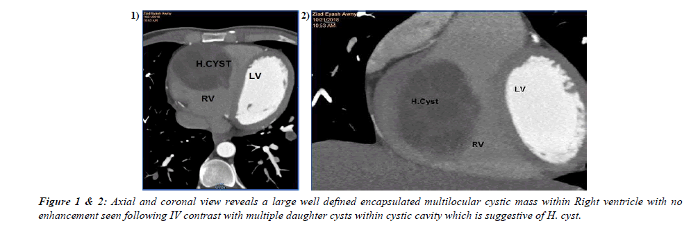
47 years old male patient presented with exertional shortness of breath. ECG was normal. The echocardiography study revealed enlarged RV size and round intracavitory mass in RV with moderate tricuspid regurgitation the patient referred for cardiac CT for further evaluation. The CT images revealed large multilocular non-enhancing cystic mass inside the mid RV measuring 70x50mm in size (figure 1&2).decision made for operation the cyst removed successfully without complications. The operative finding was consistent with the H. cyst.
Case no 2
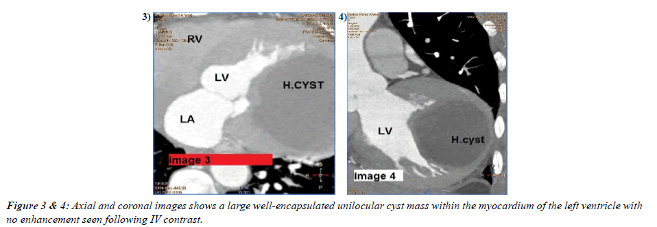
55 years old female patient presented with chest pain. ECG findings was T wave inversion in V3.V4.V5. echocardiographic study raveled mass in LV. Cardiac CT angiography revealed Large non-enhancing unilocular cystic mass localized to the lateral LV myocardial wall measuring (72mmx65mm) in caliber the patient underwent operation and the cyst removed successfully. The operative finding was consistent with the H. cyst.
Case no 3
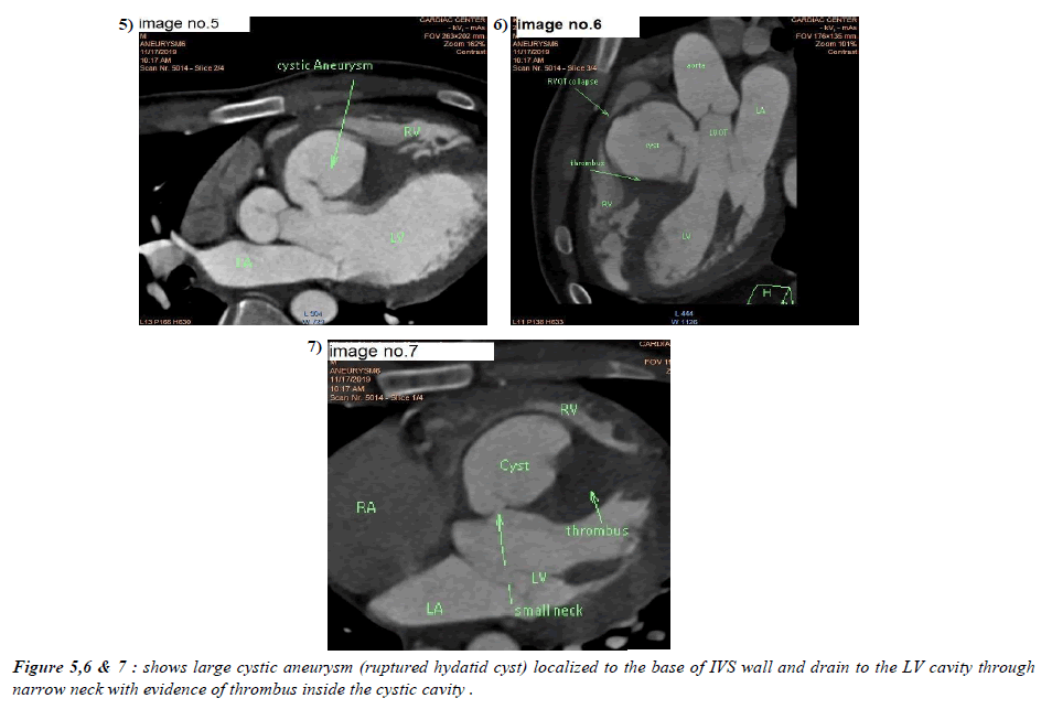
22 years old male patient presented with left side weakness and attacks of shortness of breath, brain MRI revealed ischemic stroke the patient had history of Operation for H. cyst in the liver before 2 years the patient referred for echocardiography examination to search for the underlying cardiac cause of the stroke. The echocardiographic study showed large aneurysmal pouch localized to the base of IVS wall and protruding to RV&RVOT, the mass was connected though a small neck to the LV cavity and the cyst lumen was partially filled by the thrombus. The patient referred for Cardiac CTA for further evaluation cardiac CT revealed large aneurysmal cystic mass localized to the basal portion of IVS wall and connected through small neck to LV cavity and the cystic cavity was partially filled by thrombus and out pouching toward the RV causing significant RVOT narrowing. Serological markers was positive for H. cyst the patient treated conservatively.
Discussion
Hydatid disease is highly endemic in Iraq and other Mediterranean countries cardiac involvement is rare [5]. The coronary circulation is the main pathway by which the parasitic larvae reach the heart. Because of a rich coronary blood supply, the left ventricle is the commonest site of involvement by cardiac H. cysts cardiac H. cysts are usually asymptomatic, especially in their early stages, and only 10% of patients have clinical symptoms. Symptoms depends on the location and size of the cyst [6,7]. In first case the patient presented with Dysponea the symptom most likely related to the large size of the cyst which occupying most of the RV cavity and causing RV overload with absence of other underlying causes that could be related to his dysponea, which has been excluded simultaneously by CT angiography like pulmonary embolism and other parachymal lung diseases. Findings of contrast cardiac CT was highly suggestive of H. cyst in this patient and positive serological test confirmed the diagnosis. In the second case the patient presented with chest pain because Cardiac H. cysts sometimes with intra-cavity expansion, result in local ischemia to myocardium by pressure effect and sometimes eroding into adjacent areas [8]. The findings of CT angiography was highly suggestive for H. cyst and confirmed with positive serological test for Hydatid dsease. The first 2 cases underwent open heart surgery cardiopulmonary bypass was used. All cysts were cleaned after quilting the cystic cavities, and daughter cysts were removed carefully. The cavities were closed with purse-string sutures. In all patients, mebendazole was administered postoperatively Cardiac hydatid cysts may lead to serious complications including cyst rupture anaphylactic shock, tamponade, pulmonary, cerebral or peripheral arterial embolism [9,10]. Hydatid cyst of the heart is a rare cause of embolization as its seen in the third case in which rupture of the cyst resulted cerebral embolization and stroke. Right sided cardiac hydatid cyst have a tendency to expand intracavitarily and subendocardially whereas left-sided cysts tend to grow sub epicardially and this is evident in our case reports [3].
Hydatid cyst of the interventricular septum is rare but it can lead to serious complications and this is evident in clinical history of third case report in which the patient developed stroke after rupture of the H. cyst in interventricular septum [9]. Diagnosis of H. cyst depend on the clinical history serologic test and imaging findings. The ELISA is one of the most specific serologic tests that can be used for diagnosis and a positive result for Echinococcus antibodies confirms the diagnosis [11]. Echocardiography is a non-invasive procedure which provides important findings on size and number of cysts, locations and relationships with adjacent structures [12]. CT or MRI should be used for localizing and defining the morphologic features of hydatid cysts. Specific signs include calcification of the cyst wall, presence of daughter cysts, and membrane detachment. CT best shows wall calcification, where as MRI depicts the exact anatomic location and nature of the internal and external structures in the diagnosis of these 3 case reports we depended on Cardiac CT angiography and positive serological tests [3].we preferred ECG gated cardiac CT for examination as there is less artifacts from cardiac motion and respiratory artifacts. MRI scan was not easily feasible for our patients and it has been not done. Surgery remains the treatment of choice in the management of hydatid disease. Although anthelminthic drugs have been used in the preoperative and postoperative periods since 1977, extirpation of the lesion under cardiopulmonary bypass is recommended [4,13].
Conclusion
Cardiac cyst is rare and patients may be presented with atypical symptoms or may be asymptomatic but it can present with serious complications. Diagnosis must be suspected in patients who live in endemic regions. The clinical history of our cases underlines the importance of early diagnosis and management of H. cyst and also the importance MSCT in the diagnosis as well as it is role in surgical planning and management.
Funding
The author(s) received no financial support for the research, authorship, and/or publication of this article.
Declaration of Conflicting Interests
The author(s) declared no potential conflicts of interest with respect to the research, authorship, and/or publication of this article.
Author Contributions
All authors contributed and reviewed the content of this manuscript.
References
- Chadly A, Krimi S, Mghirbi T. Cardiac hydatid cyst rupture as cause of death. Am J Fore Med Pathol. 2004;25(3):262-264.
- Eckert J, Deplazes P. Biological, epidemiological, and clinical aspects of echinococcosis, a zoonosis of increasing concern. Clin Microbo Rev. 2004;17(1):107-135.
- Dursun M, Terzibasioglu E, Yilmaz R, et al. Cardiac hydatid disease: CT and MRI findings. American J Roentg. 2008;190(1):226-232.
- Onursal E, Elmac? TT, Tirel? E, et al. Surgical treatment of cardiac echinococcosis: Report of eight cases. Surgery Today. 2001;31(4):325-30.
- Eckert J, Deplazes P. Biological epidemiological and clinical aspects of echinococcosis a zoonosis of increasing concern. Clini microbi Rev. 2004;17(1):107-35.
- Fennira S, Kamoun S, Besbes B, et al. Cardiac hydatid cyst in the interventricular septum: A literature review. Internat J Infe Dises. 2019;88:120-126.
- Niarchos C, Kounis GN, Frangides CR, et al. Large hydatic cyst of the left ventricle associated with syncopal attacks. Inter J Cardio. 2007;118(1):e24-e26.
- Ben-Hamda K, Maatouk F, Ben-Farhat M, et al. Eighteenyear experience with echinococcosus of the heart: Clinical and echocardiographic features in 14 patients. Internat J Cardio. 2003;91(3):145-151.
- Kaplan M, Demirtas M, Cimen S, et al. Cardiac hydatid cysts with intracavitary expansion. Annal Thora surg. 2001;71(5):1587-1590.
- Fennira S, Sarray H, Kammoun S, et al. A large cardiac hydatid cyst in the interventricular septum: A case report. Interna J Infec Dise. 2019;78:31-33.
- Ozer N, Aytemir K, Kuru G, et al. Hydatid cyst of the heart as a rare cause of embolization: Report of 5 cases and review of published reports. J Amer Soci Echocardi. 2001;14(4):299-302.
- Miralles A, Bracamonte L, Pavie A, et al. Cardiac echinococcosis: Surgical treatment and results. J Thor Cardi Surg. 1994;107(1):184-190.
- Gormus N, Yeniterzi M, Telli HH, et al. The clinical and surgical features of right-sided intracardiac masses due to echinococcosis. Heart Ves. 2004;19(3):121-124.
Indexed at, Google Scholar, Cross Ref
Indexed at, Google Scholar, Cross Ref
Indexed at, Google Scholar, Cross Ref
Indexed at, Google Scholar, Cross Ref
Indexed at, Google Scholar, Cross Ref
Indexed at, Google Scholar, Cross Ref
Indexed at, Google Scholar, Cross Ref
Indexed at, Google Scholar, Cross Ref
Indexed at, Google Scholar, Cross Ref
Indexed at, Google Scholar, Cross Ref
Indexed at, Google Scholar, Cross Ref


