Research Article - Journal of Trauma and Critical Care (2018) Volume 2, Issue 2
By adhering to NICE guidelines, could it be possible to avoid CT scan brain in patients presenting with head injury safely?
Syed Nouman Mohsin*57 Academy Square, Academy Street, Our Lady's Hospital, Navan, Co.Meath, Republic of Ireland
- *Corresponding Author:
- Dr. Syed Nouman Mohsin
57 Academy Square
Academy Street
Our Lady’s Hospital
Navan Co.Meath,
The Republic Of Ireland
Tel: 0892359954
E-mail: dr_syednouman31@hotmail.com
Accepted date: March 01, 2018
Citation: Mohsin SN. By adhering to NICE guidelines, could it be possible to avoid CT scan brain in patients presenting with head injury safely? J Trauma Crit Care. 2018;2(2):3-6
Abstract
Aims: An audit to assess the competency of NICE guidelines in patients presenting with head injury and if guidelines are followed is it possible to safely avoid doing unnecessary CT scan? Materials and Methods: Retrospective audit of all patients attending the hospital’s emergency department over a time period of one year with the head injury. Case notes and electronic needs were reviewed to determine whether the CT scan brain was indicated in line with the NICE guidelines. Results: Of 1187 CT scan brain request, 340 were for the head injury. 324 (95.3%) were reported as normal. 124(36.47%) were done as per criteria set by NICE for head injury and 216 (63.53%) were done against it. Conclusion: NICE guidelines are deemed competent in the management of head injury and can be safely followed. The A&E department can see a significant decrease in the number of CT scan brain request by following them.
Keywords
Audit, Head injury, Trauma, Competency.
Introduction
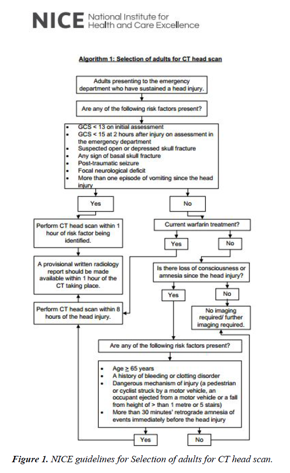
In June 2003, NICE published guidelines on Triage, Assessment, Investigation and Early Management of Head Injury in Infants, Children and Adults (Figure 1) [1,2]. The primary concern is clinically important head injury. 11 indications for CT are given, with the suggestion that CT should be completed within one hour of request in all but two of these indications. For these two exceptions, CT should be completed within eight hours of the injury. The present audit aimed to determine the competency of NICE guideline in the management of patients presenting with head injury and confident decision whether to opt for imaging (Computed tomography of brain) or discharge the patient by strictly following the guidelines as decided by national institute for clinical excellence for the head injury.
Methods
The retrospective audit was carried out at Our Lady’s Hospital Navan, Navan, Co.Meath, Ireland. The inclusion criteria was all the patients who attended the A&E with the head injury regardless of number of hours post trauma and type of head injury sustained above the age of 12. Exclusion criteria were children under the age of 12. We defined ‘‘head injury’’ as any trauma to the head except superficial injuries to the face. Scalp hematoma, age related changes, incidental finding of lesions in the brain, CT facial bone and their positive finding done and reported in conjunction with the CT brain were labeled as normal in this study as the sole purpose is to assess intracranial trauma. The audit covered a 12 months period strictly from 1st of January 2017 to 31st of December 2017. Firstly, data was collected from the radiology department of the patients who underwent CT scan brain in A&E [3]. After which, using the hospital’s medical record computer data base for the radiology (RIS) patient who underwent CT scan brain for the head injury were scanned and a generic research was conducted on age, sex, GCS at the time of presentation, Indication for undergoing CT scan brain [4,5], type of head injury sustained, whether there was any LOC or not, use of anticoagulants, time after the CT scan was reported and outcome of CT scan brain [6]. Data was entered in the Microsoft excel (Table 1) and further analysis was carried out with the help of IBM SPSS statistics trail version. We identified 340 cases and reviewed the case notes and electronic medical records to determine whether the CT had been requested within the existing NICEHI guideline.
Table 1. Headache with associated symptoms such as blurring of vision, nausea, dizziness, confusion, drowsiness, visible injuries and neck pain.
| CT scans brain that were done as per clinical indication set by NICE for head injury= 124(36.47%) | CT scans brain that were done against the criteria set by NICE for head injury=216(63.53%) |
|---|---|
| NICE 1 GCS <13 on initial assessment= 5 (1.47%) | Headache= 70 (20.59%) |
| NICE 2 GCS <15 at 2 hours after injury on assessment in the emergency department= 7(2.06%) | Headache with associated symptoms= 58(17.06%) |
| NICE 3 Suspected open or depressed skull fracture=1(0.29%) | Patients Qualified for NICE NEXT STEP in which clinical indication NICE 1 to 7 was not present but other risk factors not present as well, which include |
| NICE 4 Any sign of basal skull fracture=31(9.12%) | Age >65 years |
| NICE 5 Post-traumatic seizure=0 | A history of bleeding or clotting disorder |
| NICE 6 Focal neurological deficit=11(3.24%) | Dangerous mechanism of injury (a pedestrian or cyclist struck by a motor vehicle, an occupant ejected from a motor vehicle or a fall from height of > than 1 meter or 5 stairs) |
| NICE 7 More than one episode of vomiting since the head injury=12 (3.53%) | More than 30 minutes’ retrograde amnesia of events immediately before the head injury LOC ± 83(24.41%), Amnesia=5 (1.47%) |
| Ct Scans Brain That Were Done As Per Clinical Indication Set By NICE For Head Injury (Patients Qualified for NICE NEXT STEP in which clinical indication NICE 1 to 7 was not present, however, other risk factor present which include) | |
| Age >65 years | |
| A history of bleeding or clotting disorder | |
| Dangerous mechanism of injury (a pedestrian or cyclist struck by a motor vehicle, an occupant ejected from a motor vehicle or a fall from height of > than 1 meter or 5 stairs) | |
| More than 30 minutes’ retrograde amnesia of events immediately before the head injury | |
| LOC +ve= 22 (6.47%), Amnesia= 5(1.47%) and risk factor present such as Current Warfarin Treatment= 30 (8.82%) | |
Results
A total of 1187 CT scan brain request were made during the period reviewed, and there were 340 head injuries (male: 198 (58.25%), female: 142 (41.8%)). The age of the youngest patient who had CT scan brain due to head injury was of 14 years of age and the age of the oldest patient was 97 years old with a mean age of 46.65% and standard deviation of 22.023. 327 (96.2%) had the GCS of 15/15, 8 (2.4%) patients had the GCS of 14/15, 3 (0.9%) patients had the GCS of 13/15, 1 (0.3%) patient had the GCS of 11/15, 1 (0.3%) had the GCS of 7/15 at the time of presentation in the A&E after sustaining head injury. Out of 340 patients, 212 (62.4%) of the patients didn’t had LOC, while documented LOC was observed in 128 (37.6%) of the patients immediately after the head injury. Mechanical fall was the mechanism of head injury in 130 (38.2%) of the patients followed by RTA in 44 (12.9%) of the patients. Some of the patients sustained head injury as a result of collapsed 39 (11.5%) and as a result of assault 37 (10.9%). Head injuries due to sports were responsible for 32 (9.4%). Other less common causes include traumatic head injury due to farm animals, heavy object fall on head and seizure followed by head injury. 325 (95.5%) of the CT scan brain were reported as normal with pathologies identified in only 15 (4.41%) of the patients. Common pathologies identified were subdural hematoma in 5 (1.5%) patients, extradural hematoma in 2 (0.6%) patients, intracranial hemorrhage in 2 (0.6%) patients, surface hemorrhage in 2 (0.6%) patients, contusion in 2 (0.6%) patients and subgleal hematoma (0.3%) patients (Table 2).
Table 2.Positive yield in the number of ct scans that were done as per nice guideline, their age sex, clinical indication, loc +, -, gcs at the time of presentation, type of head injury susutained and abnormality found.
| Age | Sex | Clinical indication | Gcs at the time of presentaion | Type of head injury sustained | Loc + or - | Abnormality found |
|---|---|---|---|---|---|---|
| 25 | F | NICE 4 signs of basal skul fracture | 15/15 | sports | + | extra dural hematoma |
| 28 | M | NICE 7, more than one episode of vomiting | 15/15 | mechanical fall | - | intracranical hemorrhage |
| 87 | F | NICE 1, GCS less than 13 at initial assessment | 7/15 | mechanical fall | - | subdural hematoma |
| 36 | M | NICE 6 focal neurology | 15/15 | object fall on head | - | subgleal hematoma |
| 72 | F | NICE 7, more than one episode of vomiting | 15/15 | collapse and sustain head injury | - | hemarrahgic contusion |
| 86 | M | NICE 4, signs of basal skul fracture | 15/15 | mechanical fall | - | subdural hematoma |
| 97 | F | NICE 4, signs of basal skul fracture | 15/15 | mechanical fall | + | subdural hematoma |
| 35 | M | NICE 4, signs of basal skul fracture | 15/15 | assault | + | subdural hematoma |
| 62 | M | NICE 3, skull fracture | 15/15 | mechanical fall | - | extra dural hematoma |
| 76 | F | NICE 6 focal neurology | 15/15 | mechanical fall | + | intracranical hemorrhage |
| 18 | M | NICE 7, more than one episode of vomiting | 15/15 | assault | - | surface hemorrhage |
| 27 | F | NICE 6 focal neurology | 15/15 | mechanical fall | - | subdural hematoma |
| 38 | M | NICE 6, focal neurology | 15/15 | mechanical fall | + | depressed skull fracture |
| 18 | M | NICE 7, more than one episode of vomiting | 15/15 | assault | - | surface hemorrhage |
Discussion
In 2003, NICE issued a guideline for head injury management in the UK. These lowered the threshold for CT scanning of patients with mild head injury and sidelined the use of SXR [7,8]. A significant increase in rate of CT scanning was anticipated [9]. This study enabled us to compare the effectiveness of the NICE guideline in contrast to our current practice which render increased radiation exposure to patients and increased the economic burden on the radiology department of the hospital without any clinical justification and indication for doing the CT scan brain in head injury [10]. We divided the clinical indication of doing the CT scan brain in our study in to NICE and those that were done against the criteria set by NICE guidelines for the management of head injury and labeled as NOT NICE CRITERIA (Table 3). Both of them are further classified in to NICE 1 to 7 and if out of 7 clinical indication none of them are present so before putting the patient in to NOT NICE CRITERIA, patients were analyzed for next step in the NICE guideline and was labeled as NICE NEXT STEP, which include whether the patient is on current warfarin treatment, whether there was any loss of consciousness or not whether there was any amnesia or not and if there was whether any risk factors such as age greater than 65 years, history of bleeding or clotting disorder, more than 30 minutes retrograde amnesia or dangerous mechanism of injury is present or not. NOT NICE CRITERIA is further divided in to clinical indications headache and headache with associated symptoms [1] as these were the two major indications in NOT NICE CRITERIA for attending physicians to decide to opt for CT scan brain in patients presented with head injury [11,12].
Table 3. Positive yield in the number of ct scans that were done against nice guideline, their age sex, clinical indication, loc +, -, gcs at the time of presentation, type of head injury susutained and abnormality found.
| Age | Sex | Clinical indication | Gcs at the time of presentation | Type of head injury sustained | Loc | Abnormality found |
|---|---|---|---|---|---|---|
| 53 | M | not NICE crieteria exception | 15/15 | road traffic accident | - | hemorrhagic contusion |
Conclusion
The present audit compared the provision of CT imagining in head injuries patients. No patient with brain injury was missed with NICE guideline assuming that when it is not indicated by NICE guideline brain injury did not exist. In Our Lady’s Hospital Navan, there is clear cut evidence that by following NICE guideline in the management [13] of head injury can significantly decrease the economic burden on the hospital and can decrease the radiation exposure of patients [14,15].
Limitation
Out of 340 CT scans brains that were done in the ED of Our Lady’s Hospital Navan, there is a positive yield in one CT scan that was done against the criteria set by NICE guidelines. 53, Male presented 1 day after being involved in Road Traffic Accident in which he pounded his head with the steering of the car with headache and neck pain, LOC was negative, GCS was 15/15 underwent CT scan brain and hemorrhagic contusion was reported This is the only exception in the whole study.
References
- National Institute for Clinical Excellence. Head Injury. Triage, assessment, investigation and early management of head injury in infants, children and adults, Clinical Guideline 4. Developed by the National Collaborating Centre for Acute Care. London: NICE. 2003.
- National Institute for Health and Care Excellence. ‘Head injury’, NICE clinical guideline 176. London: National Clinical Guideline Centre. 2014.
- Stiell IG, Wells GA, Vandemheen K, et al. The Canadian CT Head Rule for patients with minor head injury. Lancet. 2001;357:1391-1396.
- Haydel MJ, Preston CA, Mills TJ, et al. Indications for computed tomography in patients with minor head injury. N Engl J Med. 2000;343:1570-1571.
- Ono K, Wada K, Takahara T, et al. Indications for computed tomography in patients with mild head injury. Neurol Med Chir (Tokyo) 2007;47(7):291-297.
- Abdul Latip LS, Ahmad Alias NA, Ariff AR, et al. CT scan in minor head injury: A guide for rural doctors. J Clin Neurosci. 2004;11:835-839.
- Cassidy JD, Carroll LJ, Peloso PM, et al. Incidence, risk factors and prevention of mild traumatic brain injury: Results of the WHO Collaborating Centre Task Force on Mild Traumatic Brain Injury. J Rehabil Med. 2004;43:28-60.
- Mack LR, Chan SB, Silva JC, et al. The use of head computed tomography in elderly patients sustaining minor head trauma. J Emerg Med. 2003;24:157-162.
- Shravat BP, Huseyin TS, Hynes KA. NICE guideline for the management of head injury: an audit demonstrating its impact on a district general hospital, with a cost analysis for England and Wales. Emerg Med J. 2006;23(2):109-113.
- Smits M, Dippel DW, de Haan GG, et al. Minor head injury: Guidelines for the use of CT – A multicenter validationstudy. Radiology. 2007;245(3):831-838.
- Miller EC, Derlet RW, Kinser D. Minor head trauma: Is computed tomography always necessary? Ann Emerg Med. 1996;27(3):290-294.
- Reinus WR, Wippold FJ 2nd, Erickson KK. Practical selection criteria for noncontrast cranial computed tomography in patients with head trauma. Ann Emerg Med. 1993;22(7):1148-1155.
- Stein SC, Ross SE. Minor head injury: A proposed strategy for emergency management. Ann Emerg Med. 1993;22(7):1193-1196.
- Miller EC, Holmes JF, Derlet RW. Utilizing clinical factors to reduce head CT scan ordering for minor head trauma patients. J Emerg Med. 1997;15(4):453-457.
- Shackford SR, Wald SL, Ross SE, et al. The clinical utility of computed tomographic scanning and neurologic examination in the management of patients with minor head injuries. J Trauma. 1992;33(3):385-394.
