Case Report - Otolaryngology Online Journal (2016) Volume 6, Issue 3
Bilateral Complete Congenital Choanal Atresia in an Adult Managed Endoscopically with Mucosal Flaps without Stenting
Sujata Apurva Gawai1* and Nishit J Shah Mail2
1Assistant Professor, Department of ENT, D.Y.Patil Medical College and Hospital, India
2Department of ENT, Bombay Hospital and Medical Research Centre, India
- *Corresponding Author:
- Sujata Apurva Gawai
Assistant Professor Department of ENT, D.Y.Patil Medical College and Hospital, Navi Mumbai, India
E-mail: drsujatagawai@gmail.com
Received date: April 09, 2016; Accepted date: May 19, 2016; Published date: May 23, 2016
Introduction
Choanal atresia is the developmental failure of the nasal cavity to communicate with the nasopharynx. Choanal atresia is a relatively rare congenital anomaly and occurs in approximately 1 in 5000 to 8000 live births, with a female to male ratio of 2:1 [1,2].
Generally, 65% to 75% of patients with choanal atresia are unilateral, whereas the rest are bilateral3. About 30% are pure bony, whereas 70% are mixed bony-membranous [2,4]. No patients were found to have a purely membranous atresia [5].
Fifty percent of all patients with choanal atresia and up to 75% of patients with bilateral disease have other associated congenital anomalies [5]. Bilateral choanal atresia is very rare in adults [6-8].
Bilateral choanal atresia will present as an acute respiratory emergency at birth as newborns are obligate nasal breathers. Placement of an appropriately sized oral airway usually resolves the distress. However, surgical correction is usually necessary early in life [5].
Case report
An 18 year old female presented to us with the complaints of total bilateral nasal blockage, bilateral nasal discharge and anosmia since birth. History revealed cyanotic attacks at rest and during feeding in childhood which were relieved on crying. Parents gave the history of evaluation of her nasal blockage and radiological investigation in neonatal period when she was diagnosed for bilateral complete choanal atresia and surgery was advised. However, parents refused to choose surgery at that time. Patient survived her neonatal period without any active management. She did not have remarkable complaints during early childhood or adolescence.
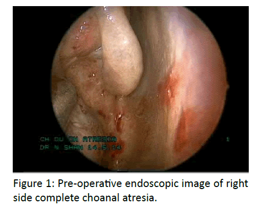
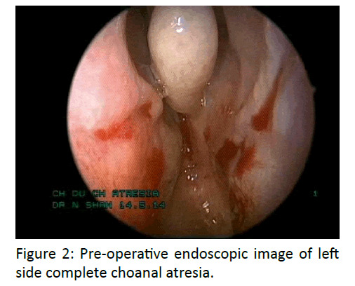
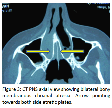
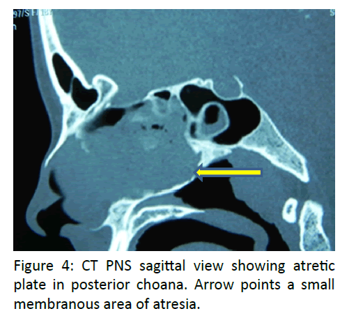
On clinical examination, thick tenacious secretion was present in both nostrils without any evidence of bubbling of nasal secretions with respirations. There was lack of movement of cotton wool wick on holding it in front of nostrils. No misting was seen on cold spatula test. Nasal endoscopy with 0 degree telescope revealed bilateral complete choanal atresia (Figures 1 and 2). Definite diagnosis of the condition was made on CT PNS, which showed a mixed bony membranous type of choanal atresia (Figures 3 and 4). There was no other associated congenital anomaly.
Procedure
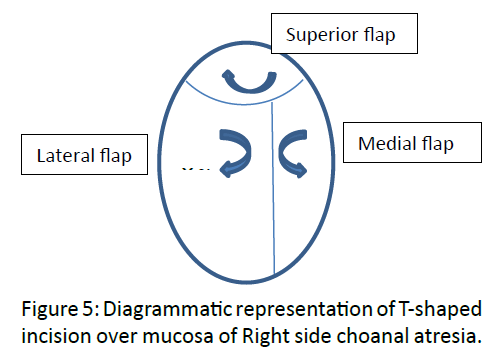
Patient underwent a trans-nasal endoscopic approach using a zero degree endoscope, under general anaesthesia with endotracheal intubation. Bilateral nasal cavities were full of thick secretions, after clearing the secretions, infiltration was given with 2% lignocaine with adrenaline on the mucosa over the anterior face of atretic area, lateral wall and posterior portion of septum. A T-shaped mucosal incision was taken with a pointed cautery tip, with the horizontal limb of the T concave upwards and vertical limb more towards septum. Mucosal flaps were raised superiorly, laterally and small flap medially (Figure 5).
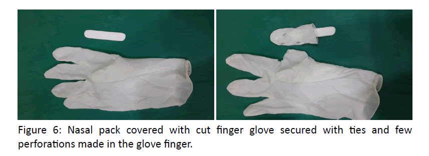
After lifting the flaps, bony atretic plate was visualised and only a small area in the centre of the atresia was membranous. The bone from the atretic plate, lateral nasal wall (medial aspect of pterygoid plate), and lateral portion of the posterior septum was drilled by microdebrider burr drill 15 degree, 3 mm rough diamond nasal burr. On the floor the bony atretic plate was drilled till the edge of hard palate. The mucosal flaps were replaced where the bony atresia was drilled so that the nasal cavity is in smooth continuity with the nasopharynx and there was no exposed bone. Same procedure was repeated on the other side. No stent or mitomycin C was applied as the nasal cavities were wide and there was no exposed bone left after replacing the raised mucosal flaps. The posterior end of septum and soft palate were normal. Bilateral glove finger nasal packs to make sure it’s easy removal were placed under endoscopic guidance taking care of mucosal flaps (Figure 6). Packs were removed after two days.
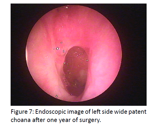
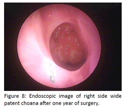
Postoperative recovery was uneventful. After pack removal patient complains of inability to speak loudly and nasal twang to voice, which was recovered in two days. Patient was adapted to breathing from nose within seven days. However, Sense of smell was absent. After endoscopic nasal cleaning, patient was taught to do nasal wash with normal saline after 15 days. She had been reviewed by endoscopy weekly for one month and thereafter, once in a month for 3 months. At the end of one year and two months bilateral choanae are as wide as it was intra operatively and well healed with normal nasal mucosa (Figures 7,8).
Discussion
There have been several theories regarding the embryogenesis of choanal atresia, but it is generally thought to be secondary to persistence of either the nasobuccal membrane of Hochstetter or the buccopharyngeal membrane from the foregut. This membrane normally ruptures between the fifth and sixth weeks of gestation to produce choanae. Failure of this membrane to rupture causes atresia of choanae [2,9].
Since a new-born child is an obligate nasal breather, respiratory distress and collapse occurs in patients with bilateral congenital choanal atresia immediately after birth. If not promptly recognized, it can lead to severe asphyxia and death. They usually present with similar pattern of cyclic cyanosis which is relieved by crying. Feeding difficulties may lead to failure to thrive. Most patients with bilateral congenital choanal atresia are detected in neonatal period. However, it can be diagnosed in adults with long-term bilateral nasal obstruction with nasal discharge [10].Very rarely neonates with complete Bilateral Choanal Atresia can gain the ability to breathe through the mouth [11].
Immediate management of bilateral choanal atresia involves training the infant to breathe through the mouth with the aid of an indwelling oral appliance such as a McGovern nipple or an oropharyngeal airway. If there is severe respiratory distress and airway cannot be established by endotracheal intubation, an emergency tracheotomy should be performed until further evaluation and treatment can be established. Nevertheless, surgical correction is usually necessary early in life [5].
Tests such as CT of the paranasal sinuses and nasal endoscopy are essential in diagnosis and therapeutic plan –to know the extent, location and distinctness of atresia bone, membrane, and mixed (bony membranous) [12].
Better instrumentation in endoscopic sinus surgery and advances in CT scans have made the trans-nasal repair the most popular method. Nasal endoscopy is beneficial in the management of choanal atresia since it helps to confirm the diagnosis, characterize the extent of lateral nasal wall contribution to the stenosis or atresia, evaluate the composition of the atresia (bony and/or membranous), and guide surgery [2].
There are several techniques: microscopic and endoscopic trans-nasal, trans-palatal, trans-septal, and dilations trans-antral intranasal [13-17].
A shift has occurred in the surgical philosophy over the last few decades away from trans-palatal and toward trans-nasal surgery5. The main advantage of the trans-nasal procedure is minimally invasive, quick (avoiding the need for prolonged anaesthetic agents), less traumatic with minimal blood loss, and provides excellent visualization and the ability to perform exact surgery on patients of all ages [2,18].
Richardson and Osguthorpe has compared transnasal repair with trans-palatal repair in 37 with congenital atresia or severe stenosis of both posterior nasal choanae. They found that trans-nasal repair allowed correction with minimal blood loss and without facial growth or occlusal abnormalities [19].
Paulo Tinoco et al in 2009, has reported a case of 34 year male managed with mucosal flaps without stent placement [20].
In our case also, we preferred to use mucosal flaps to cover the surgical area so as to prevent restenosis and to avoid stent placement. Stent and Mitomycin C were not used as the exposed bone was completely covered with the raised mucosal flaps. With the use of stent, there is a fear of foreign body reaction, skin necrosis in the columella, nasal alar erosion and requirement of intense antimicrobial therapy. We bypassed these by not using stents. At the end of one year bilateral choana are wide open with the surrounding healed mucosa.
Despite the nasal blockage was removed, and patient was breathing through nose, she continued to have anosmia. Gross-Isseroff et al. have studied the postoperative olfactory function at their case series of four of which 3 of them were Bilateral Choanal Atresia, and concluded that bilateral cases sustained olfactory deficit while unilateral case had the normal olfactory function [21].
Conclusion
Bilateral complete congenital choanal atresia is a life threatening condition with early diagnosis in new born due to its symptoms and high degree of suspicion by the paediatrician. It is very rarely found in adults. Our patient survived in neonatal period with bilateral complete congenital choanal atresia, without any management.
Careful review of the computed tomography scan and experience with endoscopic nasal surgery makes the trans-nasal endoscopic treatment a safe and effective approach for managing bilateral choanal atresia. Stenting and Mitomycin C are not necessary when the exposed bone is completely covered by raising adequate mucosal flaps. Management of choanal atresia with mucosal flaps alone has shown good results in our patient.
References
- Theogaraj SD, Hoehn JG, Hagan KF (1983) Practical management of congenital choanal atresia. Plast Reconstr Surg 72: 634-42.
- Paraya A, Choakchai M (2009) Choanal atresia. J Med Assoc Thai 92: 699-706
- Dobrowski JM, Grundfast KM, Rosenbaum KN, Zajtchuk JT (1985) Otorhinolaryngic manifestations of CHARGE association. Otolaryngol Head Neck Surg 93: 798-803.
- Brown OE, Pownell P, Manning SC (1996) Choanal atresia: a new anatomic classification and clinical management applications. Laryngoscope 106: 97-101.
- Elluru RG & Wootten CT (2010) Congenital malformation of the nose. Cummings Otolaryngology Head and Neck Surgery. 5th ed. Philadelphia: Mosby Elsevier; page 2694
- Panda NK, Simhadri S, Ghosh S (2004) Bilateral choanal atresia in an adult: is it compatible with life? J Laryngol Otol 118: 244-5.
- El-Sawy H, Siddiq MA, Anbarasu A (2006) Bilateral choanal atresia and paranasal sinus hypoplasia in an adult patient with hypogammaglobulinaemia. Eur Arch Otorhinolaryngol, 263: 1136-8.
- Yasar H, Ozkul MH (2007) Bilateral congenital choanal atresia in a 51- year-old woman. Am J Rhinol 21: 716-8.
- Dunham ME, Miller RP (1992) Bilateral choanal atresia associated with malformation of the anterior skull base: embryogenesis and clinical implications. Ann Otol Rhinol Laryngol 101: 916-9.
- Voegels RL, Chung D, Lessa MM, Lorenzetti FT, Goto EY, Butugan O (2002) Bilateral congenital choanal atresia in a 13-year-old patient. Int J Pediatr Otorhinolaryngol 65: 53-7
- Baker DC Jr, Waltner JG, Novick W (1960) Posterior choanal atresia. Ann Otol Rhinol Laryngol 69: 805-9
- Schweinfurth JM (2002) Image guidance-assisted repair of bilateral choanal atresia. Laryngoscope., 112: 2096-8.
- Lazar RH, Younes RT (1995) Transnasal repair of choanal atresia using telescopes. Arch Otolaryngol Head Neck Surg., 121: 517-20.
- Samadi DS, Shah UK, Handler SD (2003) Choanal atresia: A twenty-year review of medical comorbiditied and surgical outcomes. Laryngoscope 113: 254-8.
- Vogels RL, Chung D, Lessa MM, Lorenzetti FTM, Goto EY, Butugan O (2002) Bilateral congenital choanal atresia in a 13-year-old patient. Int J Pediatr Otorhinolaryngol. 65: 53-7.
- Josephson GD, Vickery CL, Giles WC, Gross CW (1998) Transnasal endoscopic repair of congenital choanal atresia. Arch Otolaryngol Head Neck Surg 124: 537-40.
- Cedin AC, Rocha JrFP, Deppermann MB, Manzano PAM, Murao M, Shimuta AS (2002) Transnasal endoscopic surgery of choanal atresia without the use of stents. Laryngoscope., 112: 750-52.
- Stankiewicz JA (1990) The endoscopic repair of choanal atresia. Otolaryngol Head Neck Surg 103: 931-7.
- Richardson MA, Osguthorpe JD (1988) Surgical management of choanal atresia. Laryngoscope; 98: 915-8.
- Paulo T, José Carlos Oliveira Pereira, Rodolfo Caldas Lourenço Filho. Bilateral Choanal Atresia in 34 Year-old Patients, Intl. Arch. Otorhinolaryngol., São Paulo - Brazil, v.14, n.4, p. 481-484, Oct/Nov/ December – 2010.
- Gross-Isseroff R, Ophir D, Marshak G, Ganchrow JR, Beizer M, Lancet D (1989) Olfactory function following late repair of choanalatresia. Laryngoscope 99: 1165-6.
